Clinical Significance
The prevalence of arrhythmias is expected to be 1.5% to 5% in the general population, with atrial fibrillation being the most common.[1] Arrhythmias may or may not produce any symptoms and can be paroxysmal, leading to difficulty in estimating true prevalence. The overall presence of arrhythmia is associated with higher morbidity and mortality.
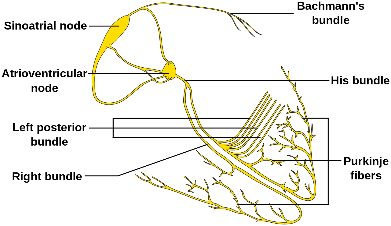
Evaluation of Arrhythmia
In patients suspected of arrhythmias, an electrocardiogram (EKG) is the first step and will usually give the diagnosis. However, at times, the patient may suffer from paroxysmal arrhythmia. The following modalities can be used for diagnosing based on the frequency of the symptoms a patient has secondary to a suspected arrhythmia.
- Ambulatory EKG monitoring for patients with frequent symptoms
- The event recorder needs to be triggered by the patient to record and will not a viable choice for patients with syncope.
- Loop event recorder records up to 2 minutes before the trigger. It is good for patients with syncope.
- Implantable loop recorder for patients with less frequent symptoms.
Tachyarrhythmia
Tachyarrhythmia is defined as an abnormal rhythm with a ventricular heart rate of 100 beats per minute or more. It can be further classified based on the origin of the arrhythmia into:
Supraventricular Tachycardia (SVT): Arrhythmia originating from above the AV node (from atrial origin or AV junction origin).
- Atrial fibrillation (AFib)
- Atrial flutter
- Atrial tachycardia
- Atrial premature complex (PAC)
- Atrioventricular nodal reentrant tachycardia (AVNRT)
- Atrioventricular reentrant tachycardia (AVRT)
- AV junctional extrasystoles
Ventricular Tachycardia (VT): The origin of the arrhythmia is below the AV node.
- Ventricular fibrillation (V-fib)
- Ventricular premature beats (PVC)
- Ventricular tachycardia (sustained or non-sustained)
Tachyarrhythmias can also be classified based on the QRS complex duration into:
Narrow QRS complex tachycardia when QRS is <120 milliseconds in duration:
- Sinus tachycardia
- Atrial tachycardia (AT)
- Atrial flutter
- Atrioventricular nodal reentrant tachycardia (AVNRT)
- Atrioventricular reentrant tachycardia (AVRT)
- Junctional ectopic tachycardia
- Sinoatrial nodal reentrant tachycardia (SANRT)
- Atrial fibrillation (irregular QRS complexes)
Wide QRS complex tachycardia (QRS ≥120 milliseconds in duration) is classified as monomorphic ventricular tachycardia, polymorphic ventricular tachycardia, or ventricular fibrillation.
Supraventricular Tachycardia Syndromes[2][3]
These are usually narrow complex tachycardias with QRS width being less than 3 mm or 120 milliseconds on the EKG strip. Supraventricular tachycardia is further classified into atrioventricular reciprocating tachycardia, atrioventricular nodal reentrant tachycardia, and atrial tachycardia based on the mechanism of tachycardia.
I. Atrioventricular reciprocating tachycardia (AVRT): As found in Wolff-Parkinson-White syndrome (the presence of delta wave without arrhythmia doesn’t require investigation or treatment).
Mechanism: Accessory pathway present outside of the AV node-Bundle of Kent. It can be further categorized into:
- Antidromic: Conduction down the accessory pathway and up to the AV node leading to the formation of a delta wave.
- Orthodromic: Conduction down the AV node into an accessory pathway with no delta wave.
Signs & Symptoms: Palpitation, shortness of breath, or syncope.
EKG Findings: Slurred upstroke of the QRS, delta wave, may give an impression of the wide QRS complex.
Management: Amiodarone or procainamide. If this fails, the next step is synchronized cardioversion.
Definitive Therapy: Ablation of the accessory pathway.
II. Atrioventricular Nodal Reentrant Tachycardia (AVNRT)
Mechanism: Slow & fast fibers present in AV node & peri-nodal tissue leading to re-entry.
Signs & Symptoms: Sudden tachycardia, palpitation, shortness of breath, chest tightness, or syncope.
EKG Findings: Narrow complex tachycardia with P waves hidden in T waves. Heart rate is in the range of 150-160 bpm.
Management:
Step 1: Carotid massage/Valsalva maneuver
Step 2: Adenosine
Step 3: Cardioversion
Step 4: Ablation or chronic suppressive therapy with beta-blockers and calcium channel blockers such as diltiazem/verapamil.
III. Atrial fibrillation: It is the most common arrhythmia in the United States. It affects more than 20% of the general population at some time in their lives.[4] There are five types based on their duration:[5]
- New-onset
- Paroxysmal: Self-terminating or intermittent
- Persistent: Fails to self-terminate within 7 days and requires treatments (medical or electrical cardioversion)
- Long-standing Persistent: Lasts for ≥ 1 year
- Permanent: Persistent for ≥ 1 year despite treatment
Mechanism: Multiple reentrant wavelets due to atrial ectopy from muscle fibers near the proximal part of the pulmonary vein.
Signs & Symptoms: It can be asymptomatic or can cause symptoms like palpitation, shortness of breath, irregularly irregular pulse, or even hypotension.
EKG Findings: Irregularly irregular narrow complex tachycardia with no discernable P-waves.
Management: The management strategy for atrial fibrillation can be classified into rate control or rhythm control. The decision to use a rate control or a rhythm-control strategy depends on the hemodynamic stability, candidacy for ablation, and the presence of co-morbidities. Patients with atrial fibrillation are at increased risk of ischemic-embolic stroke, and anticoagulation recommendations are based on the CHA2DS2VaSc score.
The CHA2DS2VaSc score is determined by the presence of the following factors: Congestive heart failure (CHF) with ejection fraction (EF) less than 40%, hypertension, age > 65 years, diabetes mellitus, history of stroke (non-hemorrhagic), or transient ischemic attack (TIA), vascular disease (peripheral vascular disease - PVD), age > 75 years, female sex. Each factor adds a point to the score, except for a history of stroke/TIA, which adds 2 points.
- If the score is 0: No anticoagulation or aspirin based on individual assessment
- If the score is 1: Aspirin or anticoagulation based on individual assessment.
- If the score is 2 or more: Anticoagulation is recommended if not at high risk of bleeding.
Rate Control Strategy: The heart rate goal is < 110 bpm in patients with chronic atrial fibrillation. It can be achieved with either beta-blockers or calcium channel blockers. Digoxin is usually used as adjuvant therapy in a patient with a "difficult to control" heart rate or in heart failure patients.
Cardioversion Strategy: Cardioversion is preferred in hemodynamically unstable patients or if rate control fails. It is also preferred in a young patient with no other co-morbidities. Cardioversion can be performed within 36 hrs of the onset of atrial fibrillation, but if the presentation is delayed or is of unknown duration, the absence of thrombi needs to be confirmed with a transesophageal echocardiogram (TEE). If a thrombus is present on an echocardiogram, the patient will need anticoagulation for at least three weeks before cardioversion can be performed. The patient needs to be on anticoagulation for at least four weeks post cardioversion. Various modalities are available for cardioversion therapy and include synchronized electric cardioversion or chemical cardioversion with medications including flecainide, propafenone, amiodarone, or dronedarone. Maze procedure is usually reserved for the patient undergoing other cardiac surgery.
Mechanism: Reentrant circuit usually around the tricuspid annulus in the right atrium.
Signs & Symptoms: Can be asymptomatic, or it can cause palpitation, shortness of breath, or hypotension.
EKG Findings: Regular tachycardia with Saw-tooth appearance of P wave with a variable degree of AV block.
Management: General goals include control of ventricular rate with AV blocking agents (beta-blockers or calcium channel blockers), but the restoration of sinus rhythm through cardioversion or ablation is preferred.
- Multifocal Atrial Tachycardia (MAT)
Mechanism: multiple automatic atrial foci due to increased sympathetic tone secondary to various causes, including hypoxemia (chronic obstructive pulmonary disease (COPD), or stimulant use.
Signs & Symptoms: Usually asymptomatic. Patients will have symptoms of the underlying illness, such as dyspnea.
EKG Findings: Three or more P wave morphologies with different PR intervals.
Management: Oxygen therapy if hypoxemic and treatment of the underlying cause.
Refractory cases: Rate Control with calcium channel blockers as the first choice in the setting of COPD followed by beta-blockers.
- Junctional tachycardia:[7] Arrhythmia originating from or near the AV node.
Mechanism: Rhythm arising from the AV node.
Risk Factors: Post cardiac surgery, myocardial ischemia (or during reperfusion), or digoxin toxicity.
Signs & Symptoms: Usually well-tolerated and asymptomatic.
EKG Findings: Inverted P Wave in the lead 2 with short PR or No P waves with a narrow complex.
Management: Treat the underlying cause.
IV. Ventricular Tachycardia: Origin is below the AV node. It is the major cause of sudden cardiac deaths in the United States.
a) Non-Sustained Ventricular Tachycardia:[8] When the rapid ventricular rhythm terminates on its own within 30 seconds.
Mechanism: Channelopathies secondary to structural abnormality, electrolyte disturbances, metabolic imbalance, and the effect of pro-arrhythmic drugs.
Risk Factors: Structural or ischemic heart disease.
Signs & Symptoms: Asymptomatic or palpitations.
EKG Findings: Monomorphic wide complex with more than three beats in a row but lasts less than three seconds.
Management: Implantable cardioverter-defibrillator (ICD) and/or medical therapy.
b) Sustained Ventricular Tachycardia
Mechanism: Presence of damaged fibers in ischemic heart disease leading to re-entry of current. Some patients do not have structural heart disease. Approximately 10% of the cases are idiopathic.
Risk Factors: Structural heart disease and post-myocardial infarction.
Signs & Symptoms: Palpitation, hypotension, or syncope.
EKG Findings: Monomorphic wide complex tachycardia.
Management: Intravenous lidocaine, amiodarone, or procainamide. Catheter ablation is an option too.
c) Ventricular Fibrillation
Mechanism: Presence of damaged fibers in ischemic heart disease leading to re-entry of current leading to disorganized high-frequency excitation. Patients with Cardiomyopathies can have Ventricular fibrillation due to an increase in end-diastolic pressure, wall tension, or the presence of abnormal channels in ventricular fibers.
Risk Factors: Structural heart disease and post-myocardial infarction.
Signs & Symptoms: Syncope and death if not treated immediately.
EKG Findings: Polymorphic fibrillatory waves.
Management: Unsynchronized cardioversion followed by amiodarone.
d) Torsades De Pointes:
Mechanism: It is usually precipitated by premature ventricular contraction leading to the “R on T phenomenon.”
Risk Factors: Congenital long QTc with hypokalemia and hypomagnesemia.
Signs & Symptoms: Syncope and death if not treated immediately.
EKG Findings: Polymorphic wide-complex tachycardia with a heart rate > 300 bpm.
Management: Intravenous magnesium or Isoproterenol, which increases heart rate and decreases QT-duration. Avoid hypokalemia and hypomagnesemia. Chronic therapy with beta-blockers in patients with long QT syndrome.
Bradyarrhythmias:[9][10] Bradyarrhythmia is defined as a heart rate below 60 beats per minute (bpm) and comprises several rhythm disorders, including atrioventricular (A-V) blocks and sinus node disorders.
Sinus Bradycardia
Mechanism: Increased vagal tone. It can be physiological in athletes.
Signs & Symptoms: Usually asymptomatic. It can lead to orthostasis or dizziness if pathological.
EKG Findings: Sinus rhythm with an upright P wave in lead II and biphasic in V1.
Management: No treatment is required unless pathological with an inadequate heart rate increase with leg raise test. Treat with isoproterenol or pacemaker if no relief.
Atrioventricular Blocks
Mechanism: Atrial impulses are conducted with a delay or not at all when an electrical impulse reaches a tissue that not excitable or is in a refractory period.
a) First Degree AV Block: Caused by increased vagal tone or conduction impairment or due to medications.
Signs & Symptoms: Generally asymptomatic but can cause dizziness.
EKG Findings: PR interval is greater than 200 milliseconds.
Management: Usually, no need to treat.
b) Second Degree AV Block: Further classified into Mobitz I block, where there is a progressive prolongation of the PR interval followed by a skipped beat, and Mobitz II block, where there is a randomly dropped QRS complex on an EKG.
Sign & Symptoms: Can be asymptomatic, dizziness, palpitations, weakness, syncope.
EKG Findings: Mobitz type I shows progressive prolongation of the PR interval followed by a dropped QRS complex or dropped beat. Mobitz type II has randomly dropped QRS complexes.
Management: Pacemaker is indicated in symptomatic Mobitz I and all of Mobitz II heart block.
c) Third Degree or complete AV Block
Mechanism: Lack of conduction of atrial impulse to ventricle leading to independent contractions.
Sign & Symptoms: Profound bradycardia, hypotension, and can lead to asystole and cardiac arrest.
EKG Findings: Bradycardia, P waves occur independently of QRS and Wide QRS for ventricular rhythm.
Management: Pacemaker placement.
Sinus Node Dysfunction
Mechanism: Senescence of the SA node, an ischemic event involving the SA node leading to impulse generation at a slower rate.
a) Sinus Pause: When the SA node has delayed impulse generation.
b) Sinus Arrest: Failure of impulse generation.
c) SA Nodal Exit Block: Failure of impulse transmission.
Sign & Symptoms: Bradycardia, dizziness, palpitation, or syncope.
EKG Findings: P wave not originating at a determined rate with regularity
Management: Symptomatic patients require pacemaker placement.