Continuing Education Activity
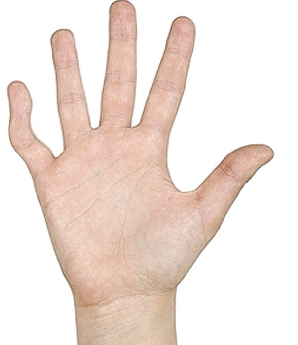
Clinodactyly is defined as a congenital curvature of a digit distal to the metacarpal phalangeal joint in the coronal plane. Curvatures with an angular deviation of fewer than 10 degrees can be seen as a normal physiologic variant. Clinodactyly is defined when the coronal angulation of the affected digit is greater than 10 degrees. The curvature arises due to the abnormal trapezoidal or triangular shape of one or more phalanges, which leads to malalignment of the associated interphalangeal joint. This abnormal shape leads to asymmetric longitudinal growth in a direction deviated from the normal longitudinal axis of the finger resulting in the visible curvature of the digit. This activity outlines the evaluation and management of clinodactyly and reviews the role of the interprofessional team in evaluating and treating patients with this condition.
Objectives:
- Identify the etiology of clinodactyly.
- Outline the appropriate and evaluation of clinodactyly.
- Review the management options available for clinodactyly.
- Describe the interprofessional team strategies for improving care coordination and communication to advance the management of clinodactyly and improve outcomes.
Introduction
Clinodactyly is defined as a congenital curvature of a digit distal to the metacarpal phalangeal joint in the coronal plane. Curvatures with an angular deviation of fewer than 10 degrees can be seen as a normal physiologic variant. Clinodactyly is defined when the coronal angulation of the affected digit is greater than 10 degrees. The curvature arises due to the abnormal trapezoidal or triangular shape of one or more phalanges, which leads to malalignment of the associated interphalangeal joint. This abnormal shape leads to asymmetric longitudinal growth in a direction deviated from the normal longitudinal axis of the finger resulting in the visible curvature of the digit.[1]
Etiology
Clinodactyly can occur in syndromic, familial, and sporadic forms.[2] The trait expresses autosomal dominant inheritance and most commonly involves the middle phalanx of the small finger. Several studies have also documented the involvement of the proximal phalanx of the index finger and thumb.[3] The curvature arises due to the abnormal trapezoidal or triangular shape of one or more phalanges, which leads to malalignment of the associated interphalangeal joint. This abnormal shape leads to asymmetric longitudinal growth in a direction deviated from the normal longitudinal axis of the finger resulting in the visible curvature of the digit.
Epidemiology
The incidence of clinodactyly is unknown; however, rates have been reported between 1% and 19.5%. It is often seen bilaterally, and males have been found to be more likely to express the trait. In has been associated with several syndromic anomalies, including Rubinstein-Taybi syndrome, Cenani-Lenz syndactyly, Klinefelter syndrome, Turner syndrome, Fanconi anemia and is present in up to 25% of children with Down syndrome. Clinodactyly is often present in the neonate; however, its presentation typically occurs in later childhood years as the deformity becomes more noticeable as longitudinal growth occurs. Patients present for treatment typically due to the abnormal appearance of the digit; however, the deformity rarely causes functional impairment due to compensatory abduction of the affected digit, which prevents the curvature from obstructing full digit flexion.
Pathophysiology
Longitudinal growth of the phalanges is driven from epiphyseal growth from the proximal portion of the phalanx. Abnormal configuration of the epiphyseal plate results in the curvature seen in clinodactyly. In clinodactyly, a C-shaped physis is often present and results in incomplete or restricted longitudinal growth on one side of the phalanx. In cases of early complete physeal plate ossification, no longitudinal growth will be possible, and a classic delta phalanx will be produced. The severity of the deformity can vary, and a classification system has been described.[4] Acquired cases of clinodactyly can be seen after traumatic injuries to the epiphyseal plate.[5] Cases of inflammatory and infectious arthritis can also cause longitudinal grown disturbances.[6]
History and Physical
History and physical exam will reveal the curvature of the affected digit in the radial/ulnar plane. It is essential to determine if the deformity affects the radial digits of the hand or if pinch function is compromised due to the deformity. Range of motion testing should be documented, and a complete history should reflect if the deformity is congenital or post-traumatic, stable or progressive.
Evaluation
Plain radiographs can aid in the diagnosis of clinodactyly. A radial or ulnar curvature will be present, and a shortened middle phalanx will often be seen, which is typically shorter on the radial rather than the ulnar side. A C-shaped physis or delta phalanx may also be present on plain radiographs.[7] Occasionally a prenatal ultrasound may identify clinodactyly.
Treatment / Management
The vast majority of cases can be treated conservatively with observation. Splinting of the affected digit has been found to be ineffective and is not recommended. Most presentations of isolated clinodactyly are due to aesthetic/cosmetic concerns, and functional concerns rarely arise. Surgery is for cosmetic correction is not advised due to the complications associated with surgical intervention, which include scarring and stiffness of the affected digit. Surgical intervention is reserved for severe clinodactyly with angulation and shortening, predominantly when there is the involvement of the thumb or radial digits, as severe deformities can often interfere with pinch and grasp.[1]
In cases of severe deformity that involve the radial digits or limit pinch, several different operative treatment options exist. Corrective osteotomy is the mainstay surgical treatment for correction of clinodactyly and, if possible, should be reserved until skeletal maturity is reached. Osteotomy can be challenging in the skeletally immature patient due to the risk of physeal injury, or excessive or incomplete correction. Options include opening or closing wedge osteotomy, reverse wedge osteotomy, and epiphyseal bracket resection and fat grafting. Opening wedge osteotomy requires bone grafting and allows for the preservation of length or the option to increase length; however, this requires a donor site that may be limited in a skeletally immature patient. Closing wedge osteotomy is an attractive option due to its simplicity; however, it typically results in some degree of shortening, which may be significant if the initial degree of shortening is substantial. The reverse wedge osteotomy is a technically challenging procedure when performed on a skeletally immature hand and requires access to both sides of the affected digit. In all cases of corrective osteotomy, the soft tissue deficit that is created with either lengthening or shortening of the digit must be addressed. Soft tissue deficits on the concave side can typically be treated with a "Z" -plasty. In more severe soft tissue deficits, rotational or advancement flaps may be necessary to gain adequate soft tissue coverage.[8]
In patients with open growth plates, epiphyseal bracket resection and fat grafting termed "Vicker's physiolysis" can be used as an alternative treatment option. This method of correction involves resecting the cartilaginous or osseous bracket on the short side of the affected digit and interposing in the created "dead space" with fatty tissue. After resection of the epiphyseal bracket and tissue interposition, this allows for the normal portion of the epiphyseal growth plate to generate and longitudinally correct the deformity over 1 to 2 years.[9] Results of Vicker's physiolysis, when compared with osteotomy, have been shown to be effective in patients with initial deformities less than 55 degrees and results in less revision surgery when compared to osteotomy alone.[10][11]
Differential Diagnosis
Differential diagnosis includes traumatic physeal injury, tumor, infection, and osteomyelitis. Clinodactlyly can be associated with several other medical syndromes including Rubinstein-Taybi syndrome, Cenani-Lenz syndactyly, and can be present in up to 25% of children with Down syndrome.
Prognosis
Expected outcomes following surgical intervention include the correction of the deformity.
Complications
Complications following surgical correction of deformity include stiffness, over and under correction of deformity, physeal arrest, skin deficits, and infection.[12]
Deterrence and Patient Education
In-depth discussion must be had with the patient and/or family explaining the risks and benefits of surgical intervention that must be reserved for patients with functional deficits, rather than aesthetic or cosmetic concerns.
Enhancing Healthcare Team Outcomes
An interprofessional team that provides a holistic and integrated approach to diagnosis and treating patients with clinodactyly is imperative. By correctly diagnosing and recognizing surgical indications for clinodactyly, primary care physicians can help avoid the added healthcare costs of possible unnecessary testing, advanced imaging studies, and consultations. The vast majority of cases of clinodactyly can be treated with observation. When more severe cases present, it is important to consult a surgical specialist for any surgical intervention that may be indicated.