Continuing Education Activity
Trendelenburg gait is an abnormal gait resulting from a defective hip abductor mechanism. The primary muscle group involved is the gluteal musculature, including the gluteus medius and minimus muscles. The weakness of these muscles causes drooping of the pelvis to the contralateral side while walking. The other causes will include any local hip pathology that may result in an impaired hip abduction and sagging of the contralateral hip.
This activity explores the multifaceted causes and underlying pathophysiology of Trendelenburg gait, showing how diverse hip and proximal femoral pathologies can contribute to this clinical presentation. Participants gain a thorough grasp of the intricacies surrounding Trendelenburg gait, gaining the knowledge needed for accurate diagnosis and effective management. Moreover, this educational activity extends beyond clinical insight, underscoring the vital role of interprofessional collaboration in managing patients with Trendelenburg gait. Healthcare professionals from various specialties must work cohesively to ensure patients receive well-coordinated care, optimizing their prospects for improved outcomes.
Objectives:
Identify and evaluate the pathophysiology of the Trendenlenburg gait.
Screen for conditions that may lead to a Trendenlenburg gait.
Assess and develop management strategies for a Trendenlenburg gait.
Implement effective collaboration and communication among interprofessional team members to improve outcomes and treatment efficacy for patients with a Trendenlenburg gait.
Introduction
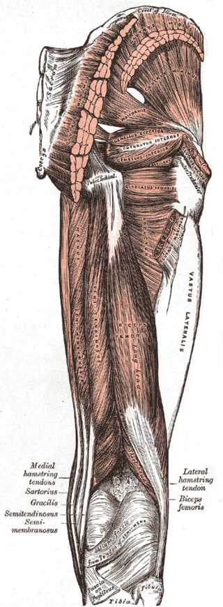
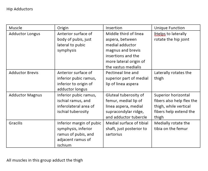
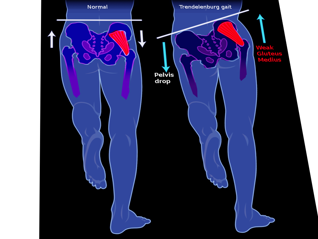
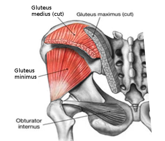
Trendelenburg gait is an abnormal gait resulting from a defective hip abductor mechanism. The primary muscle group involved is the gluteal musculature (see Image. Gluteus Muscles), including the gluteus medius and minimus muscles (see Image. Muscles of the Hip and Thigh and Image. Hip Adductors). The weakness of these muscles causes drooping of the pelvis to the contralateral side while walking. The other causes will include any local hip pathology that may result in impaired hip abduction and sagging of the contralateral hip (see Image. Trendelenburg Gait). The gait was named after a German surgeon, Friedrich Trendelenburg, who first reported the test related to this gait in 1895. The physical examination test he described helps uncover hip abductor weakness or pathology in a patient with developmental dysplasia of the hip.[1]
Etiology
The hip joint and its abductor mechanism behave like a class 1 lever with the effort and the load on opposing sides of the fulcrum.[2] Any pathology of the fulcrum, load, effort, or lever that binds all 3 will lead to a positive Trendelenburg sign and gait.
Failure of the fulcrum presents in the following conditions:
-
Osteonecrosis of hip
-
Legg-Calvé-Perthes disease
-
Developmental dysplasia of the hip
-
Chronically dislocated hips secondary to trauma
-
Chronically dislocated hips secondary to infections like tuberculosis of the hip
Failure of the lever is a feature in the following conditions:
Failure of effort (weakness of the abductor muscles) presents in the following conditions:
- Poliomyelitis
-
L5 radiculopathy
-
Superior gluteal nerve damage
-
Gluteus medius and minimus tendinitis
-
Gluteus medius and minimus abscess
-
Post-total hip arthroplasty
Epidemiology
Trendelenburg gait is a common problem in a patient who has paralysis/paresis of the hip abductors or issues with the hip joint, resulting in impaired abduction of the joint.
Pathophysiology
The center of gravity of the body passes midway through the pubic symphysis. When one foot lifts off the ground, as during the swing phase of the gait cycle, the body remains unsupported on that side, and the pelvis tends to drop to the unsupported side. To prevent the drop, the abductor muscles, mainly the gluteus medius and minimus on the supported side contract, providing stabilization of the superimposed trunk.[3] If there is any damage to the hip and its abductor mechanism due to the causes mentioned above, there will be drooping of the pelvis on the opposite side of the pathology. This drooping of the contralateral hip while standing is the positive Trendeelnburg sign. The repeated sagging of the contralateral hip during the stance phase of the abnormal side gives rise to the Trendelenburg gait.[4]
History and Physical
A patient with a Trendelenburg gait often complains of a limp. The limp can be painful or painless, depending on the etiology. If the limp is severe, there is compensatory bending or lurching to the side of the pathology to balance the body's center of gravity. This limp is called the lurching gait. Trendelenburg gait is observed when there is sagging of the contralateral (normal) hip during the stance phase of walking (swing phase of the normal sagging hip). This gait, when present, provides clues regarding specific possible etiologies, such as non-union of the neck of the femur and developmental dysplasia of the hip. When the cause is a weakness of the hip abductors and other hip muscles, as in most diffuse muscle diseases or myopathies, the pathology is more often bilateral; the pelvis sags to the unsupported side alternating with each step and is called a waddling gait.[5]
On physical gait examination, it is important to observe how a patient walks. Also watch normal walking, including the associated movements such as arm swing, the stance phase, and the swing phase. Normal walking should be followed by walking on toes, on heels, and heel-toe (tandem) walking. The patient may then be asked to stand from a squatting position, and the Romberg sign is tested at the same time as the gait. The lurching or waddling type of gait pattern should alert the physician to examine the abductor mechanism of the hip more closely.
Mild Trendelenburg gait may be difficult to appreciate while examining the patient with full clothing; therefore, it is necessary to perform a Trendelenburg test to evaluate further. To perform the test, the examiner sits or stands behind the patient. The patient is then asked to lift each foot off the ground, and the opposite side of the pelvis is elevated as high as possible alternately for at least 30 seconds.[6] This modification was suggested by Hardcastle et al and is now practiced worldwide as a standard practice. The unsupported side stays at the same level or rises slightly in healthy individuals. The pelvis drops towards the unsupported side when the abductor mechanism is weak. The patient leans towards the affected side in case of more serious weakness. This dropping of the pelvis in the standing position indicates a positive Trendelenburg test.[7][8]
The prerequisites for doing the test are as follows:
-
The patient should have a painless hip pathology. In a painful hip condition, the patient will not be able to balance, leading to spurious results.
-
The hip must not have abduction or adduction (coronal plane) deformities. Adductor deformity at the hip leads to elevation of the pelvis, leading to a false negative result. Abductor deformity at the hip leads to drooping of the pelvis on the contralateral side, leading to a false positive result.
Limitations of the Trendelenburg test:
-
Kendall et al have shown that hip abductor weakness induced by superior gluteal nerve block does not correlate with the pelvis drop, mainly in athletes and asymptomatic patients with hip pathology.[9]
-
In a patient with early stages of osteonecrosis, despite having an abductor mechanism defect, the Trendelenburg sign and gait remain masked.[10]
-
Pelvis drop can occur even in healthy individuals with normal abductor mechanisms when the abductor muscle is not working adequately.
Trendelenburg gait versus antalgic gait: patients with both Trendelenburg gait and antalgic gait walk with a limp. Differentiating the 2 from the history and physical examination is crucial. Antalgic gait is caused by pain on weight bearing of the affected side, whereas Trendelenburg is caused by impaired abductor mechanism of the hip. Antalgic gait is observed when the affected lower extremity has a shortened stance (weight-bearing) phase.
Evaluation
A detailed physical examination should be carried out to diagnose the condition leading to the abnormal gait. The main focus of the evaluation will be the hip joint, the upper femur, and the proximal hip muscles, especially the abductors. Other investigations include x-rays, ultrasonography, computed tomography scans, and magnetic resonance imaging to diagnose the primary condition. Blood tests may be performed to corroborate the radiological findings.
Treatment / Management
The main focus of the treatment after identification of the etiology involves correction of the etiological factor resulting in the Trendelenburg gait, which varies according to the pathology. Trendelenburg gait by itself wears the hip joint, and appropriate treatment is essential. Physical therapy is the mainstay treatment for gluteus medius and minimus weakness. Physical therapy involves strengthening the weakened hip abductor muscles. This involves lying on the unaffected side while abducting the affected leg towards the ceiling. A theraband can be applied on the lower limb to increase the resistance during the exercise. Other modalities include lateral side steeping and balance exercises.
Patients who have abductor weakness after an arthroplasty require specific exercises, which include:
-
Non–weight-bearing standing abduction
-
Weight-bearing standing abduction
-
Side-lying abduction
-
Resisted side-stepping exercises
Weight-bearing exercises have been shown to have a better functional recovery than non-weight-bearing exercises. Alteration of the gait pattern after gait training can also compensate for the hip abductor weakness.[11] Study results have shown that for gluteus medius tears in patients with hip abduction, manual muscle strength is less than four-fifths, along with gait dysfunction, and surgical intervention is likely indicated.[12]
Differential Diagnosis
Trendelenburg gait must be differentiated from other gait patterns, including the following:
- Functional gait disorder: this is a relatively common entity yet difficult to diagnose [13]
- Short limb gait
- Antalgic gait
- Extension lurch gait
- High stepping gait
- Stamping gait
A thorough history and physical examination are essential to rule out other abnormal gait patterns.
Prognosis
Trendelenburg gait can be treated efficiently, irrespective of etiology. Timely detection of altered gait, prompt diagnosis, and effective treatment of the primary condition are essential for a good outcome.
Complications
Chronic untreated abnormal Trendelenburg gait may lead to the development of secondary pathology at the knees or ankles over the years. This condition accelerates the process of wear and tear at the hip joint. The wear and tear usually occur at the portions of the hip, which are typically not used during normal gait, more so in athletes.[14][15] Untreated Trendelenburg gait can also lead to dynamic lower extremity valgus.[16] The mechanism behind this is that the pelvis of the unaffected side will be elevated, thereby having the trunk lean towards the affected side. The ground resultant force will, in turn, be directed to the lateral knee joint, producing a valgus knee on the affected side; this can lead to knee pain over time.
Consultations
Depending on the etiology, the following consultations may be indicated:
- Joint replacement orthopedic specialist
- Pediatric orthopedic specialist
- Occupational therapist
- Physical therapist
- Radiologist
Deterrence and Patient Education
Patients need to be aware of the Trendelenburg gait and other abnormal gait patterns and should seek medical treatment promptly, which can prevent irreversible damage to the hip joint.
Pearls and Other Issues
Other pertinent information regarding this condition includes:
- Pathology of Trendelenburg gait can be in the fulcrum, lever, or the effort arm of the hip abductor mechanism.
- The Hardcastle modification of Trendelenburg eliminates false-positive results.
- Trendelenburg gait causes wear and tear of the hip joint, so prompt diagnosis and treatment are essential.
Enhancing Healthcare Team Outcomes
A patient-centric and interprofessional approach is essential while managing a patient with a Trendelenburg gait. This team includes the primary care physician, orthopedic specialists, and nurses and may also include physical therapists and a chiropractor, all working in an interprofessional team approach. Patients must follow up with the therapist and orthopedic nurse for many months because recovery is gradual. For benign causes, full recovery is possible, but if there is permanent nerve damage, a persistent limp may develop.