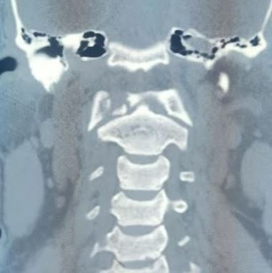
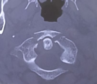
C1 Fractures

Earn CME/CE in your profession:


Continuing Education Activity

The craniocervical junction is comprised of C1 (atlas) and C2 (axis). The occipito-atlantal and atlantoaxial articulations provide 50% of the flexion and rotation in the cervical spine, respectively. Due to their high degree of motion, these bony segments are also the most often injured in adults. C1 fractures should be promptly identified and treated in all patients, although they rarely require surgery. Any bony fracture of the atlas merits a thorough examination of the ligamentous structures between O-C1 and C1-C2, which in comparison, often confer a poor prognosis. This activity reviews the etiology, presentation, evaluation, and management of C1 vertebral fractures and examines the role of the interprofessional team in evaluating, diagnosing, and managing the condition.

Objectives:

  • Describe the unique vertebral anatomy of the first cervical vertebra (atlas).
  • Discuss the components of a proper evaluation and assessment of a patient presenting with a potential first cervical vertebra fracture, including any indicated imaging studies.
  • Summarize the treatment and management options available for first cervical vertebra fractures based on the specific fracture type.
  • Explain the importance of interprofessional team strategies for improving care coordination and communication to aid in prompt diagnosis of C1 vertebral fracture and improving outcomes in patients diagnosed with the condition.

Introduction

Atlas has unique anatomy in that it sits just inferior to the occiput and, through the articulations with C2 and condyles of the occipital bone, joins the skull with the cervical spine. C1 is devoid of a vertebral body. It contains the anterior and posterior arches, which encircle the spinal cord (posteriorly) and the odontoid process (anteriorly). The arches are joined by lateral masses on either side, whose superior and inferior articular surfaces take part in the occipito-cervical and atlantoaxial joints, respectively. The atlantoaxial joint is highly mobile and is stabilized by the anterior atlantoaxial ligament (between the anterior ring of atlas and C2), transverse ligament (posterior to odontoid process), and posterior atlantoaxial ligament (between the posterior ring of atlas and C2). The transverse ligament contributes the most to the C1-C2 articulation among these ligaments. The atlas characteristically lacks a spinous process or vertebral body.[1] It behaves as an intercalated segment cradling the base of the occiput at the atlanto-occipital articulation allowing almost fifty percent of flexion-extension to occur at the neck.[2]

The craniocervical junction is comprised of the occiput, C1 (atlas), and C2 (axis). The occipito-atlantal and atlantoaxial articulations provide 50% of the flexion and rotation in the cervical spine, respectively. Due to their high degree of motion, these bony segments are also the most often injured in adults. C1 fractures should be promptly identified and treated in all patients, although they rarely require surgery. Any bony fracture of the atlas merits a thorough examination of the ligamentous structures between O-C1 and C1-C2.

Etiology

C1 fractures generally occur due to an axial loading mechanism, which can be combined with flexion/extension or rotation to create the typical fracture patterns. The area is subject to high stresses from a large moment arm of the cranium. Low-energy trauma in the elderly and high-impact trauma in young cohorts have been implicated in the pathogenesis of atlas fractures.[3][4]

Epidemiology

Atlas fracture comprises approximately 10 to 13% of all cervical fractures.[5][6] The annual incidence has been observed to increase by almost 700% and is estimated at 157 per million among elderly patients.[3] Bimodal distribution with peaks at approximately 30 and 80 years of age has been observed.[5][6] Most cases occur above 50 years of age.[3]

The median age of involvement has been observed to increase by 2.6 years annually.[5]  Only one-third of all atlas fractures are isolated.[4] C2 is the most common concomitant injury observed.[7]

Pathophysiology

Classification of atlas ring fractures as proposed by Gehweiler with further subdivision of Gehweiler type 3b fracture postulated by Dickman et al. is the most commonly utilized tool.[8]

Gehweiler classification of atlas fractures

  • Type 1: Fractures of the anterior arch of the atlas
  • Type 2: Fractures of the anterior arch of the atlas
  • Type 3: Fractures of both the anterior and posterior arches (Jefferson burst fracture)

                      3a: Ligamentous disruption

                      3b: Bony avulsion with an intact transverse atlantal ligament

  • Type 4: Fractures of the lateral mass of the atlas
  • Type 5: Isolated fractures of the transverse process of the atlas.[4]

 Dickman Classification of Transverse Atlantal Ligament (TAL) Injuries

  • Type 1: Intra-ligamentous rupture 
  • Type 1a: Central lesion
  • Type 1b: Lesion close to the lateral mass
  • Type 2: Bony avulsion injuries
  • Type 2a: Isolated bony avulsion
  • Type 2b: Bony avulsion associated with lateral mass fractures

Levine Classification of Atlas Fractures

  • Type 1: Isolated bony apophysis (transverse process) fracture
  • Type 2: Isolated posterior arch fracture
  • Type 3: Isolated anterior arch fracture
  • Type 4: Comminuted fracture of the lateral mass 
  • Type 5: Bilateral burst fracture (AKA Jefferson Fracture)

Fifty percent of atlas fractures involve a concomitant spine fracture, and there is also a 40% association with axis fractures.

History and Physical

Most atlas fractures result from significant trauma. History will often reveal an axial load injury to the cranium, including a dive into shallow water, a football tackle, or a motor vehicle collision with blunt cranial trauma. However, different patient populations, such as osteoporotic patients or patients with neuromuscular diseases, may be at increased risk.

Any cervical trauma exam must initiate with an assessment of the ABCs: airway, breathing, and circulation. If tracheal intubation is required, it should be done with manual in-line stabilization to avoid any displacement of fractures/dislocations. A thorough neurologic evaluation must be performed, starting with the Glasgow Coma Scale. All other traumatic injuries to other organs must be evaluated initially according to the Advanced Trauma Life Support (ATLS) protocol.

The physical exam must be detailed; one should take note of any axial neck pain or external signs of trauma to the cervical spine. A thorough neurologic exam should be performed, including cranial nerves, and a complete upper and lower extremity sensory and motor examination. In patients who may demonstrate signs of neurologic shock, a rectal exam and bulbocavernosus reflex should be tested.

C1 fractures usually present with axial neck pain with no evidence of neurologic dysfunction. When involved, cranial nerves in the medulla and pons are at risk due to their caudal location: cranial nerves VI to XII may result in their respective palsies. Fractures involving the C1/C2 transverse foramina can cause blunt vertebral artery injury (BVAI), resulting in basilar insufficiency; a thorough cranial neurologic exam should be performed. ROM should not be permitted prior to radiographic clearance.[9]

Evaluation

Gehweiler type three has been observed to be the most common subtype of atlas fracture.[4]

The rule of Spence showed that lateral mass displacement (LMD) over 6.9 mm on open-mouth radiographs correlated with rupture to the transverse atlantal ligament (TAL).[10][11]. LMD of more than 8.1 mm has high sensitivity and specificity for TAL injury.[12] Widening of the anterior atlanto-dental interval (ADI) to more than 3 mm in functional lateral X-rays is another important marker.[11] A C1:C2 ratio >1.10 on plain radiographs has been observed to have high sensitivity in detecting concurrent transverse ligament injury.[13]

There is a high risk of vertebral artery injury among cohorts with associated other injuries to the spine, increased atlantodental interval, and lateral mass displacement.[14] Vascular anatomy in relation to C1 is of paramount importance.[15] Fractures involving the transverse process and lateral mass impose a high risk of injury to the artery.[11]

Treatment / Management

Management strategies

  1. Surgery as a primary option
  2. Surgery as a secondary option after failed initial non-operative management
  3. Surgery involving the C1 level, with the main indication being a concomitant cervical spine fracture
  4. Non-operative strategy.[7]

The indication for surgery is mostly dictated by the concomitant subaxial cervical injuries.[4]

Gehweiler type 1, type 2, 3b, or type 5 are stable and managed non-surgically with the Minerva cast/Halo brace or collar for an average period of 8.5 weeks.[16][8]

Progressive diastasis at the fracture segment, as well as articular subluxation due to the lengthening of the transverse ligament (TAL), can occur.[17] Lateral displacement of the lateral mass with subluxation of the occipital condyle can occur and therefore needs to be assessed by periodic follow-up imaging.[4][7]

To distinguish between stable and unstable type 3 injuries, it is necessary to evaluate the integrity of the transverse atlantal ligament (TAL) with magnetic resonance imaging.[7]

Unstable atlas fractures (type 3b) with a moderately dislocated ligamentous bony avulsion of the TAL and sagittal split type 4 fractures may be treated by atlas osteosynthesis only.[11][8] Gehweiler type 4 fractures are preferentially managed by occipito-cervical instrumentation.[8]

C1 and subaxial instrumentations can be done through:

  • Lateral mass
  • C2 pars screw (less risk of vertebral injury)
  • Transarticular (Magerl)
  • Transpedicular/Pars(Harms and Goel)
  • Translaminar screws placements

C1 lateral mass-C2 translaminar and C1 lateral mass-C2/3 transarticular (C1LM-C2TL and C1LM-C2/3TACL) fixation resulted in satisfactory atlantoaxial stabilization compared with C1LM-C2 pedicular screw (Goel-Harms technique) are not feasible due to anatomical constraints.[18]

Sectioning or neurectomy of the C2 root provides better as well as minimizes operative time and blood. The subsequent occipital numbness has not been shown to significantly hinder the quality of life.[19]

The key anatomic landmark for C1 lateral mass screw placement:

  • Harms - at the inferior border of the posterior arch of C1 and the midpoint of the C1 lateral mass.[19]
  • Goel - at the center of the posterior surface of the lateral mass, 1 to 2 mm cranial to the C1-C2 facet.[20]

The screws are angled approximately 15 degrees medially and approximately 30 degrees cranially in Goel’s method. Harms method requires no angulation.[20] No clear consensus on the ideal angulation and location.[20]

Navigation facilitates accuracy and minimizes radiation exposure. However, the navigation system is costly; there is a learning curve associated with the setup and utilization.[20]

CT evaluation of the accuracy of free-hand C-1 lateral mass screws placement

  • Type I (ideal)-screw threads completely within the bone
  • Type II (safe)-less than half the diameter of the screw violates the surrounding cortex, and
  • Type III (unacceptable)-a clear violation of transverse foramen or spinal canal.[21]

97.2% of screws were rated Type I or II in one study.[21] Of the ten screws that were unacceptable, there were no known associated neurological or vascular injuries.

Anatomical landmarks, ideal trajectories, and screw lengths are the most important determinants for accurate instrumentation.[21] 3D-printed models can help in restoring Occipitocervical inclination.[22][23][24] No difference in the safety and accuracy between the free-hand and navigated techniques between the C2 pedicle and C2 pars screws has been observed.[25]

The posterior atlantoaxial or occipitocervical fusion is supposed to be the main surgical method, but it will result in a limited range of motion of the upper cervical spine.[10][26] Transoral anterior C1-ring osteosynthesis via systems such as Jefferson-fracture reduction plate (JeRP) connotes good bone union while preserving C1–C2 motion.[27][28][29] This also improves the integrity of the C0-C1-C2, the integrity of secondary stabilizers comprising the alar ligaments, facet capsule, and neck musculature, and restores the C0-C2 height.[10]

The primary indication for the JeRP system is an unstable C1 fracture (Gehweiler type I/III) with or without TAL injury (Dickman type II).[10] Similarly, posterior C1-ring osteosynthesis is superior to C1-2 fixation fusion in improving neck pain and movements.[30]This also has been shown to significantly reduce operative time, intraoperative blood loss, radiation dose, length of hospital stay, and economic burden.[30]

Differential Diagnosis

In the pediatric population, one should carefully distinguish fractures of the C1 vertebra from unfused ossification centers. Three ossification centers develop in an immature atlas, one for the anterior ring and one for each posterior neural arch. These centers appear at one year of age. The connection between anterior and posterior arches is composed of neurocentral synchondrosis, which fuses at seven years of age. The posterior arch usually closes by three years of age. These unfused ossification centers can mimic fractures and should be considered whenever children under six years present with cervical spine trauma.

Prognosis

The overall prognosis of C1 fractures is favorable, and on most occasions, conservative management provides adequate fracture healing. These fractures are associated with a very low incidence of neurological deficit, as the fracture fragments tend to be outwardly displaced rather than encroach on the neural canal. The spinal canal is also very spacious at this level. Associated bony and ligament (transverse ligament) injuries predominantly determine the prognosis and healing of these fractures.

Complications

Complications of Osteosynthesis

  • It is, however, cumbersome to reduce fractures within a deep and narrow space while undertaking the Transoral approach.
  • The end of the lateral mass screw may damage posterior the posterior pharyngeal wall, increasing the risk of wound complications or causing postoperative dysphagia.[26]

Complications of Posterior instrumentation

  • Egress of CSF following unintended durotomies.
  • Epidural and subdural cerebellar hematomas.
  • Injury to the vertebral artery and internal carotid artery.
  • Substantial bleeding from venous plexus between C1 and C2.
  • Venous air embolism.
  • Hardware complications and pseudo-arthrosis.[31]

Revision surgery was required in around 6% of cases in one, mostly due to surgical-site infection and instability.[4]

All-cause 30-day mortality was 12.2% in one study.[4] High Charlson Co-morbidity Index and higher age are associated with increased mortality, whereas concomitant C2 fracture was associated with improved survival.[6]

Postoperative and Rehabilitation Care

After applying a halo, regular X-rays are required to ensure the fracture is healing. The halo is often required for 8 to 16 weeks, depending on the rate of healing. Once the halo is removed, a collar is placed, and the patient needs to be enrolled in a rehabilitation program to regain muscle strength.

Deterrence and Patient Education

In general, a majority of C1 fractures are managed conservatively with some form of immobilization. But the patients should be aware of the need for regular follow-up as advised by the medical personnel and the possibility of needing surgical intervention if the conservative treatment fails. Smoking can prevent fracture healing and should be strictly avoided. Strict compliance with the collar is also recommended for fracture healing. 

Enhancing Healthcare Team Outcomes

C1 fractures are relatively common following trauma and involve patients of all ages. This fracture can be life-threatening, and all personnel in the emergency department or the trauma team must be aware of its presentation and management. While a neurosurgeon usually manages the primary pathology, the aftercare is generally performed by neurosurgery nurses and physiotherapists, operating as part of an interprofessional healthcare team. When promptly treated, the isolated C1 fracture has a good outcome. The small case series reveals that even after anterior plate fixation, the outcomes are good, with patients achieving a return to their pre-injury status. However, the outcomes for the elderly and young males are guarded. This population often suffers from a multiorgan injury and requires intubation and admission to the ICU. Even when discharged, some of these individuals have residual neurological deficits.[32][33]


(Click Image to Enlarge)
C1 arch fracture
C1 arch fracture
Contributed by Sunil Munakomi, MD

(Click Image to Enlarge)
C1 fracture
C1 fracture
Contributed by Sunil Munakomi, MD
Details

Author

Daniel Kim

Updated:

8/13/2023 2:54:08 AM

References


[1]

Forbes J,M Das J, Anatomy, Head and Neck, Atlantoaxial Joint StatPearls. 2022 Jan;     [PubMed PMID: 33085418]


[2]

Kaiser JT,Reddy V,Lugo-Pico JG, Anatomy, Head and Neck, Cervical Vertebrae StatPearls. 2022 Jan;     [PubMed PMID: 30969556]


[3]

Lyons JG,Mian HM, Epidemiology of atlas fractures in the United States: A 20-year analysis. Journal of craniovertebral junction     [PubMed PMID: 35386248]


[4]

Fiedler N,Spiegl UJA,Jarvers JS,Josten C,Heyde CE,Osterhoff G, Epidemiology and management of atlas fractures. European spine journal : official publication of the European Spine Society, the European Spinal Deformity Society, and the European Section of the Cervical Spine Research Society. 2020 Oct;     [PubMed PMID: 32002697]


[5]

Cloney MB,El-Tecle N,Dahdaleh NS, Traumatic atlas fracture patients comprise two subpopulations with distinct demographics and mechanisms of injury. Clinical neurology and neurosurgery. 2022 Aug 17;     [PubMed PMID: 35987045]


[6]

Matthiessen C,Robinson Y, Epidemiology of atlas fractures--a national registry-based cohort study of 1,537 cases. The spine journal : official journal of the North American Spine Society. 2015 Nov 1     [PubMed PMID: 26133259]

Level 3 (low-level) evidence

[7]

Ylönen H,Danner N,Jyrkkänen HK,Kämäräinen OP,Leinonen V,Huttunen J, Surgically Treated C1 Fractures: A Population-Based Study. World neurosurgery. 2021 Oct;     [PubMed PMID: 34284160]


[8]

Laubach M,Pishnamaz M,Scholz M,Spiegl U,Sellei RM,Herren C,Hildebrand F,Kobbe P, Interobserver reliability of the Gehweiler classification and treatment strategies of isolated atlas fractures: an internet-based multicenter survey among spine surgeons. European journal of trauma and emergency surgery : official publication of the European Trauma Society. 2022 Feb;     [PubMed PMID: 32918554]

Level 3 (low-level) evidence

[9]

Payabvash S,McKinney AM,McKinney ZJ,Palmer CS,Truwit CL, Screening and detection of blunt vertebral artery injury in patients with upper cervical fractures: the role of cervical CT and CT angiography. European journal of radiology. 2014 Mar     [PubMed PMID: 24355656]


[10]

Ma W,Xu N,Hu Y,Li G,Zhao L,Sun S,Jiang W,Liu G,Gu Y,Liu J, Unstable atlas fracture treatment by anterior plate C1-ring osteosynthesis using a transoral approach. European spine journal : official publication of the European Spine Society, the European Spinal Deformity Society, and the European Section of the Cervical Spine Research Society. 2013 Oct;     [PubMed PMID: 23775293]


[11]

Kandziora F,Scholz M,Pingel A,Schleicher P,Yildiz U,Kluger P,Pumberger M,Korge A,Schnake KJ,Spine Section of the German Society for Orthopaedics and Trauma., Treatment of Atlas Fractures: Recommendations of the Spine Section of the German Society for Orthopaedics and Trauma (DGOU). Global spine journal. 2018 Sep;     [PubMed PMID: 30210964]


[12]

Eun J,Oh Y, The relationship between radiologic parameters and transverse atlantal ligament injury obtained from MRI scans in patients with an isolated atlas burst fracture: A retrospective observational study. Medicine. 2021 Dec 10;     [PubMed PMID: 34889272]

Level 2 (mid-level) evidence

[13]

Lin P,Chuang TC,Baker JF, C1:C2 ratio is a potential tool assessing atlas fracture displacement and transverse ligament injury. Journal of craniovertebral junction     [PubMed PMID: 31772425]


[14]

Cloney M,Kim H,Riestenberg R,Dahdaleh NS, Risk Factors for Transverse Ligament Disruption and Vertebral Artery Injury Following an Atlas Fracture. World neurosurgery. 2021 Feb;     [PubMed PMID: 33309644]


[15]

Rusconi A,Peron S,Roccucci P,Stefini R, The internal carotid artery and the atlas: anatomical relationship and implications for C1 lateral mass fixation. Surgical and radiologic anatomy : SRA. 2021 Jan;     [PubMed PMID: 32734343]


[16]

Tomaszewski R,Sesia SB,Studer D,Rutz E,Mayr JM, Conservative treatment and outcome of upper cervical spine fractures in young children: A STROBE-compliant case series. Medicine. 2021 Apr 2;     [PubMed PMID: 33787631]

Level 2 (mid-level) evidence

[17]

Starnoni D,Ecker T,Barges-Coll J, Navigation-Assisted Posterior Open Reduction and Internal Fixation in a C-CLAMP Fashion for an Isolated C1 Fracture. Journal of neurological surgery. Part B, Skull base. 2021 Feb;     [PubMed PMID: 33692933]


[18]

Du YQ,Li T,Ma C,Qiao GY,Yin YH,Yu XG, Biomechanical evaluation of two alternative techniques to the Goel-Harms technique for atlantoaxial fixation: C1 lateral mass-C2 bicortical translaminar screw fixation and C1 lateral mass-C2/3 transarticular screw fixation. Journal of neurosurgery. Spine. 2020 Jan 17;     [PubMed PMID: 31952043]

Level 2 (mid-level) evidence

[19]

Conroy E,Laing A,Kenneally R,Poynton AR, C1 lateral mass screw-induced occipital neuralgia: a report of two cases. European spine journal : official publication of the European Spine Society, the European Spinal Deformity Society, and the European Section of the Cervical Spine Research Society. 2010 Mar;     [PubMed PMID: 19856190]

Level 3 (low-level) evidence

[20]

Butt BB,Gagnet P,Piche J,Patel R,Park P,Aleem IS, Lateral mass screw placement in the atlas: description of a novel surgical technique, radiographic parameters, and review of the literature. Journal of spine surgery (Hong Kong). 2021 Sep;     [PubMed PMID: 34734138]


[21]

Hu Y,Kepler CK,Albert TJ,Yuan ZS,Ma WH,Gu YJ,Xu RM, Accuracy and complications associated with the freehand C-1 lateral mass screw fixation technique: a radiographic and clinical assessment. Journal of neurosurgery. Spine. 2013 Apr;     [PubMed PMID: 23373564]


[22]

Rajasekaran S,Soundararajan DCR,Shetty AP,Kanna RM, Motion-Preserving Navigated Primary Internal Fixation of Unstable C1 Fractures. Asian spine journal. 2020 Aug;     [PubMed PMID: 32050311]


[23]

Barrenechea IJ,Márquez L,Bruna AE, Unilateral C1 split fracture osteosynthesis using a patient-specific three-dimensional-printed guide: Technique report. Journal of craniovertebral junction     [PubMed PMID: 35068828]


[24]

Wu C,Deng JY,Li T,Zeng BF,Hu HG,Zhu YF,Wei Q, 3D-Printed Screw-Rod Auxiliary System for Unstable Atlas Fractures: A Retrospective Analysis. Orthopaedic surgery. 2021 May;     [PubMed PMID: 33826254]

Level 2 (mid-level) evidence

[25]

Azimi P,Yazdanian T,Benzel EC,Aghaei HN,Azhari S,Sadeghi S,Montazeri A, Accuracy and safety of C2 pedicle or pars screw placement: a systematic review and meta-analysis. Journal of orthopaedic surgery and research. 2020 Jul 20;     [PubMed PMID: 32690035]

Level 1 (high-level) evidence

[26]

Zou X,Ouyang B,Ma X,Chen Y,Ge S,Zhang S,Ni L,Xia H,Wu Z, [Progress in treatment of unstable atlas fracture]. Zhongguo xiu fu chong jian wai ke za zhi = Zhongguo xiufu chongjian waike zazhi = Chinese journal of reparative and reconstructive surgery. 2020 Jun 15;     [PubMed PMID: 32538574]


[27]

Zou X,Ouyang B,Wang B,Yang H,Ge S,Chen Y,Ni L,Zhang S,Xia H,Wu Z,Ma X, Motion-preserving treatment of unstable atlas fracture: transoral anterior C1-ring osteosynthesis using a laminoplasty plate. BMC musculoskeletal disorders. 2020 Aug 12;     [PubMed PMID: 32787814]


[28]

Tu Q,Chen H,Li Z,Chen Y,Xu A,Zhu C,Huang X,Ma X,Wang J,Zhang K,Yin Q,Xu J,Xia H, Anterior reduction and C1-ring osteosynthesis with Jefferson-fracture reduction plate (JeRP) via transoral approach for unstable atlas fractures. BMC musculoskeletal disorders. 2021 Aug 30;     [PubMed PMID: 34461878]


[29]

Shin JW,Suk KS,Kim HS,Yang JH,Kwon JW,Lee HM,Moon SH,Lee BH,Park SJ,Park SR,Kim SK, Direct Internal Fixation for Unstable Atlas Fractures. Yonsei medical journal. 2022 Mar;     [PubMed PMID: 35184429]


[30]

Yan L,Du J,Yang J,He B,Hao D,Zheng B,Yang X,Hui H,Liu T,Wang X,Guo H,Chen J,Wang S,Ma S,Dong S, C1-ring osteosynthesis versus C1-2 fixation fusion in the treatment of unstable atlas fractures: a multicenter, prospective, randomized controlled study with 5-year follow-up. Journal of neurosurgery. Spine. 2022 Feb 11;     [PubMed PMID: 35148517]

Level 1 (high-level) evidence

[31]

Macki M,Hamilton T,Pawloski J,Chang V, Occipital fixation techniques and complications. Journal of spine surgery (Hong Kong). 2020 Mar;     [PubMed PMID: 32309653]


[32]

Bhimani AD,Chiu RG,Esfahani DR,Patel AS,Denyer S,Hobbs JG,Mehta AI, C1-C2 Fusion Versus Occipito-Cervical Fusion for High Cervical Fractures: A Multi-Institutional Database Analysis and Review of the Literature. World neurosurgery. 2018 Nov     [PubMed PMID: 30071333]


[33]

Purvis TE,De la Garza-Ramos R,Abu-Bonsrah N,Goodwin CR,Groves ML,Ain MC,Sciubba DM, External fixation and surgical fusion for pediatric cervical spine injuries: Short-term outcomes. Clinical neurology and neurosurgery. 2018 May     [PubMed PMID: 29505977]