Ingrown Toenails

Earn CME/CE in your profession:


Continuing Education Activity

An ingrown toenail is a common nail problem that occurs when the nail plate grows into the periungual skin and causes inflammation and infection. If left untreated, it can cause considerable pain, discomfort, and disability. This activity will review the etiology, pathophysiology, presentation, and management options for ingrown toenails.

Objectives:

  • Identify the etiology of ingrown toenails.

  • Assess the different stages of ingrown toenails.

  • Determine the treatment and management options available for ingrown toenails.

  • Communicate interprofessional team strategies to improve coordination and communication between health care professionals, improving patient outcomes.

Introduction

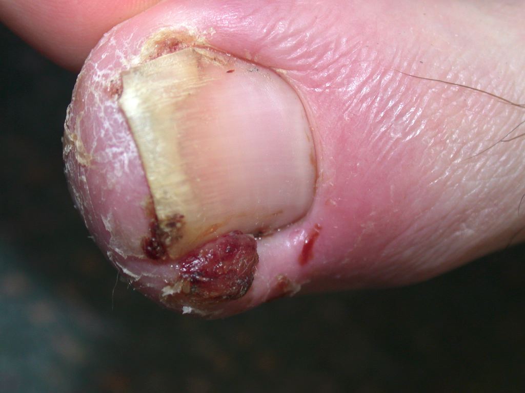
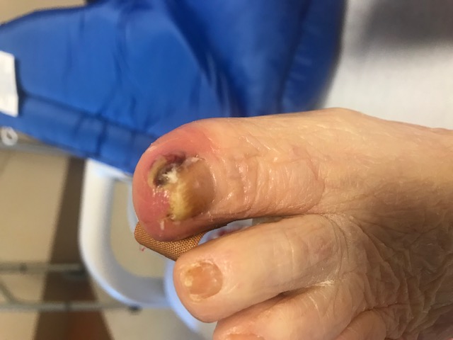
Ingrown toenail, also known as onychocryptosis or unguius incarnatus, is the most common nail problem encountered in podiatry, general family practice, and dermatology.[1]  An ingrown toenail occurs when the nail plate grows into the periungual skin and causes inflammation and infection. It causes considerable pain, discomfort, and disability if left untreated. An ingrown toenail may present at any age, but most commonly, it affects teenagers and young adults (See Image. Ingrown Toenail). The hallux nails are the most frequent location.[2] Management options range from conservative treatments to extensive surgical approaches, depending on the severity and the stage of the condition (See Image. Ingrowing Nail, Medial Border of the Left Hallux).

Etiology

Improper nail trimming appears to be the most common etiology of ingrown toenails, as it may lead to a nail spike that traumatizes adjacent soft tissue.[2][3] Other predisposing factors for ingrown toenails include tight-fitting shoes, bad foot hygiene, hyperhidrosis, trauma, and some medications, especially epidermal growth factor receptor inhibitors (gefitinib, cetuximab).[4]

The possible involvement of intrinsic risk factors in the pathogenesis of ingrown toenails, such as abnormal nail shape and anatomical abnormalities, has been widely debated. Some studies have found that pincer-nail deformity, wide nail plates, congenital malalignment of the toenails, and thickening of the nail plate represent possible risk factors for ingrown toenails.[3][5]

Other studies proved that bone abnormalities that increase the internal pressure in the ankle, foot, or toe might play a role in developing ingrown toenails.[1][6] However, more recent reports suggest no difference in anatomical abnormalities between patients with ingrown toenails and controls.[7]

Epidemiology

There have been few investigations of the epidemiology of ingrown toenails. Previous reports have noted that the prevalence was 2.5% to 5% [6]. The incidence and prevalence of ingrown toenails have increased in recent years, probably due to increased health knowledge, and may also be related to lifestyle changes such as important physical activities. Incidence peaks in adolescents and young adults with a clear male predominance (male/female ratio is approximately 2 to 1). Ingrown toenail affects almost exclusively the hallux toenails. It can occur in 1 or both nail edges. Involvement of the lateral toe edge occurs twice as much as on the medial side.[6][8]

Pathophysiology

Many theories have been proposed to explain the onset of ingrown toenails. One assumption is that it is related to excess skin surrounding the nail, which is the real culprit. It is explained by wide lateral tissue tending to bulge around the nail leading to pressure and necrosis.[9] However, the most accepted theory is that an ingrown toenail occurs when the nail plate edge grows into the overlapping lateral nail fold, causing painful inflammation and forming granulation tissue.[8][9] Reactive ground forces due to athletic activity, obesity, and/or constricting footwear aid this penetration.[2]

History and Physical

An ingrown toenail is a straightforward diagnosis. Almost all patients present with toe pain. This pain may cause different levels of discomfort and disability, ranging from simple difficulty with walking to a complete inability to ambulate.[8] Physical examination findings may vary depending on the stage of the disease.

The initial presentation, corresponding to stage 1, is characterized by signs of inflammation in the affected toe: pain, swelling, and erythema. The initial stage is followed by an acute infection with seropurulent drainage and ulceration of the nail fold, causing more edema and tenderness (stage 2). Chronic infection leads to the formation of hypertrophic granulation tissue, which increases the compression and thus adds to the swelling and discharge (stage 3).[10][11]

Evaluation

The diagnosis of an ingrown toenail is classically based on clinical features and does not require any laboratory or radiographic tests.

If physical examination reveals a subungual nodule, an X-ray examination may be needed to rule out subungual exostosis. In such cases, it reveals a subungual bony proliferation. 

Treatment / Management

Treatment methods for ingrown toenails range from medical measures to surgical interventions. Indications for the treatment depend mainly on the stage of the condition, prior treatment modalities in case of recurrence, and other factors, including allergies to local anesthetics, pregnancy, and bleeding disorders. Conservative measures are generally recommended in mild to moderate lesions (stages 1 and 2), whereas severe lesions causing disability require surgical methods (stage 3).[8][9][10][12]

General Measures

General measures for ingrown toenails include proper footwear and correct nail trimming; this includes avoiding curved cutting off the lateral margins of the nail plate. General measures should also include management of the underlying factors (hyperhidrosis, onychomycosis). Soaking the affected toe in warm, soapy water or Epsom salt water for several minutes, followed by topical antibiotic ointment, may give relief. The application of topical steroids to hypertrophic granulation tissue may decrease inflammation.[9][13]

Conservative Techniques

  • Cotton-wick insertion under the nail's corner:  Cotton wisp or pledget are placed under the ingrown lateral groove corner using a nail elevator.[14]
  • Dental floss technique: This is an alternative to cotton wisps. A string of dental floss is inserted under the ingrown nail to separate it from the lateral fold.[15]
  • The gutter splint or sleeve technique: Gutter strips are prepared by cutting to size vinyl intravenous infusion tube from top to bottom. The lateral edge of the nail plate gets splinted with this sterilized splint plastic tube and then attached with adhesive tape or strips, giving instant pain relief.[16][17]
  • Taping procedure: One end of the tape gets placed against the side of the ingrown toenail, with the rest twisted around the toe. The aim is to pull the side of the nail fold away from the nail to decrease pressure. Taping is the safest and least painful procedure among conservative options.[18]
  • Nail wiring: Two holes are made at the distal edge of the nail, and an elastic wire is inserted and bent forward. The wire's elasticity may correct the deformity of the ingrown toenail. 
  • Others: slit tape-strap procedure, acrylic nails, nail braces.[8][9][19]

Surgical Techniques

Surgical procedures for ingrown toenails are performed under local anesthesia (LA). There are various techniques for LA, including digital block, metatarsal block, or transthecal anesthesia. Any local anesthetic can be used (lidocaine, ropivacaine, mepivacaine, or prilocaine) in 0.25% to 2% concentrations. Indications for LA depend mainly on the type of surgery and the physician’s preference.

Surgical options for ingrown toenails are: 

  • Spicule excision and partial matricectomy: It consists of excising the affected portion of the nail with a partial mechanical matricectomy.[20]
  • Chemical partial matricectomy: Chemical matricectomy is commonly performed using phenol. It demonstrates a higher success rate and is less painful than mechanical matricectomy. Its success depends on good hemostasis. Other chemical agents, such as sodium hydroxide and trichloroacetic acid, can be employed.[21]
  • Wedge resection of the toenail and nail fold: This approach consists of the excision of the affected portion of the nail plate, partial matricectomy, and wedge dissection of the nail bed and the hypertrophic nail fold. Clinicians should generally avoid this technique. 
  • Excision of the affected nail and total matricectomy: This is a more radical solution to ingrown toenails. It consists of the affected nail and nail bed excision and a total matricectomy (chemical or mechanical). It is indicated for stage IV ingrown toenails, onychogryphosis, and onychodystrophy.
  • Soft-tissue nail fold excision technique: This procedure does not touch the nail as its basis is the theory that the nail is not the causative factor in developing ingrown toenails. It consists of a wide excision of the soft tissue enveloping elliptically.[22][23]
  • Other techniques: Newer techniques, including electrocautery, radiofrequency ablation, and carbon dioxide laser ablation, have become the newest form of ingrown toenail management.[3][9][24]

Thus, surgical treatment options for ingrown toenails are numerous, and there is no consensus on the technique of choice. The ideal procedure should lead to the best functional and aesthetic outcome, as well as a low rate of recurrence. Many studies have proven that simple nail avulsions lead to high recurrence rates, while phenol matricectomy has shown greater success.

Differential Diagnosis

Differential diagnosis includes mainly subungual exostosis and tumors of the nail bed (benign and malignant). If suspected, an X-ray examination that reveals the subungual bony proliferation can confirm the diagnosis of subungual exostosis. Malignant tumors to consider are subungual melanoma and subungual squamous cell carcinoma, which can be locally aggressive.[2][9]

Prognosis

Recurrences can occur following all the procedures mentioned above. They result from incomplete matricectomy and regrowth of a spicule from the lateral horn. Simple nail avulsion has the highest recurrence rate (70%). Chemical matricectomy and laser matricectomy showed lower recurrence rates than mechanical matricectomy.[11]                                                                             

Complications

Secondary infection of the nail fold, paronychia, and scarring of the nail fold are essential to consider and treat.

Enhancing Healthcare Team Outcomes

Ingrown toenails are usually encountered and managed by a wide variety of healthcare professionals, including general practitioners, dermatologists, general and orthopedic surgeons, and podiatrists. Specialty clinician involvement generally involves changing dressings, monitoring healing progress, and ensuring the patient uses appropriate accompanying medications (eg, topical antibiotics).  The foot and nail nurse should also assist the clinician with educating the patient and family about appropriate nail-trimming techniques. An interprofessional team approach is thus, essential to render the best patient care and achieve optimal outcomes.


(Click Image to Enlarge)
<p>Ingrown Toenail

Ingrown Toenail. An ingrown toenail occurs when the nail plate grows into the periungual skin and causes inflammation and infection. It can cause considerable pain, discomfort, and disability if left untreated.


DermNet New Zealand


(Click Image to Enlarge)
<p>Ingrowing Nail, Medial Border of the Left Hallux

Ingrowing Nail, Medial Border of the Left Hallux. The image shows an ingrowing nail on the medial border of the left hallux.


Contributed by MA Dreyer, DPM, FACFAS

Details

Updated:

9/18/2022 8:45:43 PM

References


[1]

Bryant A, Knox A. Ingrown toenails: the role of the GP. Australian family physician. 2015 Mar:44(3):102-5     [PubMed PMID: 25770573]


[2]

DeLauro NM, DeLauro TM. Onychocryptosis. Clinics in podiatric medicine and surgery. 2004 Oct:21(4):617-30, vii     [PubMed PMID: 15450901]


[3]

Park DH, Singh D. The management of ingrowing toenails. BMJ (Clinical research ed.). 2012 Apr 3:344():e2089. doi: 10.1136/bmj.e2089. Epub 2012 Apr 3     [PubMed PMID: 22491483]


[4]

Wollina U. Systemic Drug-induced Chronic Paronychia and Periungual Pyogenic Granuloma. Indian dermatology online journal. 2018 Sep-Oct:9(5):293-298. doi: 10.4103/idoj.IDOJ_133_18. Epub     [PubMed PMID: 30258794]


[5]

Langford DT, Burke C, Robertson K. Risk factors in onychocryptosis. The British journal of surgery. 1989 Jan:76(1):45-8     [PubMed PMID: 2917259]


[6]

Cho SY, Kim YC, Choi JW. Epidemiology and bone-related comorbidities of ingrown nail: A nationwide population-based study. The Journal of dermatology. 2018 Dec:45(12):1418-1424. doi: 10.1111/1346-8138.14659. Epub 2018 Sep 28     [PubMed PMID: 30264897]


[7]

Kose O, Celiktas M, Kisin B, Ozyurek S, Yigit S. Is there a relationship between forefoot alignment and ingrown toenail? A case-control study. Foot & ankle specialist. 2011 Feb:4(1):14-7. doi: 10.1177/1938640010382293. Epub 2010 Oct 4     [PubMed PMID: 20921151]

Level 2 (mid-level) evidence

[8]

Ezekian B, Englum BR, Gilmore BF, Kim J, Leraas HJ, Rice HE. Onychocryptosis in the Pediatric Patient. Clinical pediatrics. 2017 Feb:56(2):109-114. doi: 10.1177/0009922816678180. Epub 2016 Dec 8     [PubMed PMID: 27941086]


[9]

Khunger N, Kandhari R. Ingrown toenails. Indian journal of dermatology, venereology and leprology. 2012 May-Jun:78(3):279-89. doi: 10.4103/0378-6323.95442. Epub     [PubMed PMID: 22565427]


[10]

Gera SK, PG Zaini DKH, Wang S, Abdul Rahaman SHB, Chia RF, Lim KBL. Ingrowing toenails in children and adolescents: is nail avulsion superior to nonoperative treatment? Singapore medical journal. 2019 Feb:60(2):94-96. doi: 10.11622/smedj.2018106. Epub     [PubMed PMID: 30843080]


[11]

Romero-Pérez D, Betlloch-Mas I, Encabo-Durán B. Onychocryptosis: a long-term retrospective and comparative follow-up study of surgical and phenol chemical matricectomy in 520 procedures. International journal of dermatology. 2017 Feb:56(2):221-224. doi: 10.1111/ijd.13406. Epub 2016 Oct 12     [PubMed PMID: 27734499]

Level 2 (mid-level) evidence

[12]

Freiberg A, Dougherty S. A review of management of ingrown toenails and onychogryphosis. Canadian family physician Medecin de famille canadien. 1988 Dec:34():2675-81     [PubMed PMID: 20469491]


[13]

Daniel CR 3rd, Iorizzo M, Tosti A, Piraccini BM. Ingrown toenails. Cutis. 2006 Dec:78(6):407-8     [PubMed PMID: 17243428]


[14]

Fishman HC. Practical therapy for ingrown toenails. Cutis. 1983 Aug:32(2):159-60     [PubMed PMID: 6617254]


[15]

Woo SH, Kim IH. Surgical pearl: nail edge separation with dental floss for ingrown toenails. Journal of the American Academy of Dermatology. 2004 Jun:50(6):939-40     [PubMed PMID: 15153897]


[16]

Erdem O, Dağtaş BB, Koku Aksu AE, Göktay F. Suture-secured modified gutter splint method for the conservative treatment of ingrown toenails. Journal of the American Academy of Dermatology. 2023 Jun:88(6):e301. doi: 10.1016/j.jaad.2021.02.021. Epub 2021 Feb 12     [PubMed PMID: 33582261]


[17]

Taheri A, Mansoori P, Alinia H, Lewallen R, Feldman SR. A conservative method to gutter splint ingrown toenails. JAMA dermatology. 2014 Dec:150(12):1359-60. doi: 10.1001/jamadermatol.2014.1757. Epub     [PubMed PMID: 25188750]


[18]

Geizhals S, Lipner SR. Clinical pearl: kinesiology tape for onychocryptosis. Cutis. 2019 Apr:103(4):197     [PubMed PMID: 31116812]


[19]

Watabe A, Yamasaki K, Hashimoto A, Aiba S. Retrospective evaluation of conservative treatment for 140 ingrown toenails with a novel taping procedure. Acta dermato-venereologica. 2015 Sep:95(7):822-5. doi: 10.2340/00015555-2065. Epub     [PubMed PMID: 25669233]

Level 2 (mid-level) evidence

[20]

Ergün T, Korkmaz M, Ergün D, Turan K, Muratoğlu OG, Çabuk H. Treatment Of Ingrown Toenail with a Minimally Invasive Nail Fixator: Comparative Study with Winograd Technique. Journal of the American Podiatric Medical Association. 2022 Aug 29:():1-16. doi: 10.7547/22-024. Epub 2022 Aug 29     [PubMed PMID: 36040860]

Level 2 (mid-level) evidence

[21]

Vinay K, Narayan Ravivarma V, Thakur V, Choudhary R, Narang T, Dogra S, Varthya SB. Efficacy and safety of phenol-based partial matricectomy in treatment of onychocryptosis: A systematic review and meta-analysis. Journal of the European Academy of Dermatology and Venereology : JEADV. 2022 Apr:36(4):526-535. doi: 10.1111/jdv.17871. Epub 2022 Jan 7     [PubMed PMID: 34913204]

Level 1 (high-level) evidence

[22]

DeBrule MB. Operative treatment of ingrown toenail by nail fold resection without matricectomy. Journal of the American Podiatric Medical Association. 2015 Jul:105(4):295-301. doi: 10.7547/13-121.1. Epub     [PubMed PMID: 26218152]


[23]

Livingston MH, Coriolano K, Jones SA. Nonrandomized assessment of ingrown toenails treated with excision of skinfold rather than toenail (NAILTEST): An observational study of the Vandenbos procedure. Journal of pediatric surgery. 2017 May:52(5):832-836. doi: 10.1016/j.jpedsurg.2017.01.029. Epub 2017 Jan 29     [PubMed PMID: 28190555]

Level 2 (mid-level) evidence

[24]

Haneke E. Controversies in the treatment of ingrown nails. Dermatology research and practice. 2012:2012():783924. doi: 10.1155/2012/783924. Epub 2012 May 20     [PubMed PMID: 22675345]