Anatomy, Shoulder and Upper Limb, Hand Palmaris Tendon


Introduction

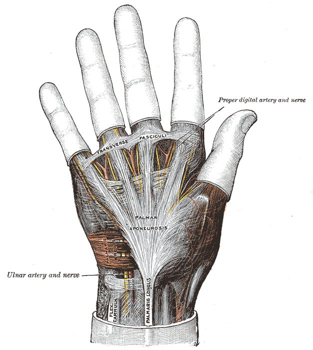
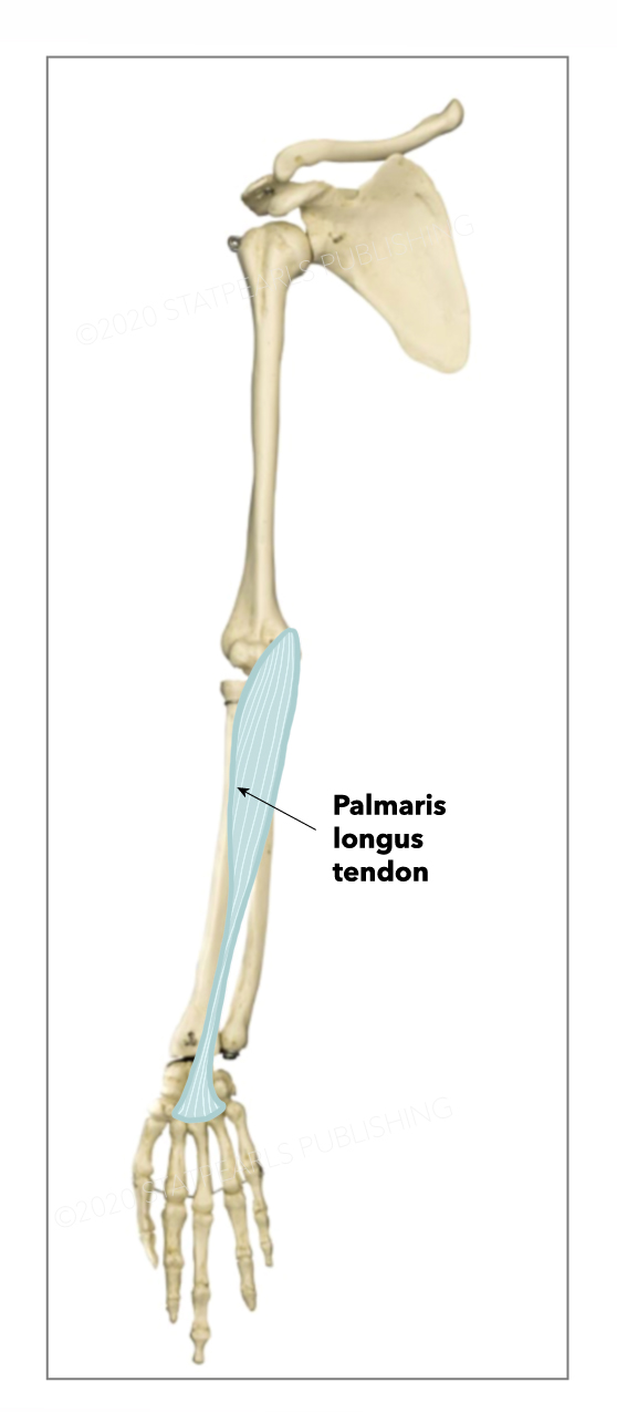
The palmaris longus is a small, fusiform-shaped muscle located on the anterior forearm of the human upper extremity. This muscle belongs to the superficial forearm flexor group, with a most common proximal attachment at the medial epicondyle of the humerus via the common forearm flexor tendon and a most common distal attachment into the connective tissue fibers of the palmar aponeurosis and the flexor retinaculum, a ligamentous structure forming the roof of the carpal tunnel and containing the median nerve and digital flexor tendons.[1] The palmaris longus can be morphologically quite variable but most commonly has a tendinous proximal attachment, a mid-length, spindle-shaped muscle belly, and a long and thin tendinous distal portion. The majority of fibers in the palmaris longus tendon pass superficially to the flexor retinaculum, the tendon broadens into a flattened collection of fibers, and the fibers interweave with the palmar aponeurosis.[1] 

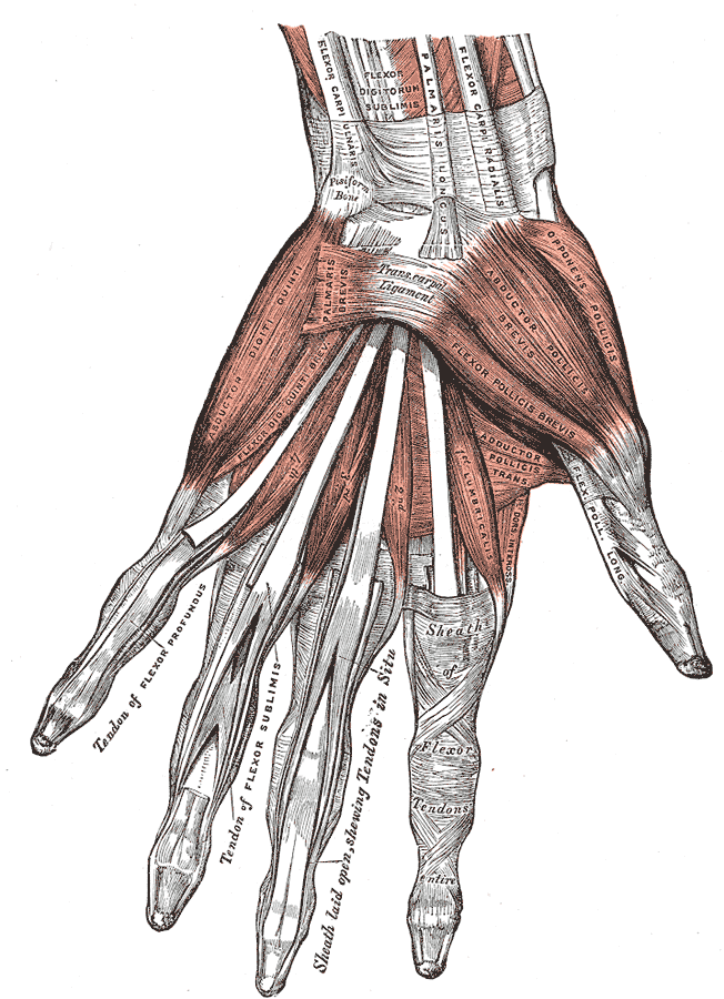
The functional contribution of the palmaris longus appears to be minimal, but it has clinical significance due to its frequent intraoperative harvest for many surgical procedures, often involving tendon repair in the upper extremity. The thin distal tendinous portion, superficial location, and lack of functional significance make the muscle easily accessible and ideal for intraoperative harvest for tendon reconstruction and other surgical procedures.[1] The palmaris longus muscle is commonly present but may be absent in a small percentage of the population, ranging from 2.5% to 26% of individuals, depending on the studied population.[1],[2] The palmaris longus tendon is located near the anatomical center of the anterior wrist, medial to the tendon of flexor carpi radialis, and lateral to the tendon of flexor carpi ulnaris.[1] The muscle’s presence can be assessed clinically by several physical exam maneuvers, with the Schaeffer test being the most commonly used. To perform the Schaeffer test, ask the patient to touch the pads of the thumb and little finger together while flexing the wrist, and if present, the tendon of palmaris longus should protrude anteriorly at the wrist joint.[3] Congenital absence of the muscle is common but has demonstrated decreased grip strength or any other functional deficit.[4]

Structure and Function

The palmaris longus belongs to the anterior forearm flexor group in the human upper extremity. The muscle attaches proximally to the medial humeral epicondyle and distally to the palmar aponeurosis and flexor retinaculum. The muscle is long and slender with variable morphology but commonly arises and inserts as a narrow tendinous structure with a widened muscular belly in the middle third. The muscle belly is spindle-shaped. Anatomically, the muscle crosses the elbow and wrist joints and consequently contributes to flexion at these joints; however, the muscle is small, and the actual contribution is likely minimal as an accessory flexor at these joints. The palmaris longus commonly sends a slip of distal tendon that inserts into the abductor pollicis brevis muscle of the thenar muscle group and may contribute to thumb abduction.[1] Congenital absence of the palmaris longus does not affect grip or pinch strength contributed to by wrist flexion.[4] The palmaris longus also appears to contribute to tensing the palmar aponeurosis and improves the stability of the overlying palmar skin.

Embryology

Mesenchyme (an early mesoderm-derived connective tissue) condenses into sets of dermatomes and myotome complexes. Myotomes migrate into the developing limb buds and give rise to myoblasts. Elongation of the limb buds, along with muscle formation from myoblasts, compartmentalizes the muscles into their respective muscle groups, including the forearm flexor group containing the palmaris longus.[5] Embryologic origins of congenital unilateral or bilateral absence of the palmaris longus have not been a subject of investigation in humans.

Blood Supply and Lymphatics

The blood supply to the palmaris longus muscle is via the ulnar artery, a branch of the brachial artery in the human upper extremity. The venous drainage of the muscle is by the cephalic and basilic veins in the upper extremity.[1] Compromise of the arterial blood supply, impaired venous outflow, and compartment syndrome can lead to ischemia of the palmaris longus and other muscles in the forearm flexor group. Prolonged reduction of effective arterial supply or venous drainage can lead to permanent ischemic Volkmann flexion contracture deformity of the unilateral wrist joint.[6]

Nerves

The palmaris longus muscle receives its innervation via branches of the median nerve containing nerve roots C5-T1. Median nerve injury at or above the elbow joint (including brachial plexus and nerve root injury) can lead to deficits in the palmaris longus and other forearm flexor muscles, leading to weakened elbow flexion, wrist flexion, radial deviation, finger flexion, thumb opposition, flexion, and abduction, in addition to the loss of sensory function in the distribution of the median nerve.[1]

Physiologic Variants

Palmaris longus is one of the most variable muscles in the human body. The morphology of the palmaris longus includes several common and uncommon anatomical variants and may be symmetric or asymmetric when assessed in cadavers. The most commonly reported atypical morphology includes unilateral and bilateral congenital absence, double muscles, split tendons, digastric muscle bellies, and varied distal insertions. Commonly reported distal insertions include the palmar aponeurosis, flexor retinaculum, forearm fascia, hypothenar muscles/fascia, abductor pollicis brevis, metacarpophalangeal joints, tendon of flexor carpi ulnaris, and the pisiform and scaphoid bones.[1] Large studies have shown differences in morphological prevalence in gender, with absence more common in females, and combined bilateral or unilateral congenital absence as frequent as 26.6% of the population, with bilateral absence being more common.[2][7] The muscle may bifurcate or trifurcate with multiple tendinous insertions distally.[8]

Surgical Considerations

The palmaris longus tendon is a commonly harvested structure for autogenous tendon grafting and other surgical procedures due to the length of the muscle's tendon, its superficial location and ease of access in the upper extremity, and its limited action as a wrist flexor and lack of functional impairment in the wrist and forearm function after harvest.[1] Surgeons can harvest the palmaris longus tendon for procedures including digital flexor and extensor tendon repair, flexor pollicis longus repair, chronic mallet finger repair, severe carpal tunnel syndrome, and carpometacarpal joint arthroplasty.[9]

Clinical Significance

Normal anatomic or aberrant palmaris longus muscles can infrequently cause median and ulnar nerve compression, leading to carpal or Guyon canal compression-like clinical presentations. The muscle can also be mistaken for soft tissue tumors of the forearm. While extremely rare, aberrant palmaris longus muscles can also cause compression of additional surrounding structures such as the ulnar artery.[1][10]

Other Issues

Surgeons commonly harvest the palmaris longus muscle for tendon grafting due to its superficial location.[1] Accidental median nerve harvesting is an infrequent complication that can be reduced by preoperative ultrasound evaluation.[11]


(Click Image to Enlarge)
<p>Hand Ligaments and Fascia

Hand Ligaments and Fascia. This anterior view shows the transverse fasciculi, palmar aponeurosis,  flexor carpi ulnaris tendon, palmaris longus tendon, and palmar aponeurosis. Other structures include the palmaris brevis, ulnar artery and nerve, and proper digital artery and nerve.


Henry Vandyke Carter, Public domain, via Wikimedia Commons


(Click Image to Enlarge)
<p>Muscles and Ligaments of the Hand; Anterior View, Flexor Carpi Ulnaris, Flexor Digitorum Sublimis, Palmaris Longus, Flexor

Muscles and Ligaments of the Hand; Anterior View, Flexor Carpi Ulnaris, Flexor Digitorum Sublimis, Palmaris Longus, Flexor Carpi Radialis, Abductor digit quinti, Flexor Digiti quinti Brevis, Palmaris brevis, Transcarpal Ligament, Flexor Pollicis Brevis, Abductor Pollicis Brevis, Opponens Pollicis, Adductor Pollicis Transversus, Flexor pollicis Longus, Sheath of Flexor Tendons


Henry Vandyke Carter, Public domain, via Wikimedia Commons


(Click Image to Enlarge)
Palmaris longus tendon
StatPearls Publishing Illustration
Details

Editor:

Bracken Burns

Updated:

8/28/2023 9:42:08 PM

References


[1]

Georgiev GP, Iliev AA, Dimitrova IN, Kotov GN, Malinova LG, Landzhov BV. Palmaris Longus Muscle Variations: Clinical Significance and Proposal of New Classifications. Folia medica. 2017 Sep 1:59(3):289-297. doi: 10.1515/folmed-2017-0035. Epub     [PubMed PMID: 28976893]


[2]

Cetin A, Genc M, Sevil S, Coban YK. Prevalence of the palmaris longus muscle and its relationship with grip and pinch strength: a study in a Turkish pediatric population. Hand (New York, N.Y.). 2013 Jun:8(2):215-20. doi: 10.1007/s11552-013-9509-6. Epub     [PubMed PMID: 24426922]


[3]

Sadeghifar A, Kahani AK, Saied A, Rasayi E. Interobserver and intraobserver reliability of different methods of examination for presence of palmaris longus and examination of fifth superficial flexor function. Anatomy & cell biology. 2018 Jun:51(2):79-84. doi: 10.5115/acb.2018.51.2.79. Epub 2018 Jun 27     [PubMed PMID: 29984051]


[4]

Sebastin SJ, Lim AY, Bee WH, Wong TC, Methil BV. Does the absence of the palmaris longus affect grip and pinch strength? Journal of hand surgery (Edinburgh, Scotland). 2005 Aug:30(4):406-8     [PubMed PMID: 15935531]


[5]

Tickle C. How the embryo makes a limb: determination, polarity and identity. Journal of anatomy. 2015 Oct:227(4):418-30. doi: 10.1111/joa.12361. Epub 2015 Aug 7     [PubMed PMID: 26249743]


[6]

Agrawal H, Dokania G, Wu SY. Neonatal volkmann ischemic contracture: case report and review of literature. AJP reports. 2014 Nov:4(2):e77-80. doi: 10.1055/s-0034-1382257. Epub 2014 Jun 24     [PubMed PMID: 25452886]

Level 3 (low-level) evidence

[7]

Yong MW, Yusof N, Rampal L, Arumugam M. Prevalence of Absence of Palmaris Longus and Its Association with Gender, Hand Dominance and Absence of FDS Tendon to Little Finger Among Malay Population. The journal of hand surgery Asian-Pacific volume. 2017 Dec:22(4):484-489. doi: 10.1142/S021881041750054X. Epub     [PubMed PMID: 29117832]


[8]

Sunil V, Rajanna S, Gitanjali, Kadaba J. Variation in the insertion of the palmaris longus tendon. Singapore medical journal. 2015 Jan:56(1):e7-9     [PubMed PMID: 25640108]


[9]

Lee KH, Jo YH, Kim SJ, Choi WS, Lee CH, Kim JH. Clinical Results of Autogenous Palmaris Longus Tendon Graft for Ruptures of Multiple Extensors in Rheumatoid Hands. The Journal of hand surgery. 2018 Oct:43(10):947.e1-947.e9. doi: 10.1016/j.jhsa.2018.02.012. Epub 2018 Mar 16     [PubMed PMID: 29551342]


[10]

Sato K, Murakami K, Mimata Y, Kikuchi Y, Oikawa R, Doita M. Superficial Ulnar Artery Crossing Over the Palmaris Longus Tendon at the Wrist in a Cadaver: A Case Report. The journal of hand surgery Asian-Pacific volume. 2018 Mar:23(1):137-139. doi: 10.1142/S2424835518720074. Epub     [PubMed PMID: 29409408]

Level 3 (low-level) evidence

[11]

Johnson CC, Vutescu ES, Miller TT, Nwawka OK, Lee SK, Wolfe SW. Ultrasound determination of presence, length and diameter of the palmaris longus tendon. The Journal of hand surgery, European volume. 2018 Nov:43(9):948-953. doi: 10.1177/1753193418778990. Epub 2018 Jun 7     [PubMed PMID: 29879859]