Anatomy, Back, Nucleus Pulposus


Introduction

The spine is the primary support of the body. It helps to connect other bony structures. The spine is made up of individual bone segments (vertebra), ligaments, and discs. There usually are 33 vertebrae in the spine, seven cervical, 12 thoracics, five lumbar, five fused sacral, and four fused coccygeal. In between these vertebrae are the intervertebral discs.[1] T

here are normally 25 discs in the spine: 7 in the cervical region, 12 thoracics, five lumbar, and one sacral. Each disc is made up of 3 main components. These components include the nucleus pulposus (NP), the annulus fibrosis (AF) and the cartilaginous endplates (CEP)[2] The annulus fibrosis is a structure that wraps around the nucleus pulposus and is made up of collagen-rich tissue.[3] The cartilaginous endplates are composed of a small amount of hyaline cartilage that is located between the vertebral endplate and the NP. These cartilaginous endplates play multiple roles such as acting as a mechanical barrier and supporting nutrient transport for the intervertebral disc.[4]

The NP is the inner gel-like portion of the intervertebral disc. It is essential in giving the spine its mechanical flexibility and strength. It is mainly composed of water (66%-86%) and type II collagen. Also, it has proteoglycans as well as small cartilage-like cells interspersed throughout. These proteoglycans are helpful in retaining water in the NP which helps create its gel-like consistency.[2]

Structure and Function

The makeup of the nucleus pulposus allows it to play an essential role in the flexibility and stability of the spine. It is made up of water, type II collagen, chondrocyte-like cells, and proteoglycans (sulfated glycosaminoglycans). These constituents help the NP to be elastic, allow it to be flexible under stress, and resist compression.[5] The high amount of sulfated glycosaminoglycans gives the nucleus pulposus a high charge density which causes the NP to absorb water and swell, which gives it the ability to act like both a solid and a liquid in mechanical situations.[6] This gelatinous mechanical consistency allows the nucleus pulposus to take the stress placed on the spine and redistribute it to the annulus fibrosis and the cartilaginous endplates.[5]

Embryology

The notochord is a structure that all vertebrates have during their embryonic development which helps to orient the entire axial skeleton. In addition, the notochord helps to form the vertebral bodies of the spine and the intervertebral discs.[2]

The development of the intervertebral disc (IVD) begins around the 3rd week of embryonic development. During this week gastrulation occurs allowing cells to shape the three germ layers (the ectoderm, mesoderm, and endoderm). Also, the mesoderm subdivides into axial mesoderm (notochord), paraxial mesoderm (the somites), and intermediate and lateral plate mesoderm.[7]

During the 4th week of embryogenesis, the notochord induces the somites to travel either peripherally or towards the notochord. The somites that travel peripherally later become the dermatomyotome and somites that migrate toward the notochord will become the sclerotome. During weeks 5 and 6 of development, the somites continue to either condense more around the notochord or travel further away from the midline. By the end of the 7th week, the sclerotomal cells show distinct segmentation with interspersed looser areas. By week 10, the looser areas of cells around the notochord push the notochord from the center and will eventually turn into the vertebral bodies. The distinctly segmented areas become the annulus fibrosis.[7] After this, the intervertebral disc (IVD) slowly forms from the annulus fibrosis (AF) and the notochord. The notochord eventually helps to develop the nucleus pulposus (NP) of the IVD.

At birth, the cells of the NP are still like the cells of the notochord. Shortly after birth, these notochordal-like cells begin to disappear, and smaller non-vacuolated cells form in the NP which is thought to be due to the unique environment within the fully formed nucleus pulposus causing the newer cells to develop from the older notochordal cells.[8] By the end of the first decade after birth, these new smaller cells are the predominant type in the NP.[7]

Blood Supply and Lymphatics

The nucleus pulposus is poorly vascular as its cells express Fas ligand which causes apoptosis of vascular endothelial cells, which in turn creates poor blood vessel growth. Also, a few factors are released by notochordal cells during development that can suppress vascular endothelial growth factor (VEGF). However, the blood vessels that supply nutrients to the nucleus pulposus are located mainly in the longitudinal ligaments that travel next to the disc as well as in cartilaginous endplates(CEP). Most of the nutrients used by the intervertebral discs are from diffusion via the CEP or the blood supply in the outer annulus fibrosis.[5]

Nerves

The sinuvertebral nerves, as well as the gray rami communicantes, are the main nerves that supply innervation to the intervertebral discs and thus the nucleus pulposus.[5] These nerves are sympathetic nerves which supply innervation not only to the IVDs but also the ventral portion of the dura mater and both the anterior and posterior longitudinal ligaments.[9]

Clinical Significance

Disc Herniation:

Intervertebral disc herniation is a common problem in the lumbar and cervical spine that can cause varying symptoms such as pain, numbness, and weakness of both the upper and lower extremities. Intervertebral disc herniation is defined as a condition in which the nucleus pulposus is protruding past the annulus fibrosus. This herniation can be on a spectrum of partial to complete depending upon how much of the NP herniates through the AF.[10]

Many significant changes in an intervertebral disc can occur that cause a disc to weaken and the NP to herniate through the AF. Of these changes, the most common is an increase in type I collagen and a decrease in type II collagen in the NP and inner AF, less water retention by the NP, and degeneration of various constituents of the IVD such as other types of collagen as well as the extracellular matrix. Additionally, specific systems activate that contribute to the degeneration of the IVD. These include apoptosis, inflammatory pathways, and upregulation of matrix metalloproteinase expression.[11]

While the biological causes of disc herniation itself are well known, the biological causes of the symptoms of disc herniations are still under investigation. Current thought is that while mechanical compression of spinal nerves is an important cause of pain in disc herniation, another possible factor in the perception of pain is the effect of the nucleus pulposus on nervous tissue.[12] The theory is that the leakage of the NP can cause an inflammatory reaction that in turn causes changes in the nerve roots themselves.

A recent study showed that when peripheral nerves and dorsal root ganglia suffered exposure to the contents of the NP, there was a considerable increase in neuronal activity in these neuronal tissues starting at 9 hours and lasting for up to a couple of days. Similar changes also showed in the spinal cord in this same study. This study also revealed that when applying the nucleus pulposus in the absence of mechanical compression, the neural activity in the ventral posterolateral (VPL) nucleus of the thalamus increased quickly.[12] The VPL is an endpoint for the spinothalamic tract which sends pain signals from the periphery to the brain. These neurons are excited in response to painful mechanical stimuli as well as visceral nociception.[13] This study helps to show that the painful symptoms of disc herniation have two primary contributing factors: mechanical compression and inflammatory response to the nucleus pulposus.


(Click Image to Enlarge)
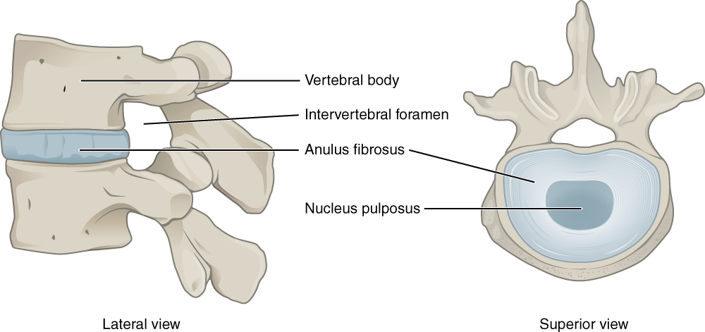
<p>Intervertebral Disk

Intervertebral Disk. This lateral view shows the disc between two vertebrae. The superior view shows the annulus fibrosis at the outer layer and the nucleus pulposus in the inner layer.


Illustration from Anatomy & Physiology, Connexions Web site. http://cnx.org/content/col11496/1.6/, Jun 19, 2013. This file is licensed under the Creative Commons Attribution 3.0 Unported license.

Details

Author

Vamsi Reddy

Editor:

Gurdeep Singh

Updated:

8/7/2023 11:52:09 PM

References


[1]

DeSai C, Reddy V, Agarwal A. Anatomy, Back, Vertebral Column. StatPearls. 2023 Jan:():     [PubMed PMID: 30247844]


[2]

Waxenbaum JA, Reddy V, Futterman B. Anatomy, Back, Intervertebral Discs. StatPearls. 2023 Jan:():     [PubMed PMID: 29262063]


[3]

Nosikova YS, Santerre JP, Grynpas M, Gibson G, Kandel RA. Characterization of the annulus fibrosus-vertebral body interface: identification of new structural features. Journal of anatomy. 2012 Dec:221(6):577-89. doi: 10.1111/j.1469-7580.2012.01537.x. Epub 2012 Jul 3     [PubMed PMID: 22747710]


[4]

Moon SM, Yoder JH, Wright AC, Smith LJ, Vresilovic EJ, Elliott DM. Evaluation of intervertebral disc cartilaginous endplate structure using magnetic resonance imaging. European spine journal : official publication of the European Spine Society, the European Spinal Deformity Society, and the European Section of the Cervical Spine Research Society. 2013 Aug:22(8):1820-8. doi: 10.1007/s00586-013-2798-1. Epub 2013 May 15     [PubMed PMID: 23674162]


[5]

Chen S, Fu P, Wu H, Pei M. Meniscus, articular cartilage and nucleus pulposus: a comparative review of cartilage-like tissues in anatomy, development and function. Cell and tissue research. 2017 Oct:370(1):53-70. doi: 10.1007/s00441-017-2613-0. Epub 2017 Apr 17     [PubMed PMID: 28413859]

Level 2 (mid-level) evidence

[6]

Nerurkar NL, Elliott DM, Mauck RL. Mechanical design criteria for intervertebral disc tissue engineering. Journal of biomechanics. 2010 Apr 19:43(6):1017-30. doi: 10.1016/j.jbiomech.2009.12.001. Epub 2010 Jan 18     [PubMed PMID: 20080239]


[7]

Rodrigues-Pinto R, Richardson SM, Hoyland JA. An understanding of intervertebral disc development, maturation and cell phenotype provides clues to direct cell-based tissue regeneration therapies for disc degeneration. European spine journal : official publication of the European Spine Society, the European Spinal Deformity Society, and the European Section of the Cervical Spine Research Society. 2014 Sep:23(9):1803-14. doi: 10.1007/s00586-014-3305-z. Epub 2014 Apr 29     [PubMed PMID: 24777668]

Level 3 (low-level) evidence

[8]

McCann MR, Tamplin OJ, Rossant J, Séguin CA. Tracing notochord-derived cells using a Noto-cre mouse: implications for intervertebral disc development. Disease models & mechanisms. 2012 Jan:5(1):73-82. doi: 10.1242/dmm.008128. Epub 2011 Oct 25     [PubMed PMID: 22028328]


[9]

Raoul S, Faure A, Robert R, Rogez JM, Hamel O, Cuillère P, Le Borgne J. Role of the sinu-vertebral nerve in low back pain and anatomical basis of therapeutic implications. Surgical and radiologic anatomy : SRA. 2003 Feb:24(6):366-71     [PubMed PMID: 12647025]


[10]

Dydyk AM, Ngnitewe Massa R, Mesfin FB. Disc Herniation. StatPearls. 2023 Jan:():     [PubMed PMID: 28722852]


[11]

Amin RM, Andrade NS, Neuman BJ. Lumbar Disc Herniation. Current reviews in musculoskeletal medicine. 2017 Dec:10(4):507-516. doi: 10.1007/s12178-017-9441-4. Epub     [PubMed PMID: 28980275]


[12]

Nilsson E, Brisby H, Rask K, Hammar I. Mechanical compression and nucleus pulposus application on dorsal root Ganglia differentially modify evoked neuronal activity in the thalamus. BioResearch open access. 2013 Jun:2(3):192-8. doi: 10.1089/biores.2012.0281. Epub     [PubMed PMID: 23741630]


[13]

Ab Aziz CB, Ahmad AH. The role of the thalamus in modulating pain. The Malaysian journal of medical sciences : MJMS. 2006 Jul:13(2):11-8     [PubMed PMID: 22589599]