Continuing Education Activity
Kaposi varicelliform eruption, also called eczema herpeticum, refers to a disseminated skin infection due to a virus that usually leads to localized vesicular eruptions in a patient with an underlying cutaneous disease. Although rare, it is a potentially life-threatening disorder. Herpes simplex virus is considered the main causative agent. The most commonly reported cases occur in patients with atopic dermatitis. However, it has been described in association with other skin conditions such as pemphigus foliaceus, ichthyosis vulgaris, bullous pemphigoid, Darier disease, Grover disease, Hailey-Hailey disease, dyskeratosis follicularis, mycosis fungoides, Sezary syndrome, psoriasis, pityriasis rubra pilaris, rosacea, seborrheic dermatitis, contact dermatitis (both allergic and irritant), second-degree burns and skin grafts. Clinical features of Kaposi varicelliform eruption include widespread clusters of umbilicated vesicles and pustules that evolve into crusted skin erosions. The most frequently affected sites are the trunk, neck, and head. The diagnosis of Kaposi varicelliform eruption is made primarily on clinical findings. The Tzanck smear, viral cultures, skin biopsy, or detection of viral DNA by Polymerase Chain Reaction may be helpful in doubtful cases. Antiviral therapy has been effective but should be started as soon as possible after diagnosis to reduce morbidity and mortality. This activity reviews the evaluation and treatment of Kaposi varicelliform eruptions and the role of the interprofessional team in evaluating and treating this condition.
Objectives:
Assess the morbidity and mortality associated with Kaposi varicelliform eruptions.
Evaluate the co-morbid conditions associated with Kaposi varicelliform eruptions.
Identify how the diagnosis of Kaposi varicelliform eruptions is usually made.
Communicate the importance of the medical team in evaluating and managing patients with Kaposi varicelliform eruptions.
Introduction
Kaposi varicelliform eruption, also called eczema herpeticum, refers to a disseminated skin infection due to a virus that usually leads to localized vesicular eruptions in a patient with an underlying cutaneous disease. Although rare, it is a potentially life-threatening disorder. Herpes simplex virus is considered the main causative agent.[1][2][3]
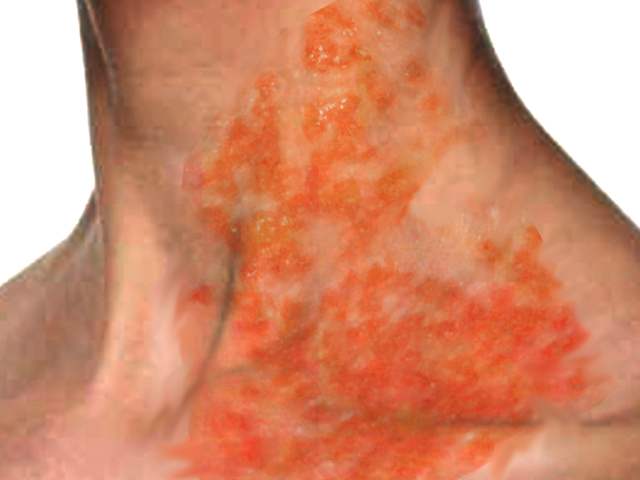
The most commonly reported cases occur in patients with atopic dermatitis. However, it has been described in association with other skin conditions such as pemphigus foliaceus, ichthyosis vulgaris, bullous pemphigoid, Darier disease, Grover disease, Hailey-Hailey disease, dyskeratosis follicularis, mycosis fungoides, Sezary syndrome, psoriasis, pityriasis rubra pilaris, rosacea, seborrheic dermatitis, contact dermatitis (both allergic and irritant), second-degree burns and skin grafts.[4] Clinical features of Kaposi varicelliform eruption include widespread clusters of umbilicated vesicles and pustules that evolve into crusted skin erosions (See Image. Kaposi Varicelliform Eruption).
- The most frequently affected sites are the trunk, neck, and head.
- The diagnosis of Kaposi varicelliform eruption is made primarily on clinical findings.
- The Tzanck smear, viral cultures, skin biopsy, or detection of viral DNA by Polymerase Chain Reaction may be helpful in doubtful cases.
- Antiviral therapy has been effective but should be started as soon as possible after diagnosis to reduce morbidity and mortality.
Etiology
Kaposi varicelliform eruption is caused mainly by herpes simplex virus type 1; however, other viral agents such as herpes simplex virus type 2, Coxsackie A16, vaccinia, varicella zoster, and smallpox also have been involved in its pathogenesis.
Epidemiology
Kaposi varicelliform eruption is a rare condition first described in 1887 by Moritz Kaposi. It occurs more frequently in children due to its relationship with atopic dermatitis. However, adult cases have been reported. The incidence is not precisely known due to its rarity and the lack of a large case series.[5][6] The disease equally affects men and women and does not appear to have a specific ethnic predominance.
Pathophysiology
Kaposi varicelliform eruption was initially assumed to be secondary to a fungal infection. However, the finding of cytoplasmic inclusion bodies in the histological examination suggested a viral origin.
The mechanisms underlying the pathogenesis of viral reactivation in Kaposi varicelliform eruption remain incompletely understood. It is held that a defective skin barrier acting in conjunction with immune deficiencies seems to lead to the development of the disease. Both cell-mediated and humoral immunity dysfunction are implicated. In addition, the Th-2 cytokine environment found in atopic dermatitis seems crucial.[7] Many recent studies suggest a genetic contribution to the pathogenesis of Kaposi varicelliform eruption.
Histopathology
A skin biopsy is not required to confirm a diagnosis, but if it is performed, histological findings include intra-epidermal blister, acantholysis, multinuclear giant cells with intranuclear inclusion, and ballooning degeneration of the keratinocytes.
History and Physical
Patients with Kaposi varicelliform eruption present with a sudden skin eruption of painful clusters of umbilicated vesicles and pustules. Vesiculopustules often evolve into crusted, hemorrhagic, and punched-out skin erosions that may enlarge and coalesce to form extensive denuded areas more likely to get a bacterial infection.
The distribution of affected skin reflects the crucial role of skin barrier impairment since Kaposi varicelliform eruption begins in areas of underlying dermatosis. This inaugural topographic distribution may lead to a delayed diagnosis because the eruption is often confused with the pre-existing condition.
Kaposi varicelliform eruption may be associated with systemic symptoms such as malaise, high temperature, and swollen lymph nodes. In addition, the disease can be complicated by multiple organ involvements, mainly of the central nervous system, liver, lungs, gastrointestinal tract, and adrenal glands.
Evaluation
The diagnosis of Kaposi varicelliform eruption is mainly based on a clinical examination, although several laboratory tests can be helpful.[8][9] A Tzanck smear is performed by scraping the floor of an opened vesicle and staining the material with Wright-Giemsa stain, demonstrating multinucleated giant cells. It is inexpensive, easily applicable, and quick to perform. However, this method suffers from low sensitivity and does not differentiate between herpes simplex virus 1 and 2 or between herpes simplex virus and varicella-zoster virus. Viral culture and direct fluorescence antibody staining on Tzanck smear are the most reliable techniques for herpes simplex virus detection. A skin biopsy or polymerase chain reaction may be performed in case of atypical, equivocal, or old lesions. A histological examination may confirm a diagnosis that may not have been thought of clinically, whereas polymerase chain reaction detects viral DNA by Polymerase Chain Reaction.
Treatment / Management
Treatment of Kaposi varicelliform eruption must be instituted with no delay since it is a potentially life-threatening disease. Antiviral therapy is effective in reducing morbidity and preventing complications. Nucleoside analogs are the antiviral agents most commonly used since they inhibit viral DNA replication.[10][11][12]
Acyclovir is the most widely studied and prescribed drug for Kaposi varicelliform eruption. High-dose intravenous acyclovir is often necessary for disease control. Most patients achieve resolution of the skin lesions over several days. Prophylactic treatment with systemic antibiotics is recommended to prevent secondary bacterial infection.
Differential Diagnosis
Kaposi varicelliform eruption may be confused with various conditions:
- Chickenpox
- Impetigo
- Allergic contact dermatitis
Prognosis
Kaposi varicelliform eruption is a serious condition that may have fatal outcomes. The disease may occur as a primary or a recurrent type of infection. The primary form mainly concerns children and is usually disseminated and associated with systemic symptoms and life-threatening complications such as bacterial sepsis, viremia, and multiple organ involvement. The recurrent type occurs in adulthood and is usually a milder and more localized form, generally presenting without viremia. Septicemia resulting from secondary bacterial infection of cutaneous lesions also increases morbidity and mortality. The most common species isolated from patients with Kaposi varicelliform eruption are Staphylococcus aureus, group A beta-hemolytic streptococcus, Peptostreptococcus, and Pseudomonas aeruginosa. A risk of ocular involvement exists when herpes simplex virus-associated Kaposi varicelliform eruption affects the face. Ocular anomalies include uveitis, conjunctivitis, keratitis, and blepharitis. The most serious ophthalmological sequela is herpetic keratitis, which may lead to vision loss resulting from corneal scarring.
Pearls and Other Issues
Kaposi varicelliform eruption should be diagnosed accurately since it may have fulminant outcomes. Although there is no consensual therapeutic approach, the early use of antiviral therapy associated with systemic antibiotics is crucial.
Enhancing Healthcare Team Outcomes
Kaposi varicelliform eruption may present to any interprofessional team member; the most important thing is to refer these patients immediately to the dermatologist. The diagnosis is clinical, but the general practitioner may not have the clinical expertise to make this diagnosis. If the eye is affected, an ophthalmology consult should be made
Treatment of Kaposi varicelliform eruption must be instituted with no delay since it is a potentially life-threatening disease. Antiviral therapy is effective in reducing morbidity and preventing complications. Acyclovir is the most widely studied and prescribed drug for Kaposi varicelliform eruption. High-dose intravenous acyclovir is often necessary for disease control, so a pharmacist should be involved to verify dosing and perform medication reconciliation. Most patients achieve resolution of the skin lesions over several days. Prophylactic treatment with systemic antibiotics is recommended to prevent secondary bacterial infection. Clinicians administer these drugs and need to be aware of the signs of adverse drug reactions, as well as monitor the progress of treatment. These patients need close monitoring until the lesions have resolved, and management by an interprofessional team is the optimal approach.[13]