Neuroanatomy, Dural Venous Sinuses


Introduction

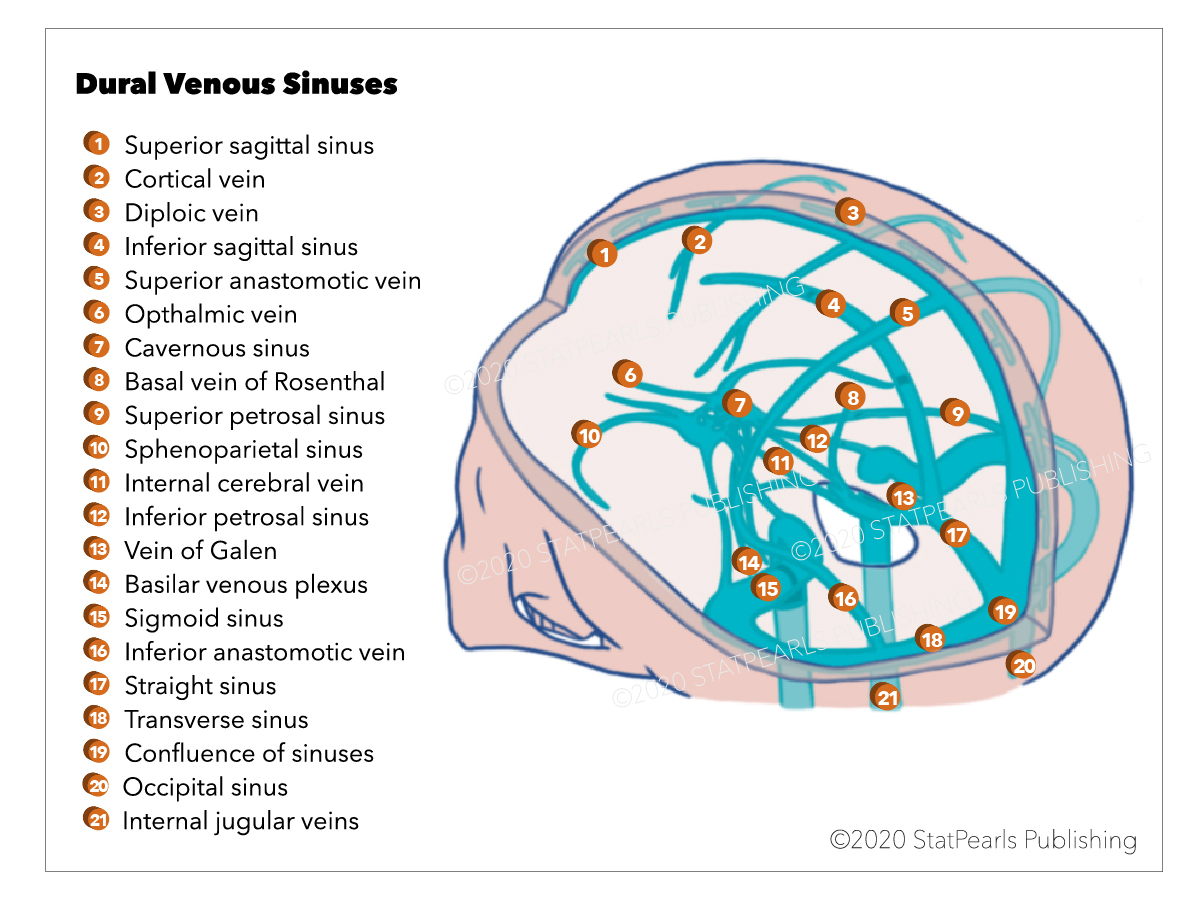
Dural venous sinuses are a group of sinuses or blood channels that drains venous blood circulating from the cranial cavity. It collectively returns deoxygenated blood from the head to the heart to maintain systemic circulation. There are seven major dural venous sinuses located within the cranial cavity, specifically between the periosteal and meningeal layer of the dura mater: superior sagittal, inferior sagittal, straight, transverse, sigmoid, cavernous, and superior petrosal sinuses. Most of these sinuses are found adjacent to the falx cerebri and tentorium cerebelli. The cavernous sinus is clinically the most important dural venous sinus. Characteristic structures of venous sinuses are vital to physicians and healthcare providers, especially in cases of possible thrombosis and infection.[1][2][3]

Structure and Function

The primary function of the dural venous sinuses is to drain all venous blood within the cranial cavity and deliver it back to the cardiovascular circulation via the internal jugular vein below the jugular foramen, which will further drain into the superior vena cava before reaching the heart. Aside from draining venous blood, superior sagittal sinus also participates in the cerebrospinal fluid (CSF) circulation by serving as the main location for CSF return after passing through arachnoid granulations.[4]

Embryology

Development of the dural venous sinuses is mainly related to the development of the meningeal layers of the brain, specifically the dura mater. Major dural venous sinuses, such as the superior petrosal sinus, derive from pro-otic veins, while cavernous sinus originated from the vena capitis medialis.

Blood Supply and Lymphatics

The majority of the venous blood drained into the dural venous sinuses come from a variety of tributaries found within the cranium. Structures which act as tributaries to the venous sinus flow include the following:

  • Cerebral and cerebellar veins which cross the subdural space to drain into the superior sagittal vein, hence, the name “bridging veins”
  • Emissary veins that progress through the skull allowing communication between intracranial and extracranial contents
  • Diploic veins which drain blood between the inner and outer layer flat bones of the skull (diploe)
  • Meningeal veins collect blood from the meninges
  • Arachnoid granulations for CSF returning to the venous circulation

Physiologic Variants

Major dural venous sinuses differ in their specific functions depending on their location within the cranium and its associated structures that pass through the sinuses. The superior sagittal sinus is midsagittal and superior to the falx cerebri. It collects blood from cerebral and cerebelli veins going to the confluence of sinuses (torcular herophili). The inferior sagittal sinus lies within the inferior aspect of the falx cerebri and connects with the great cerebral vein forming the straight sinus. The straight sinus drains contents from inferior sagittal sinus and great cerebral vein and terminates into the confluence of sinuses. It can also drain into the transverse sinus. The occipital sinus is in the posterior aspect of the tentorium cerebelli and is a small channel draining contents from the occiput into the confluence of sinuses. The transverse sinuses are located bilaterally in the tentorium cerebelli. It forms as an attachment into the occipital bone transporting venous blood from the confluence of sinuses to the left and right sigmoid sinuses. Sigmoid sinuses are also paired sinuses known for their S-shape sinus found in the base of the cranial fossa posteriorly. Left and right sigmoid sinuses collectively drain venous blood into the internal jugular vein, which exits at the jugular foramen. The cavernous sinus is located on the cavity of the sphenoid bone surrounding the pituitary gland, which drains contents from the ophthalmic veins (orbit) and pterygoid venous plexus (deep face). Cavernous sinuses further drain into both petrosal sinuses superiorly (into the sigmoid sinus) and inferiorly (into the internal jugular vein). Left and right cavernous sinus connect via the intercavernous sinuses, which is considered as the most clinically important venous sinus because of its association with the other significant structures in the head including cranial nerves III (oculomotor), IV (trochlear), ophthalmic and maxillary branches of cranial nerve V (trigeminal), and cranial nerve VI (abducens) which lies below the internal carotid artery. For these reasons, the cavernous sinus is also known as the “anatomic jewel box.” The confluence of sinuses drains blood from superior sagittal, straight, and occipital sinuses and lies along the occipital bone posteriorly; this is where these three sinuses meet to transmit venous blood to the left and right transverse sinuses.

Clinical Significance

All dural venous sinuses are valveless; thus, they can promote a forward or backflow of blood to other associated structures, allowing pathogens and neoplastic cells to travel to different parts of the brain. The dangerous triangle of the face is a triangular area bounded by the medial angle of the eyes, sides of the nose and the upper lip as its boundaries. This anatomy is important because it serves as the passageway of pathogens from the face into the brain via the cavernous sinuses. Infection coming from the superficial and deep layers of the face can easily travel into the cavernous sinuses leading to the development of a thrombus, a condition termed as cavernous sinus thrombosis. This condition may result in the swelling of the affected sinuses and pertinent damage to the associated cranial nerves. Patients may develop internal strabismus caused by the lesion in the cranial nerve VI, which is usually the first cranial nerve affected, which is then followed by damage to further movement and sensation of the eyes and skin of the face as well as the scalp, respectively. Ophthalmoplegia or paralysis of the eye muscles can also be manifestations of other damaged cranial nerves. Pulsatile proptosis characterized by a combination of pulsation and protrusion of the eyeball can also present in patients with affected venous sinuses.[4][5][6][7]

Other Issues

Endoscopic anatomical studies were done in the past, showing that cavernous sinuses are easily reachable through the sphenoid sinuses, which provided ways to study brain lesions better. A 3-dimensional atlas offers a means to identify lesions within the brain, including the venous sinuses, and at the same time to describe corresponding disorders for each lesion. Anatomic and scanning electron microscopy studies into the falx cerebri revealed regional differences between a complex structure of falcine venous plexus and its communication with the structures of the venous sinuses. This information is vital, especially to neurosurgeons performing surgical interventions to the brain.


(Click Image to Enlarge)
Dural Venous Sinuses graphic
Graphic by Emma Gregory
Details

Author

Vamsi Reddy

Updated:

8/8/2023 12:56:23 AM

References


[1]

Karakas AB, Govsa F, Ozer MA, Eraslan C. 3D Brain Imaging in Vascular Segmentation of Cerebral Venous Sinuses. Journal of digital imaging. 2019 Apr:32(2):314-321. doi: 10.1007/s10278-018-0125-4. Epub     [PubMed PMID: 30242780]


[2]

Ngnitewe Massa R, Minutello K, Mesfin FB. Neuroanatomy, Cavernous Sinus. StatPearls. 2023 Jan:():     [PubMed PMID: 29083662]


[3]

Graffeo CS, Perry A, Copeland WR 3rd, Raghunathan A, Link MJ. Histology of the distal dural ring. Clinical anatomy (New York, N.Y.). 2017 Sep:30(6):742-746. doi: 10.1002/ca.22916. Epub 2017 Jun 12     [PubMed PMID: 28514500]


[4]

Patel CR, Fernandez-Miranda JC, Wang WH, Wang EW. Skull Base Anatomy. Otolaryngologic clinics of North America. 2016 Feb:49(1):9-20. doi: 10.1016/j.otc.2015.09.001. Epub     [PubMed PMID: 26614826]


[5]

Kiyofuji S, Perry A, Graffeo CS, Giannini C, Link MJ. The dangers of the "Head Down" position in patients with untreated pituitary macroadenomas: case series and review of literature. Pituitary. 2018 Jun:21(3):231-237. doi: 10.1007/s11102-017-0851-5. Epub     [PubMed PMID: 29236218]


[6]

Taniguchi D, Nakajima S, Hayashida A, Kuroki T, Eguchi H, Machida Y, Hattori N, Miwa H. Deep cerebral venous thrombosis mimicking influenza-associated acute necrotizing encephalopathy: a case report. Journal of medical case reports. 2017 Sep 26:11(1):281. doi: 10.1186/s13256-017-1444-7. Epub 2017 Sep 26     [PubMed PMID: 28950885]

Level 3 (low-level) evidence

[7]

Choi KY, Yang CJ. A case report of cavernous sinus thrombosis after trauma. International journal of pediatric otorhinolaryngology. 2017 Apr:95():101-103. doi: 10.1016/j.ijporl.2017.02.007. Epub 2017 Feb 13     [PubMed PMID: 28576515]

Level 3 (low-level) evidence