Hyperbaric Treatment of Central Retinal Artery Occlusion

Earn CME/CE in your profession:


Continuing Education Activity

Central retinal artery occlusion (CRAO) is a relatively rare but potentially devastating ocular emergency. Patients present with sudden, painless monocular vision loss. The prognosis for visual recovery is poor, and until recently, no treatment could restore any degree of functional vision to the affected eye. Central retinal artery occlusion leads to retinal hypoxia, and the retinal tissue is incredibly intolerant of hypoxia. In 2006, hyperbaric oxygen therapy was approved for treating central retinal artery occlusion. However, it remains a vastly underutilized modality, partially due to insufficient availability and awareness in the ophthalmology community. This activity describes the pathophysiology, evaluation, and management of central retinal artery occlusion and highlights the role of the interprofessional team in caring for affected patients.

Objectives:

  • Identify the etiology of central retinal artery occlusion.

  • Differentiate the presentation of a patient with central retinal artery occlusion.

  • Determine the treatment and management options available for central retinal artery occlusion.

  • Summarize a well-coordinated interprofessional team approach to care for patients affected by central retinal artery occlusion effectively.

Introduction

Central retinal artery occlusion (CRAO) is a relatively rare emergency and potentially devastating eye condition. Patients present with sudden, painless monocular vision loss. The prognosis for visual recovery is poor, and up until recently, no treatment could be offered to restore at least some functional vision to the affected eye. The retina has the highest oxygen consumption rate of any organ in the body at 13 mL/100 gm per minute. Retinal tissue is not tolerant of hypoxia. Until 2006, when hyperbaric oxygen therapy was approved for central retinal artery occlusion treatment, ophthalmologists had no treatment options for these patients. It is still a vastly underutilized modality, partially due to a lack of availability and knowledge in the ophthalmology community.[1][2][3][4]

Etiology

Patients at increased risk for developing central retinal artery occlusion are those with:

  • Giant cell arteritis
  • Atherosclerosis
  • Thromboembolic disease
  • Vasospasm[5]

Arterial supply to the eye is via the ophthalmic artery, a branch of the cavernous portion of the internal carotid artery. The central retinal artery enters the globe along with the optic nerve and serves the inner layers of the retina through its many branches. Fifteen percent to 30% of the population have a cilioretinal artery, part of the choroidal arterial supply. This supplies the area of the retina around the macula, which is the central vision area. Patients with a cilioretinal artery may still have preserved central vision because this portion of the retina is still being perfused.

In central retinal artery occlusion, the inner retinal layers normally served by the retinal circulation will die. This is what causes the vision loss. However, these layers may receive enough oxygen via diffusion from the choroidal circulation to maintain viability if the patient is exposed to increased FiO2. Normally, the choroidal circulation supplies most of the oxygen to the retina.[6][7][8]

Epidemiology

CRAO tends to occur more often in men. It occurs in about 1 out of 50,000-100,000 with a mean age of 60-65.[9]

CRAO due to arteritis is most commonly caused by giant cell arteritis, but systemic lupus er­ythematosus, polyarteritis nodosa, and other vasculitides can also lead to CRAO. However, most CRAOs are nonarteritic. In fact, 90% are nonarteritic. Carotid artery disease is the most common cause of retinal artery occlusion, accounting for about 70% of cases.[10] Other non-inflammatory causes include emboli (e.g., atrial fibrillation), hypercoagulable disorders, hematological cancers, and sickle cell anemia.

Central retinal artery occlusion is a rare, emergent eye condition that should be suspected in any patient with unilateral, sudden, painless vision loss. These patients usually present with vision loss limited to finger counting or light perception. If there is no light perception, the ophthalmic artery may be occluded, which causes permanent and debilitating vision loss that would not be amenable to HBO2 treatment as this would preclude any ciliary collateral circulation, which could maintain retinal viability with HBO2. However, it may be addressed by thrombolysis.

Pathophysiology

If the level of occlusion is at the ophthalmic artery, there may be no treatment options available to the patient. This is because posterior ciliary circulation is blocked, and there is no collateral circulation to the inner layers of the retina.[11][12][13]

Therapy must be started before the tissue is irreparably damaged.

The degree and location of occlusion will affect the response to treatment.

The adequate partial pressure of oxygen must be maintained long enough for the retinal vessels to recanalize. This usually occurs within 72 hours.

The most extensive published case series (Hayreh) showed the natural progression of disease without treatment with hyperbaric oxygen.[14]

  • Some patients had transient symptoms for minutes to hours that resolved without treatment, and they were left with minimal visual impairment.
  • Some patients have a variant retinal vascular supply, the cilioretinal artery. This occurs in almost half the population and about one-third of all eyes. There is much variability in how much of the retina is supplied by such arteries.[15] These arteries can aid in sparing some of the central retinal function.
  • Without HBO2 therapy, the natural outcomes for patients without cilioretinal arteries are poor, with less than 2% ending up with recovery of baseline vision and most (80%) ending up with light perception, motion detection, or finger counting at best.[14]  
  • Patients with cilioretinal arteries had much better outcomes.

History and Physical

CRAO presents as sudden onset, painless loss of vision. Usually, there is only light perception, hand motion detection, or finger counting, and visual acuity is often worse than 20/200.

A patient's lack of light perception may indicate an ophthalmic artery occlusion; therefore, no blood flows to choroidal vessels.

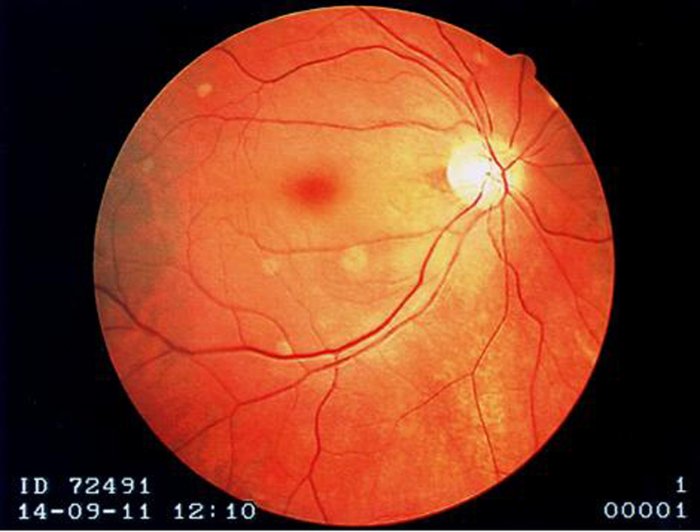
If central visual acuity is spared and fundoscopy is consistent with central retinal artery occlusion, the patient probably has a cilioretinal artery blood supply. The fundoscopy may show a pale yellow ischemic retina. A cherry-red spot may be present where the macula is, but this is not a consistent or reliable finding. The cherry red spot is associated with poor outcomes.[16]

Evaluation

This is a "stroke of the eye" and should be treated with the same urgency as an evolving stroke or myocardial infarction. Oxygen by non-rebreather mask should be initiated immediately while performing the evaluation. Fundoscopy should be performed and documented immediately. An ophthalmologist should be consulted, preferably a vitreoretinal specialist. The presence of pain, a history of trauma, findings of flashers or floaters, and age younger than 40 years are more common with ocular trauma, retinal detachment, or vitreal hemorrhage and make central retinal artery occlusion less likely.  

Treatment / Management

General Treatment

  • Provide immediate supplemental oxygen at the highest normobaric FiO2 available.
  • Consult ophthalmology, but do not delay treatment if readily available.
  • Baseline complete blood count (CBC), ESR, C-reactive protein (CRP), prothrombin time (PT), partial thromboplastin time (PTT), INR if on coumadin
  • ECG
  • Carotid ultrasound, MRI, and echocardiogram can be done later. These are not urgent studies for these patients but are part of the complete assessment for risk factor modification, just as in stroke.
  • If the onset of symptoms is within 24 hours of presentation, and the patient has no response to normobaric hyperoxia after 15 minutes, refer for emergency hyperbaric medicine consultation.[17]

Hyperbaric Oxygen Treatment Protocol[18]

Indicated for central retinal artery occlusion 

Initial treatment:

  • Compress to 2 atmosphere absolute (ATA).
  • If vision improves significantly at 2 ATA, remain at this depth for 90 minutes.
  • If vision fails to improve after 30 minutes at 2 ATA, then compress to 2.4 ATA; if vision improves at this depth, treat at this depth for 90 minutes. If there is no improvement at 2.4 ATA, compress to 2.8 ATA and perform the US Navy Treatment Table No. 6.

If there is no improvement after the initial US Navy Table No. 6, options are:

  • Discontinue treatment
  • Continue with normobaric oxygen therapy
  • Give two additional treatments at 2.8 ATA for 90 minutes with 5-minute air breaks every 30 minutes 2 times per day, and reassess after 4 to 6 days. Most patients will reach a plateau, after which they will not experience any further improvement in visual acuity, and treatment can then be discontinued. Some patients require eight treatments before improving visual acuity, but few, if any, patients will respond beyond this point.

A more straightforward protocol with good results treats at 2.5 ATA for 90 minutes with 5-minute air breaks every 30 minutes 2 times daily for 2-3 weeks.[19]   

Fluorescein angiography can be a safe and valuable tool to evaluate these patients for successful recanalization of the central retinal artery.[20]

Differential Diagnosis

The differential for acute painless vision loss includes central retinal vein occlusion, vitreous hemorrhage, retinal detachment, posterior circulation stroke, ischemic optic neuropathy, and amaurosis fugax.

Pertinent Studies and Ongoing Trials

An unfavorable meta-analysis was published in 2002, which failed to show a significant improvement in CRAO outcomes with HBO2 despite most of them being treated within 12 hours of symptom onset.[21] Another equivocal study compared outcomes between two hospitals, one of which used HBO2 and the other which did not. Although outcomes were better with HBO2, the clinical significance was minimal, and no significant difference was seen for those achieving visual acuity better than 20/200.[22]

There is an exploration of hyperbaric oxygen for central retinal vein occlusions, as there is little literature support beyond case reports and small series. However, case reports of good resolution despite delays in treatment in CRVO patients are encouraging.[23]

Hyperbaric Chamber for Central Retinal Vein Occlusion (CRVO) Patients.ClinicalTrials.gov ID NCT02405741Sponsor Assaf-Harofeh Medical Center

Prognosis

Patients treated with hyperbaric oxygen within 8 hours of symptom onset have an 83% chance of improving visual acuity of 3 lines or better on the Snellen chart.[24] A report on 39 patients with CRAO noted a 72% improvement with an average of 5 lines of improvement on a modified Snellen chart.[22]  

Complications

Neovascularization after CRAO can occur, particularly in people with diabetes.  This can lead to long-term visual loss, neovascular glaucoma, and vitreous hemorrhage, even in the face of initial salvage of vision with HBO2.[25][12] It is unknown whether treatment with HBO2 affects this process.[25]

HBO2 is usually well tolerated, although some will have issues with confinement anxiety and otic barotrauma.  Other complications are rare.  

Deterrence and Patient Education

Immediate presentation to healthcare is a must, as CRAO is a very time-sensitive disease.  Visual salvage is dependent on rapid initiation of HBO2 and potentially thrombolytics.  Patients at risk should be advised to present directly to an emergency department should they develop sudden visual loss.  

Pearls and Other Issues

Recovery or improvement of visual acuity during the initial treatment of central retinal artery occlusion with Hyperbaric Oxygen (HBO2) indicates retinal viability and the potential for return of vision despite the ischemic period suffered before treatment.

A primate study demonstrated no retinal damage if retinal ischemia was stopped within 90 minutes. Damage ensued shortly after that, with 240 min­utes producing irreversible damage.[26]

Animal models have shown reduced cell loss from 58% to 30% in animals treated with HBO2 after experimental central retinal artery occlusion.[27]

Acute central retinal artery obstruction, even when treated promptly, often results in severe, permanent vision loss.[2]

Recent studies are looking at using thrombolytics and surgical removal of clots, and while this is promising, outcomes have been disappointing.[28][29][30]

Supplemental O2 is a mainstay in the treatment of central retinal artery occlusion. Retinal blood flow may be reestablished in 72 hours via recanalization. However, if ischemia and hypoxia have resulted in cell death and necrosis of the inner retinal layers usually supplied by the retinal artery, vision may not return with recanalization. The ischemic penumbra is tissue capable of recovery within a specific timeframe.[17]

Central retinal artery occlusion should be treated as a stroke of the eye and admitted to the hospital for close monitoring, testing, and risk factor modification.[31]

Hyperbaric oxygen for central retinal artery occlusion is a level IIB recommendation of the Undersea and Hyperbaric Medical Society (American Heart Association classification).[32] It is one of the only treatments we can offer to patients who suffer from this debilitating condition.

Enhancing Healthcare Team Outcomes

CRAO is best managed by an interprofessional team that includes ER triage nurses. Clinicians need to know that CRAO is a medical emergency. There were no treatments for this condition in the past, but now numerous series indicate that HBO therapy may help. Thus, triage and rapid transfer of these patients to an HBO chamber is paramount if vision is to be saved. Fundoscopy should be performed and documented immediately. An ophthalmologist should be consulted, preferably a vitreoretinal specialist. Ideally, patients with CRAO are evaluated by a stroke team (neurology).  

Patients with CRAO with symptom duration of less than 6 to 8 hours have the best outcomes after HBO therapy.[4][33][4]


(Click Image to Enlarge)
<p>Central Retinal Artery Occlusions

Central Retinal Artery Occlusions. Fundoscopic exam findings in central retinal artery occlusions, including a cherry red fovea, optic disk pallor, and boxcar segmentation of the retinal veins.


Contributed by Evan J Kaufman, OD, FAAO. University of Virginia, Department of Ophthalmology.

Details

Updated:

2/1/2024 12:47:03 PM

References


[1]

Guler Alis M, Acikalin B, Alis A, Ucal YO. Transient Retinal Artery Occlusion After Uncomplicated Rhinoplasty. The Journal of craniofacial surgery. 2019 May/Jun:30(3):e221-e224. doi: 10.1097/SCS.0000000000005180. Epub     [PubMed PMID: 30730513]


[2]

Kim SH, Cha YS, Lee Y, Kim H, Yoon IN. Successful treatment of central retinal artery occlusion using hyperbaric oxygen therapy. Clinical and experimental emergency medicine. 2018 Dec:5(4):278-281. doi: 10.15441/ceem.17.271. Epub 2018 Dec 31     [PubMed PMID: 30571907]


[3]

Bagli BS, Çevik SG, Çevik MT. Effect of hyperbaric oxygen treatment in central retinal artery occlusion. Undersea & hyperbaric medicine : journal of the Undersea and Hyperbaric Medical Society, Inc. 2018 Jul-Aug:45(4):421-425     [PubMed PMID: 30241121]


[4]

Wu X, Chen S, Li S, Zhang J, Luan D, Zhao S, Chu Z, Xu Y. Oxygen therapy in patients with retinal artery occlusion: A meta-analysis. PloS one. 2018:13(8):e0202154. doi: 10.1371/journal.pone.0202154. Epub 2018 Aug 29     [PubMed PMID: 30157206]

Level 1 (high-level) evidence

[5]

Naravane AV, Miller HV, Abel AS, Davies JB. Retinal Vasospasm-Induced Central Retinal Artery Occlusion and the Possible Role for Hyperbaric Oxygen Treatment. Journal of neuro-ophthalmology : the official journal of the North American Neuro-Ophthalmology Society. 2023 Nov 6:():. doi: 10.1097/WNO.0000000000002028. Epub 2023 Nov 6     [PubMed PMID: 37938116]


[6]

Butler FK, Hagan C, Van Hoesen K, Murphy-Lavoie H. Management of central retinal artery occlusion following successful hyperbaric oxygen therapy: case report. Undersea & hyperbaric medicine : journal of the Undersea and Hyperbaric Medical Society, Inc. 2018 Jan-Feb:45(1):101-107     [PubMed PMID: 29571239]

Level 3 (low-level) evidence

[7]

Elder MJ, Rawstron JA, Davis M. Hyperbaric oxygen in the treatment of acute retinal artery occlusion. Diving and hyperbaric medicine. 2017 Dec:47(4):233-238. doi: 10.28920/dhm47.4.233-238. Epub     [PubMed PMID: 29241233]


[8]

Youn TS, Lavin P, Patrylo M, Schindler J, Kirshner H, Greer DM, Schrag M. Current treatment of central retinal artery occlusion: a national survey. Journal of neurology. 2018 Feb:265(2):330-335. doi: 10.1007/s00415-017-8702-x. Epub 2017 Dec 13     [PubMed PMID: 29236169]

Level 3 (low-level) evidence

[9]

Rumelt S, Dorenboim Y, Rehany U. Aggressive systematic treatment for central retinal artery occlusion. American journal of ophthalmology. 1999 Dec:128(6):733-8     [PubMed PMID: 10612510]

Level 1 (high-level) evidence

[10]

Babikian V, Wijman CA, Koleini B, Malik SN, Goyal N, Matjucha IC. Retinal ischemia and embolism. Etiologies and outcomes based on a prospective study. Cerebrovascular diseases (Basel, Switzerland). 2001 Aug:12(2):108-13     [PubMed PMID: 11490104]


[11]

Soares A, Gomes NL, Mendonça L, Ferreira C. The efficacy of hyperbaric oxygen therapy in the treatment of central retinal artery occlusion. BMJ case reports. 2017 May 12:2017():. pii: bcr-2017-220113. doi: 10.1136/bcr-2017-220113. Epub 2017 May 12     [PubMed PMID: 28500127]

Level 3 (low-level) evidence

[12]

Tang PH, Engel K, Parke DW 3rd. Early Onset of Ocular Neovascularization After Hyperbaric Oxygen Therapy in a Patient With Central Retinal Artery Occlusion. Ophthalmology and therapy. 2016 Dec:5(2):263-269     [PubMed PMID: 27613631]


[13]

Olson EA, Lentz K. Central Retinal Artery Occlusion: A Literature Review and the Rationale for Hyperbaric Oxygen Therapy. Missouri medicine. 2016 Jan-Feb:113(1):53-7     [PubMed PMID: 27039492]


[14]

Hayreh SS, Zimmerman MB. Central retinal artery occlusion: visual outcome. American journal of ophthalmology. 2005 Sep:140(3):376-91     [PubMed PMID: 16138997]


[15]

Justice J Jr, Lehmann RP. Cilioretinal arteries. A study based on review of stereo fundus photographs and fluorescein angiographic findings. Archives of ophthalmology (Chicago, Ill. : 1960). 1976 Aug:94(8):1355-8     [PubMed PMID: 949278]


[16]

Hadanny A, Maliar A, Fishlev G, Bechor Y, Bergan J, Friedman M, Avni I, Efrati S. Reversibility of retinal ischemia due to central retinal artery occlusion by hyperbaric oxygen. Clinical ophthalmology (Auckland, N.Z.). 2017:11():115-125. doi: 10.2147/OPTH.S121307. Epub 2016 Dec 29     [PubMed PMID: 28096655]


[17]

Celebi ARC. Hyperbaric Oxygen Therapy for Central Retinal Artery Occlusion: Patient Selection and Perspectives. Clinical ophthalmology (Auckland, N.Z.). 2021:15():3443-3457. doi: 10.2147/OPTH.S224192. Epub 2021 Aug 13     [PubMed PMID: 34413628]

Level 3 (low-level) evidence

[18]

Murphy-Lavoie H, Butler FK, Hagan C. Arterial insufficiencies: Central retinal artery occlusion. Undersea & hyperbaric medicine : journal of the Undersea and Hyperbaric Medical Society, Inc. 2022 Fourth Quarter:49(4):533-547. doi: 10.22462/07.08.2022.12. Epub     [PubMed PMID: 36446298]


[19]

Di Vincenzo H, Kauert A, Martiano D, Chiabo J, Di Vincenzo D, Sozonoff I, Baillif S, Martel A. Efficacy and safety of a standardized hyperbaric oxygen therapy protocol for retinal artery occlusion. Undersea & hyperbaric medicine : journal of the Undersea and Hyperbaric Medical Society, Inc. 2022 Fourth Quarter:49(4):495-505. doi: 10.22462/07.08.2022.9. Epub     [PubMed PMID: 36446295]


[20]

Chiabo J, Kauert A, Casolla B, Contenti J, Nahon-Esteve S, Baillif S, Arnaud M. Efficacy and safety of hyperbaric oxygen therapy monitored by fluorescein angiography in patients with retinal artery occlusion. The British journal of ophthalmology. 2023 Sep 18:():. pii: bjo-2023-323972. doi: 10.1136/bjo-2023-323972. Epub 2023 Sep 18     [PubMed PMID: 37722767]


[21]

Rosignoli L, Chu ER, Carter JE, Johnson DA, Sohn JH, Bahadorani S. The Effects of Hyperbaric Oxygen Therapy in Patients with Central Retinal Artery Occlusion: A Retrospective Study, Systematic Review, and Meta-analysis. Korean journal of ophthalmology : KJO. 2022 Apr:36(2):108-113. doi: 10.3341/kjo.2021.0130. Epub 2021 Nov 8     [PubMed PMID: 34743490]

Level 1 (high-level) evidence

[22]

Masters TC, Westgard BC, Hendriksen SM, Decanini A, Abel AS, Logue CJ, Walter JW, Linduska J, Engel KC. CASE SERIES OF HYPERBARIC OXYGEN THERAPY FOR CENTRAL RETINAL ARTERY OCCLUSION. Retinal cases & brief reports. 2021 Nov 1:15(6):783-788. doi: 10.1097/ICB.0000000000000895. Epub     [PubMed PMID: 31306292]

Level 2 (mid-level) evidence

[23]

Johnson DR, Cooper JS. Retinal Artery and Vein Occlusions Successfully Treated with Hyperbaric Oxygen. Clinical practice and cases in emergency medicine. 2019 Nov:3(4):338-340. doi: 10.5811/cpcem.2019.7.43017. Epub 2019 Sep 25     [PubMed PMID: 31763582]

Level 3 (low-level) evidence

[24]

Beiran I, Goldenberg I, Adir Y, Tamir A, Shupak A, Miller B. Early hyperbaric oxygen therapy for retinal artery occlusion. European journal of ophthalmology. 2001 Oct-Dec:11(4):345-50     [PubMed PMID: 11820305]


[25]

John Blegen HM 4th, Reed DS, Giles GB, Wedel ML, Hobbs SD. Long-Term Outcomes After Central Retinal Artery Occlusion Treated Acutely With Hyperbaric Oxygen Therapy: A Case Series. Journal of vitreoretinal diseases. 2021 Mar-Apr:5(2):142-146. doi: 10.1177/2474126420951989. Epub 2020 Sep 17     [PubMed PMID: 37009086]

Level 2 (mid-level) evidence

[26]

Hayreh SS, Kolder HE, Weingeist TA. Central retinal artery occlusion and retinal tolerance time. Ophthalmology. 1980 Jan:87(1):75-8     [PubMed PMID: 6769079]


[27]

Gaydar V, Ezrachi D, Dratviman-Storobinsky O, Hofstetter S, Avraham-Lubin BC, Goldenberg-Cohen N. Reduction of apoptosis in ischemic retinas of two mouse models using hyperbaric oxygen treatment. Investigative ophthalmology & visual science. 2011 Sep 29:52(10):7514-22. doi: 10.1167/iovs.11-7574. Epub 2011 Sep 29     [PubMed PMID: 21873680]


[28]

Raber FP, Gmeiner FV, Dreyhaupt J, Wolf A, Ludolph AC, Werner JU, Kassubek J, Althaus K. Thrombolysis in central retinal artery occlusion: a retrospective observational study. Journal of neurology. 2023 Feb:270(2):891-897. doi: 10.1007/s00415-022-11439-7. Epub 2022 Oct 28     [PubMed PMID: 36305969]

Level 2 (mid-level) evidence

[29]

Ferreira D, Soares C, Tavares-Ferreira J, Fernandes T, Araújo R, Castro P. Acute phase treatment in central retinal artery occlusion: thrombolysis, hyperbaric oxygen therapy or both? Journal of thrombosis and thrombolysis. 2020 Nov:50(4):984-988. doi: 10.1007/s11239-020-02072-0. Epub     [PubMed PMID: 32166539]


[30]

Huang L, Wang Y, Zhang R. Intravenous thrombolysis in patients with central retinal artery occlusion: a systematic review and meta-analysis. Journal of neurology. 2022 Apr:269(4):1825-1833. doi: 10.1007/s00415-021-10838-6. Epub 2021 Oct 9     [PubMed PMID: 34625849]

Level 1 (high-level) evidence

[31]

Lee KE, Tschoe C, Coffman SA, Kittel C, Brown PA, Vu Q, Fargen KM, Hayes BH, Wolfe SQ. Management of Acute Central Retinal Artery Occlusion, a "Retinal Stroke": An Institutional Series and Literature Review. Journal of stroke and cerebrovascular diseases : the official journal of National Stroke Association. 2021 Feb:30(2):105531. doi: 10.1016/j.jstrokecerebrovasdis.2020.105531. Epub 2020 Dec 10     [PubMed PMID: 33310593]


[32]

Murphy-Lavoie H, Butler F, Hagan C. Central retinal artery occlusion treated with oxygen: a literature review and treatment algorithm. Undersea & hyperbaric medicine : journal of the Undersea and Hyperbaric Medical Society, Inc. 2012 Sep-Oct:39(5):943-53     [PubMed PMID: 23045923]


[33]

St Peter D, Na D, Sethuraman K, Mathews MK, Li AS. Hyperbaric oxygen therapy for central retinal artery occlusion: Visual acuity and time to treatment. Undersea & hyperbaric medicine : journal of the Undersea and Hyperbaric Medical Society, Inc. 2023 Third Quarter:50(3):253-264     [PubMed PMID: 37708058]