Continuing Education Activity
Traumatic lumbar spondylolisthesis, also known as traumatic lumbar locked facet syndrome, is an acute anterior shift of a lumbar vertebral body (L1 and L5) over another. This rare injury arises from complex trauma and high-energy mechanisms resulting in pathoanatomical changes at the intervertebral articulations. Usually, lumbar spondylolisthesis is encountered in degenerative diseases. Moreover, reported cases of traumatic lumbar spine injuries commonly involve either the thoracolumbar or lumbosacral region. This activity reviews the anatomy of the lumbar vertebrae emphasizing the intervertebral joints and the mechanisms of injury as well as management options. This activity highlights the role of the interprofessional team in the care of affected patients.
Objectives:
- Identify the causes of lumbar spondylolisthesis.
- Outline the presentation of a patient with a lumbar spondylolisthesis.
- Summarize the treatment options for lumbar spondylolisthesis.
- Explain the importance of improving care coordination amongst interprofessional team members to optimize outcomes for patients with lumbar spondylolisthesis.
Introduction
Traumatic lumbar spondylolisthesis, also known as traumatic lumbar locked facet syndrome, is an acute anterior shift of a lumbar vertebral body (L1 – L5) over another. This rare injury arises from complex trauma and high-energy mechanisms resulting in pathoanatomical changes at the intervertebral articulations. Usually, lumbar spondylolisthesis is encountered in degenerative diseases. Moreover, reported cases of traumatic lumbar spine injuries commonly involve either the thoracolumbar or lumbar sacral region. Herein, we review the anatomy of lumbar vertebras emphasizing the intervertebral joints and the mechanism of injuries. Next, we discuss radio-clinical presentations as well as management options.
Etiology
The lumbar vertebrae consist of the vertebral body, two pedicles, two transverses process, articular processes (two superiors and two inferiors), lamina, and spinous process. It can be divided into three functional components or columns. The vertebral body represents the anterior column. It is a large block of bone; its flat superior and inferior surfaces are dedicated to fulfilling the weight-bearing function of the vertebra. It is estimated to bear approximated 80% of the weight load. Next, the pedicles are attached to the posterosuperior aspect of the vertebral body and represent the middle column. It acts as a bridge, transmitting tension and bending forces from the posterior column to the anterior column hence contributing to the stability of the spine. [1][2][3][4]
The posterior column refers to the transverse, the spinous, the lamina, and the posterior articular processes (zygapophyseal). The transverse and spinous processes are respectively lateral and medial processes which serve as attachment sites for the para-spinal muscles. The laminas are traditionally described as protective bones covering the neural contents of the vertebrae. They are short, broad, and extend from the spinous process to the posterior articular processes laterally where they become the pars interarticularis. The pars interarticularis divides the posterior articular processes into superior and inferior articular processes. The vertebra articulates with each other by an anterior intervertebral joint, mediated by the intervertebral disc and the posterior zygapophyseal joints. At this site, the lumbar zygapophyseal joints are formed by the articulation of the inferior articular processes of one lumbar vertebra with the superior articular processes of the next vertebra. [5][6][7]
The orientation of the zygapophyseal at the lumbar level is critical to understanding the pathologic translation that occurs in traumatic spondylolisthesis. Anatomically, the anterior portions of the lumbar facets orient coronally (promoting side-bend forces). The posterior facets face sagittal and resist rotation and side-bend forces. This configuration depicts, on a transversal plane, a “C” or “J” shape whereas, from a posterior view, it appears as the straight surface. Broadly speaking, lumbar facet joints are arranged in the sagittal plane; in the meantime, the more sagittally-oriented the facet joints, the less they can resist forward slippage.
Numerous ligaments contribute to stabilizing the lumbar spine by restraining the free motion. We categorize them according to their function. Forward flexion is limited by ligament flavum, supra-spinous, infra-spinous, and posterior longitudinal ligaments. The extension is limited by the anterior longitudinal ligament, and contro-lateral flexion is limited by inter-transverse ligaments. Moreover, Ilio-lumbar ligaments provide additional resistance against anterior translation of L5 S1.
Additional stabilization of the lumbar spine is sustained by para-spinal muscles: erector spinae, psoas major, and quadratus lomborum.
Considering the above-mentioned anatomical environment, the incidence of traumatic lumbar spondylolisthesis suggests a high-velocity mechanism able to disrupt the musculo-ligamentous structures. [8][9]
Epidemiology
Traumatic lumbar spondylolisthesis is a result of high energy trauma. It is encountered in a car crash or job-related accidents in factories where the lumbar spine is hit by a heavy object. Males are predominantly involved with an age ranging from 35 to 55 years old.
Pathophysiology
Lumbar spine spondylolisthesis can be either dysplastic, isthmic, degenerative, traumatic, or pathologic. Of these subtypes, the traumatic spondylolisthesis remains unusual as the lumbar spine is located deep beneath the thick muscular layers. A high-energy trauma is necessary to achieve this injury type. A lumbar transverse process fracture is a hint for the clinician to look for a possible traumatic spondylolisthesis. The mechanism advocated in the literature for spondylolisthesis with bilateral facet dislocation is a hyperflexion associated with varying degrees of distraction. The inferior articular facet of the superior vertebra is displaced and “locked “anterior to the superior articular facet of the vertebra below.
Hyperflexion alone can produce either pure dislocation or fracture-dislocation in the lumbar spine. However, facet joint disruption also can be unilateral. When it occurs, a rotational component is added to the flexion-distraction mechanism. The superior and inferior articular processes of the joint are displaced relative to each other due to injuries of the above-mentioned ligaments. This injury type tends to occur at the junction of rigid and mobile parts of the spine such as the thoracolumbar or lombo-sacral junction. The “seatbelt injury” is the classical example which occurs with improper use of a three-point seat belt. The lap belt holds the lower part of the spine immobile while the upper segment is hyper-flexed and moves anteriorly, resulting in facet joint disruption. It has been evoked particularly in L4, L5, and L5 S1 listhesis.[10][11]
Toxicokinetics
The severity of the neurologic deficit is according to the narrowing of the spinal canal. Hence, the symptoms range from a low back pain to a cauda equina syndrome. However, signs of a spinal cord and conus medullaris injuries such as paraplegia, complete anesthesia, and urinary retention especially can be observed at the thoracolumbar junction T12, L1 L2.
Evaluation
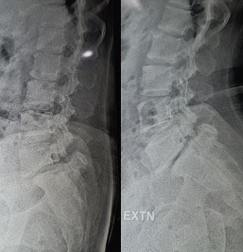
Radiologic Investigations
When the facet joint is disrupted, the normally posteriorly located inferior articular process moves anteriorly; this anterior movement is to the point that the inferior articular process is riding on the top of the superior articular process, a situation termed “perched facet.” For a more violent shear force, the inferior articular process moves more forward and becomes anchored anteriorly to the superior articular process. This phenomenon is referred to as a locked facet. The disruption of the facet joint leads to a misalignment of the vertebral bodies, narrowing the spinal canal and its neural content. This injury pattern leads to cauda equina injury and/or spinal cord injury if it affects the thoracolumbar junction.[12]
These disruptive changes to the spinal column architecture are readily observed on CT scan with 3D reconstruction as well as on MRI. For visualization of trauma to the spinal cord or cauda equina, MRI is the imaging modality of choice as it also highlights the para-spinal muscles injuries. The traumatic strain and stretch results in muscle edemas, which is well demonstrated as hyper-intense signal change on STIR images. The key features in imaging assessments are the loss of apposition at facet joints and the increased inter-spinous distance. Nonetheless, disc assessment is mandatory in all cases because severe disc injuries requiring fusion may be found even in the absence of an anterior slip.
A commonly adopted method of grading spondylolisthesis is the Meyerding classification which is based on the percentage of the distance the anteriorly translated vertebral body has moved forward relative to the superior endplate of the body. Grades using this system are as follows:
- Grade I: 0% to 25% (or low grade)
- Grade II: 26% to 50%
- Grade III: 51% to 75%
- Grade IV: 76% to 100%
- Grade V: greater than 100% (also known as spondylosis)
Treatment / Management
The goal of the surgery is to decompress the spinal canal and restore the stability of the spine. The decompression is achieved by laminectomies, the reduction of the listhesis often requires manual traction with facetectomies. The stabilization is performed by posterior lumbar interbody and fusion by open or minimally invasive surgery with satisfactory postoperative imaging.
Enhancing Healthcare Team Outcomes
Lumbosacral bony injuries are best managed by an interprofessional team that includes orthopedic nurses. The key is to restore function and minimize pain. Rehabilitation is often required to regain strength and muscle function. The outcomes depend on the cause, severity of the injury and presence of neurological deficit at time of presentation.