Continuing Education Activity
An ileostomy is a procedure in which the lumen of the ileum, part of the small bowel, is brought through the abdominal wall via a surgically-created opening called a stoma. The purpose of an ileostomy is to evacuate stool from the body via the ileum instead of the usual route via the anus. This activity reviews the indications, contraindications, and potential benefits of the ileostomy and highlights the role of the interprofessional team in the management of patients with a stoma.
Objectives:
- Describe the indications for an ileostomy.
- Outline the contraindications to ileostomy.
- Review the technique involved in creating an ileostomy.
- Explain the importance of enhancing care coordination among interprofessional team members to improve outcomes for patients undergoing ileostomy.
Introduction
An ileostomy is when the lumen of the ileum (small bowel) is brought through the abdominal wall via a surgical opening (created by an operation). This can either be temporary or permanent, an end or a loop. The purpose of an ileostomy is to evacuate stool from the body via the ileum rather than the usual route of the anus. The output from an ileostomy consists of loose or porridge-like stool consistent with that expected to pass through the small bowel (as it is the large bowel that is responsible for making the stool more solid dependent upon water absorption). The output from an ileostomy can vary but typically ranges from 200 to 700 ml per day, and an Ileostomy is typically formed on the right side of the abdomen.
Anatomy and Physiology
An ileostomy is formed from a section of ileum which is part of the small intestine. The small intestine begins at the pylorus of the stomach and is composed of three adjoining sections: the duodenum proximally, jejunum, and the ileum distally. The jejunum and ileum are intraperitoneal structures whereas the duodenum has a retroperitoneal component to it. The jejunum and duodenum are attached to the small bowel mesentery which are peritoneal folds containing blood vessels, lymphatics, and nerves. The small intestine is approximately 6 to 7 meters in length with a varying luminal diameter between 3 and 5 cm. It is has multiple functions including food digestion, secretion of enzymes and proteins, and nutrient absorption [1]. The wall of the intestine consists of the mucosa, the submucosa, muscularis propria (the muscular layer), the subserosa, and finally the serosa [2]. The ileum terminates at the ileocaecal junction in a valve at the superior aspect of the caecum before it goes on to form the ascending colon. The caecum can be identified at this point of the bowel where the tinea converge.
The anatomy of the anterior abdominal wall is important to be aware of when forming the trephine incision for the ileostomy. The layers encountered are the skin, subcutaneous fat, Scarpa's and Camper's fascia, anterior rectus sheath, muscle, posterior rectus sheath (if above the arcuate line) and the peritoneum. The muscles include the external and internal obliques, the transverse abdominis and the rectus abdominis. The obliques and transverse abdominis attach at varying levels to the lower ribs and the iliac crests, whereas the rectus abdominis arises from the costal margin and xiphoid process before extending down to the symphysis pubis. The abdominal muscles are wrapped in fascia but also have dense tendons called aponeuroses that converge at the midline to form the linea alba.
An ileostomy should be brought through the rectus muscle and sheath to reduce the risk of laterparastomal hernia formation, which occurs when the abdominal content pushes through the weakness created by the incision.
Indications
There are different indications for forming an ileostomy but essentially arrive at the same result of diverting stool out of the body without it ever entering the colon.
A loop ileostomy is when a distal loop of the ileum is brought out to the skin with 2 lumens draining into the stoma bag and is commonly used as a temporary diversion of stool usually to protect a distal anastomosis such as a colonic anastomosis in segmental colonic resections. The reason to protect such distal anastomoses is to reduce the risk of an anastomotic leak from when stool passes through the join of the two ends of the bowel [3]. Once the distal anastomosis has healed, both limbs of the loop ileostomy can be joined back together thereby restoring continuity to the gastrointestinal tract, which allows stool to pass through into the colon. With loop ileostomies, the proximal limb is the one that passes out the stool, and the distal limb usually acts as a mucous fistula, draining out the secretions produced within the mucosal lining from the lumen to the caecum. However, the distal limb does not drain out colonic secretions if the ileocaecal valve is competent, and therefore, does not decompress the colon. This is important to note if there is a colonic obstruction as then the patient would be at risk of perforation from a large bowel obstruction. This is because the colon is unable to decompress either proximally or distally to the obstructing source, causing secretions and flatus to build up under tension in an essentially closed loop of bowel. At a later date, usually between three and six months, this temporary ileostomy can be "reversed" or re-joined back together to re-establish continuity of the bowel.
An end ileostomy is when there is nothing distal to the proximal emptying limb, in other words, there is no bowel to be re-attached to this "end" at a later stage. The formation of an end ileostomy is usually considered following permanent removal of the entire colon, and therefore the patient manages their stoma for the rest of their life.
In brief, the indications for forming an ileostomy include:
- To defunction the rest of the bowel in order to protect a distal anastomosis
- To evacuate stool from the body if the entire colon has been removed such as in colorectal cancer, Crohn’s disease, ulcerative colitis, and familial adenomatous polyposis
- Relieve bowel obstruction
Contraindications
There are no absolute contraindications to ileostomy formation, but the relative ones include:
- Short mesentery that disables the ileum from being exteriorized through the abdominal wall to the skin without tension. This, unfortunately, is more common in obese patients.
- Carcinomatosis that prevents full mobilization of the ileum
The ileostomy should be formed as distal as possible to allow enough bowel length for absorption of nutrients.
A high output ileostomy can lead to electrolyte disturbances (particularly important to monitor for and treat in patients with renal impairment), as well as malabsorption leading to malnutrition.
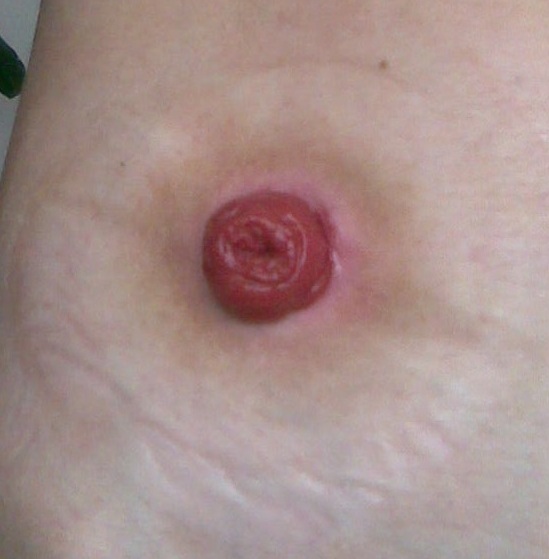
During ileostomy formation, it is important to spout the stump to get the effluent away from contact with the skin.
An ileostomy should be sited away from scars, skin creases, and bony prominences to allow placement of the stoma appliance and avoid leakage.
Equipment
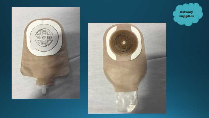
The equipment required can be split into the operative stage and the maintenance stage. In the operative stage, the formation of the trephine ileostomy involves the utilization of many instruments that are discussed below in the technique section. The maintenance stage is based on stoma education and how the patient manages their ileostomy. This includes the use of stoma bags, perhaps extra adhesive fixings such as spray, powder, paste and rings, as well as belts, adhesive removal spray, wet wipes, and waste disposal bags.
Preparation
This includes both physical and psychological elements of preparation; the stoma nurses are again invaluable here to help support the patient through this process. Physical preparation varies somewhat on the nature of the operation involved and whether it is an emergency procedure or a planned elective operation.
Points to consider include:
- Shaving of hair on the abdominal wall
- Body mass index (BMI) of patients having a planned ileostomy in an elective setting: They may be asked to lose weight prior to the operation to not only improve their anesthetic suitability but also reduce the distance the small bowel mesentery has to traverse to be bought to the skin without being under tension.
- Previous operative scars/deformities of the abdominal wall. A previous operation increases the extent of adhesions
- The presence of herniae
- Also in a planned setting, one would consider the effect of smoking and diabetic control on wound healing
- Most importantly would be the positioning of the stoma site, which is usually on the right side of the abdomen at the lateral edge of the rectus muscle, at a level where the patient can see it, access it easily and not have it interfering with belts or skin folds. It should also ideally avoid the costal margin and umbilicus.
- A nasogastric tube in cases of obstruction/perforation or if anticipating a post-operative ileus
- Adequate fluid and electrolyte resuscitation
Technique or Treatment
Ideally, the site would be marked pre-operatively with indelible ink or an "X" scored into the skin. This is so that the site can still be seen at the end of a long operation when the antiseptic prep or blood may have distorted the skin. If a laparotomy has been formed then the linea alba, which is the cut edge of the abdominal wall, is grasped with Kocher clamps or Littlewood clamps and retracted toward the midline to approximate the two wound edges together. As this will be the anatomically correct position of closure of the abdominal wall, it will also help identify where the ileostomy will be sited once the wound is closed. If a loop ileostomy is being made laparoscopically, then under vision the ileum can be grasped with a pair of Johan atraumatic graspers and bought towards the anterior abdominal wall to sit in a position where it is not under tension.
A 2.5 to 3 cm circle or ellipse of skin is excised using monopolar diathermy (it may be helpful to lift the skin upward using an Alice clamp or Littlewood clamp). The tissue is then dissected down through the subcutaneous fat to the anterior fascial sheath of the rectus muscle, which is then opened through a cruciate incision. The rectus muscle is spread or retracted medially; however, caution must be taken to avoid injuring the epigastric vessels that run deeply in the center of this muscle. Once the muscle is retracted, the posterior sheath is seen underlying this which is usually closed attached to the peritoneum on its under-surface. Another cruciate incision is made to the posterior sheath, and then two Kelly clips are used to grasp the peritoneum and lift it up. Using dissection scissors, a cut is made in the peritoneum between the two clips which will gain access into the peritoneal cavity. The surgical defect is stretched to allow two fingers to traverse through it thus ensuring enough room for the small bowel to be bought up to form the ileostomy. The next step is to gently pass the selected segment of terminal ileum (which has been checked to have enough length, mobilization and is tension-free) through the trephine that you just formed. If a loop ileostomy is being formed, it is the loop of ileum that is bought up through the abdominal wall defect to the skin. If an end ileostomy is being formed, then it is simply the stapled off the end of ileum that is bought up. The ileum should be positioned such that the proximal limb is cephalad and is at 12 o’clock. It should also protrude approximately 5 cm above the skin before a seromuscular absorbable stay suture (e.g., 3.0 monocryl or vicryl rapide) is placed to the skin to prevent the ileum from slipping back inside. This then allows you to perform your final checks (checking orientation, controlling hemostasis, washout, rectus sheath catheters, drains) prior to closing up the abdomen and protecting the wounds with dressings before focussing on the ileostomy formation. It is considered common practice to close any abdominal wounds prior to the formation of the ileostomy so as to prevent faecal contamination of the wound with stool from the ileum.
Loop Ileostomy Formation
The distal limb is opened transversely for two-thirds of its diameter in a position about halfway up from the skin level. Submucosal bleeding can be controlled with bipolar cautery. Interrupted absorbable sutures are placed at the 3, 9 and 12 o’clock position taking seromuscular bites at the lumen of the proximal limb as well as approximately 4 cm down the loop before taking a subcuticular bite of the skin at the trephine skin edge. The positions here are away from the supplying mesentery. A Langenbeck retractor can then be used to help evert the lumen so that the limb is now spouting. The interrupted sutures are then tied in place using square knots. The distal limb is also everted similarly, however, will be less spouted as there is less protrusion of the distal limb above skin level. Interrupted absorbable sutures are applied circumferentially around both limbs, taking care not to compromise the vascular supply of the mesentery.
End Ileostomy
The formation of the trephine is the same here as mentioned above. Once you pull the stapled end of ileum through the abdominal wall defect and apply the stay suture, you still proceed to carry out the usual checks and close the wounds as previously mentioned. Then taking the monopolar diathermy, you excise the staple line from the ileal end and discard it. Some surgeons may choose to cut the staple line off using dissecting scissors. However, this increases the risk of bleeding from the cut edges of the bowel which can be troublesome at times to control. Once the staple line has been excised, open up the lumen and then apply the 3 interrupted absorbable sutures at the 3, 9 and 12 o’clock position taking seromuscular bites. Again using a Langenbeck retractor, evert the mucosa of the lumen so that the limb is now spouting. The interrupted sutures are then tied in place using square knots.
Complications
These can be classified as immediate, early or late or as procedure specific and general complications. It is important to note that complications following the creation of an intestinal stoma are experienced by 20% of the patients. General complications vary depending on the type of operation being undertaken for an ileostomy to be necessary. Procedure-specific complications include the following [4]:
- Stenosis
- Ischemia/Necrosis
- Hemorrhage
- Infection/Abscess
- A parastomal hernia
- Retraction/Prolapse
- Electrolyte imbalance due to the high output of the effluent from the ileostomy
- Dehydration
- Renal impairment
- Hematoma/Seroma
- Obstruction
- Fistula formation
- Skin irritation
Clinical Significance
The formation of an ileostomy, whether in an emergency or elective setting, can be considered an adjunct to a life-saving operative technique and this is a procedure that is performed in the best interests of the patient to either improve their quality of life or to help save their life. It is critical to stress to patients that it is still possible to live a normal life and continue with their usual activities of daily living despite having an ileostomy [5].[6][7][8]
Enhancing Healthcare Team Outcomes
While an ileostomy is usually done by a general surgeon, pediatric surgeon or a colorectal surgeon, the management of the ileostomy is by a stoma nurse. Ileostomy formation can be a result of either emergency or elective surgery. It is important that in both situations, the risk of formation and its implications are discussed in detail. Key personnel involved in this process include the surgeon who has the responsibility of consenting the patient regarding the details of the procedure as well as the post-operative course involved. The stoma nurses are very helpful in helping to identify where the ileostomy is best placed as well as in stoma education and psychological support. During the operation, other key members include the scrub nurse and the surgical assistant where appropriate. It is important to seek a mental health consultant for patients with an ileostomy, because many patients do not fully realize what it entails. [9][10][11](Level V)