Continuing Education Activity
Psoriasis is a chronic inflammatory disorder that presents as well-defined, scaly, erythematous plaques and, occasionally, sterile pustules. Guttate psoriasis is a distinct variant of psoriasis that is classically triggered by streptococcal infection (pharyngitis or perianal) and is more common in children and adolescents than adults. Patients present with several small drop-like lesions that respond well to topical and phototherapies. This activity describes the pathophysiology and presentation of guttate psoriasis and highlights the role of the interprofessional team in its management.
Objectives:
Describe the pathophysiology of guttate psoriasis.
Outline the presentation of guttate psoriasis.
Identify the treatment and management options available for guttate psoriasis.
Discuss interprofessional team strategies for improving care coordination and outcomes in patients with guttate psoriasis.
Introduction
Psoriasis is a chronic inflammatory disorder that presents as well-defined, scaly, erythematous plaques and, occasionally, sterile pustules. Several clinical variants of psoriasis exist, including plaque psoriasis, guttate psoriasis, and pustular variants. Psoriasis frequently involves the joints, leading to psoriatic arthritis. Nail involvement is found in 10-80% of patients with psoriasis.[1] Psoriasis displays a spectrum of clinical manifestations, and several variants may be present simultaneously in the same patient.
Guttate psoriasis is a distinct variant of psoriasis that is classically triggered by streptococcal infection (pharyngitis or perianal) and is more common in children and adolescents than adults. Patients present with several small “drop-like” lesions that respond well to topical and phototherapies.[2]
Etiology
Risk factors for developing guttate psoriasis include recent streptococcal infection (oropharynx or perianal) or upper respiratory infection. Infections typically occur 1 to 3 weeks before the onset of guttate lesions. Guttate psoriasis lesions have also been described following TNF-alpha therapy.[3]
Epidemiology
Approximately 2% of the world population is afflicted with psoriasis; in the United States and Canada, the prevalence is as high as 4%. In general, psoriasis has a bimodal peak of onset. Onset can peak at 20 to 30 years of age and 50 to 60 years of age. Guttate psoriasis accounts for less than 30% of all total cases of psoriasis. It occurs equally in both genders and is more common in children and adolescents than adults over the age of 30.[1]
Pathophysiology
The interplay between genetic and environmental factors is implicated in the pathogenesis of psoriasis. Psoriasis is primarily a disorder of T cells, namely CD*+ T cells in the epidermis and a mixture of CD4+ and CD8+ cells in the dermis.[4] Th1 cytokines (interferon-gamma [IFN-] and interleukin [IL-]2), IL-1, I-6, and tumor necrosis factor-alpha (TNF-alpha) are upregulated, and Th2 cytokines (IL-10) are downregulated. Much of the success of biologic therapy is owed to the role of IL-23 in the pathogenesis of psoriasis. Dendritic cells release IL-23, which promotes Th17 stimulation, causing IL-17 and IL-22 release, leading to dermal inflammation and keratinocyte replication. Understanding this complex interplay between interleukins has allowed targeted biologic therapy for psoriasis.
The most important gene implicated in the pathogenesis of psoriasis is the PSORS1 gene on chromosome 6p, containing the HLA-Cw6 allele. The HLA-Cw6 allele is strongly linked to both early onset plaque psoriasis and guttate psoriasis. HLA B-13 and HLA B-17 are also strongly associated with guttate psoriasis (and erythrodermic) psoriasis.
Histopathology
Psoriasis is the model of the psoriasiform reaction pattern, defined as epidermal hyperplasia with elongation of the rete ridges. In addition to hyperplasia of the epidermis, there is an elongation of the dermal papillae, dilated superficial blood vessels, hypergranulosis, and parakeratosis.
Remnants of neutrophils in the stratum corneum are referred to as microabscesses of Munro. A discrete collection of neutrophils (spongiform pustule of Kogoj) can also be found in the upper stratum spinosum.
History and Physical
Eliciting a thorough history can be valuable in the diagnosis of guttate psoriasis. Assessing the presence of risk factors can aid in diagnosing guttate psoriasis. Guttate psoriasis classically follows a preceding streptococcal infection, typically pharyngitis or perianal streptococcus. The patient should be questioned regarding the recent use of TNF-alpha inhibitors, as they have been implicated in guttate psoriasis.
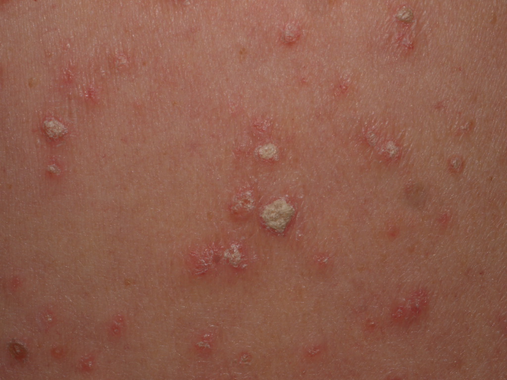
On physical exam, guttate psoriasis manifests as numerous, small, scattered papules and plaques. These are often referred to as “drop-like” and typically manifest as 2 to 6 mm papules. These lesions exhibit an isomorphic response or the Koebner phenomenon. The Koebner phenomenon refers to the appearance of lesions in areas of trauma (e.g., excoriations, sunburn, lesions of other eruptions such as Varicella or Pityrosporum folliculitis), as wound healing triggers hyperproliferative changes in the skin. Although psoriasis is the prototype of koebnerization, this response can also be seen in pityriasis rubra pilaris, lichen planus, verruca plana (flat warts), lichen nitidus, vitiligo, lichen sclerosus, elastosis perforans serpiginosa, systemic lupus erythematosus, and other dermatological conditions.
In addition to the Koebner phenomenon, all variants of psoriasis share certain characteristics that are reflected on histology, for example, erythema (elongated and dilated capillaries on histology), thickness (acanthosis with cellular infiltrates on histology), and silver scale (reflects abnormal keratinization). All lesions of psoriasis exhibit Auspitz sign, or pinpoint bleeding when the surface of a psoriatic lesion is removed. This reflects elongated vessels in the dermal papillae, together with thinning of the epidermis.
Evaluation
A diagnosis of guttate psoriasis can usually be made on history and clinical grounds alone. Skin biopsy is typically not necessary; however, lesions should demonstrate the histopathological findings discussed above. An elevated antistreptolysin O, anti-DNase B, or streptozyme titer can indicate a recent streptococcal infection, further guiding the diagnosis towards guttate psoriasis.
Treatment / Management
For mild psoriasis, topical corticosteroids are first-line treatment.[5] Topical steroids are available in several forms, including ointments, creams, lotions, gels, foams, sprays, and shampoos. Ointment formulations are the most efficacious mainly secondary to their lipophilic properties. Corticosteroids are contraindicated in cases of bacterial, viral, and mycotic infections, and atrophy of the skin. Second-line therapy for mild psoriasis is anthralin (dithranol) as monotherapy or in combination with topical corticosteroids. Other topical steroids include vitamin D analogs (calcipotriene), topical retinoids (tazarotene), topical calcineurin inhibitors, and salicylic acid.[5]
For moderate to severe psoriasis, phototherapy is first-line treatment as monotherapy or in combination. This includes phototherapy with broadband or narrowband ultraviolet B (UVB) and photochemotherapy with ultraviolet A (UVA) following psoralen ingestion or application (PUVA). Broadband UVB is the best phototherapy option for guttate psoriasis, in contrast to plaque psoriasis, which is better treated by narrowband UVB. Excimer laser (308 nm) can be used for localized psoriatic plaques.
Several biologic treatment options are indicated for plaque psoriasis. However, they have not been well-studied for guttate psoriasis. Currently, targeted biologic therapy is best reserved for the 40% of guttate lesions that progress to plaque type.
Differential Diagnosis
The differential diagnoses of guttate psoriasis include tinea corporis, secondary syphilis, nummular eczema, and pityriasis rosea. Guttate psoriasis can be distinguished from these entities by history and physical exam, although further studies such as potassium hydroxide (KOH) scrapings and serologies may be helpful in ruling out the other disorders in the differential diagnoses.
Prognosis
Forty percent of patients with guttate psoriasis convert to plaque psoriasis.
Enhancing Healthcare Team Outcomes
Psoriasis is a chronic inflammatory disorder that is challenging to evaluate and treat. It is more common in children and adolescents than adults. An interprofessional team of clinicians, nurses, and pharmacists managing patients with this disease will lead to the best outcomes. [Level 5]