IgA Nephropathy (Berger Disease)

Earn CME/CE in your profession:


Continuing Education Activity

IgA nephropathy is a common form of glomerulonephritis caused by the deposition of IgA immunoglobulins in the glomerular basement membrane. Immune-mediated damage to the basement membrane results in hematuria and renal insufficiency. Pathologically, a spectrum of glomerular lesions may be seen; however, the most commonly observed change is the mesangial proliferation with prominent IgA deposition. It also carries the name Berger disease. Although IgA nephropathy is a common disease, the data on prevalence is not very accurate because a renal biopsy is necessary to establish the diagnosis. This activity reviews the etiology, pathogenesis, evaluation, and management of IgA nephropathy and explains the role of the interprofessional team in evaluating and treating patients with this condition.

Objectives:

  • Identify the epidemiology of IgA nephropathy.

  • Describe the evaluation of IgA nephropathy.

  • Summarize the management options available for IgA nephropathy.

  • Explain interprofessional team strategies for improving care coordination and communication to improve outcomes for patients affected by IgA nephropathy.

Introduction

Immunoglobulin A (IgA) nephropathy is one of the leading causes of glomerulonephritis and renal failure.[1][2] It is characterized by the deposition of IgA immunoglobulin in the glomerular mesangium. Immune-mediated damage to the basement membrane results in hematuria and renal insufficiency.[3] Berger and Hinglais were the first to describe the disease in 1968, so it also carries the name Berger disease.[4] Pathologically, a spectrum of glomerular lesions may be seen; however, the most commonly observed change is the mesangial proliferation with prominent IgA deposition.

Recent international collaborative efforts have made important discoveries leading to a better understanding of some of the key features of the immunopathogenesis of IgA nephropathy. Furthermore, establishing multicenter networks has helped in the holistic design and execution of clinical trials providing important insights about immunotherapy in IgA nephropathy.

Etiology

IgA nephropathy is an autoimmune disease causing antibody-mediated destruction of the glomerular basement membrane.[5] Usually, there is an infectious disease preceding the nephropathy, which leads to the dysregulated immune response, but IgA nephropathy per se is not of an infectious etiology. There has been no evidence to suggest that IgA nephropathy is secondary to any specific infectious agents. The following are the possible etiologies underlying IgA nephropathy:

Familial

  • Less than 10% of cases are due to familial IgA nephropathy
  • Genome-wide linkage analysis revealed an association of IgA nephropathy to 6q22-23 and gene locus IGAN1[6]
  • There are no obvious genes within the linked interval
  • It is unclear that genetic findings in these families will have a direct bearing on typical sporadic cases of IgA nephropathy

Sporadic/idiopathic

  • More than 90% of cases are sporadic, and the underlying etiology is unknown[7]
  • No evidence to suggest the involvement of a particular infectious agent despite the association between macroscopic hematuria and mucosal inflammation
  • No evidence to point toward hypersensitivity to food antigens, with the exception of a small group of patients with celiac disease
  • Abnormal O-glycosylation of the IgA1 hinge region promotes the formation of circulating IgA immune complexes leading to mesangial cell activation and deposition[7]
  • IgA nephropathy may also be seen in hepatitis, cirrhosis, and human immunodeficiency virus (HIV) infection[5][8]

Epidemiology

Although IgA nephropathy is a common disease, the data on prevalence is not very accurate because a renal biopsy is necessary to establish the diagnosis. Not all patients undergo a biopsy to confirm the diagnosis and instead receive conservative management. About 10% of the renal biopsies in the United States show IgA nephropathy. Forty percent of renal biopsies in Asia and 20% of renal biopsies in Europe show IgA nephropathy.[4] The high prevalence is thought to be due to the early detection of hematuria during screening and aggressive treatment plans. The disease is common in children and young adults with a male predominance.[9]

The prevalence varies geographically, and variations in disease burden are seen depending on whether biopsy registry data was used or dialysis registries were utilized to obtain these estimates. Despite these caveats, organ replacement and biopsy registries indicate geographic variation in disease estimates with a higher burden in Pacific Asian regions. Biopsy registry data is sometimes inaccurate as patients with mild disease may never undergo a biopsy, and the disease may not be detected. A systematic study of biopsy-based literature from multiple countries reveals an overall incidence of over 2.5 per 100,000.[10] 

Most studies in this review were from Europe and North America, except one from Japan. In a Japanese study of the pediatric population, the incidence in the biopsy registry where broad screening programs were carried out was eight-fold greater than in a Tennessee program where the biopsy was not part of routine screening and was only done in certain cases (4.5 versus 0.57 per 100,000/year).[11] In another study, IgA nephropathy is more common in Asian individuals (45 cases per million population/year in Japan) than in Whites (31 cases per million population/year in France).[12]

Compelling data suggests a higher burden of IgA nephropathy in East and Pacific Asian countries. These reports parallel geospatial differences in the prevalence of genetic susceptibility loci observed in global genome-wide association studies.[13]

There are regional variations in the progression of the disease; however, these are difficult to establish given lead-time biases introduced by differences in biopsy practice. In a study of 669 patients, the multivariable analysis revealed that people of Pacific Asian origin have a higher risk of end-stage renal disease (ESRD).[14]

Pathophysiology

The current understanding is that IgA nephropathy occurs due to a multi-hit mechanism.[15] The first ‘hit’ is a genetically susceptible host predisposed to developing a dysregulated immune response. The next ‘hit’ is a precipitating factor producing the immunological attack. Infections are potential precipitants of IgA nephropathy.[5] Trivial mucosal infections, chronic exposure to pathogens, and abnormal handling of commensals in the gut have all been hypothesized to trigger the abnormal immune response in IgA nephropathy.[5] The damage to the basement membrane results in the ultrafiltration of larger molecules and produces hematuria. The pathophysiology of how some develop asymptomatic hematuria while some develop rapidly progressive glomerulonephritis, culminating in renal failure, is poorly understood.[16]

Susceptibility to IgA nephropathy is dependent on many genetic and environmental factors. The pathogenesis of this disease is a multi-“hit” process.[15] These “hits” are understood from the IgA moieties found in biopsies and the circulation of patients with IgA nephropathy. A central finding in patients with IgA nephropathy is the presence of immune complexes in circulation and glomeruli comprised of galactose-deficient IgA1, which is an IgG autoantibody against C3 and the hinge region O-glycans. The presence of abnormally glycosylated IgA1 is a heritable trait.[17] In a quarter of blood relatives of IgA nephropathy patients, galactose-deficient IgA1 levels are elevated, and segregation analysis reveals a major dominant gene with a polygenic background.[18] A recent study outlines the cellular mechanisms causing IgA glycosylation.[19]

The levels of galactose-deficient IgA1 could also be influenced by environmental factors. For instance, these antibodies are prone to bacteria-derived proteases.[20] Recent studies suggest that anti-glycan autoantibodies could target the IgA VH gene segment due to somatic hypermutation and not sequences found in the host germline.[21] Glomerular inflammation and mesangial proliferation are thought to occur because these immune complexes are nephritogenic. Activation of the renin-angiotensin and complement systems also leads to glomerulosclerosis and tubulointerstitial fibrosis, causing deranged renal function. Other risk factors, such as smoking and hypertension, contribute to disease progression through microvascular injury.[22] Glmoerulomegaly and maladaptive hyperfiltration injury attributed to obesity may also be implicated in the nonimmunologic progression of the disease.[23]

Experimental studies in mice suggest that exposure to bacteria is needed for excess IgA production, which is enabled by the mediators of B cell differentiation and proliferation. Although the application of this theory to IgA nephropathy in humans must be made cautiously, this idea is further aided by genome-wide association studies and studies of disease progression.[24]

It is hypothesized that cytokine APRIL (a proliferation-inducing ligand) contributes to IgA nephropathy by propagating B cell class switch to IgA-producing plasma cells through actions on the TACI receptor. APRIL gene polymorphism confers IgA nephropathy susceptibility, and various risk alleles linked to IgA nephropathy are also associated with several diseases of mucosal immunity.[25]

Activation of complement is described as a significant pathogenic contributor to IgA nephropathy, particularly the lectin pathway. Polymeric IgA1 can activate this pathway, and the mannose-binding lectin pathway is detected in glomerular deposits.[26][27] Immune complexes contain C3, as seen in the immunofluorescence study of kidney biopsies.[28] Complement factor H (CFH) and properdin are also observed in immune deposits (as observed by Gharavi et al.).[29] Genome-wide association studies report an allele localized to the CFH gene conferring protection against the development of IgA nephropathy. Further analysis indicates that the deletion of complement factor H–related (CFHR) genes is in linkage disequilibrium with the observed risk allele. The CFHR1 and CFHR3 titrate CFH activity, and their absence leads to altered CFH levels and increased CFH activity.[30]

Histopathology

Histologically, IgA nephropathy is characterized by the following: 

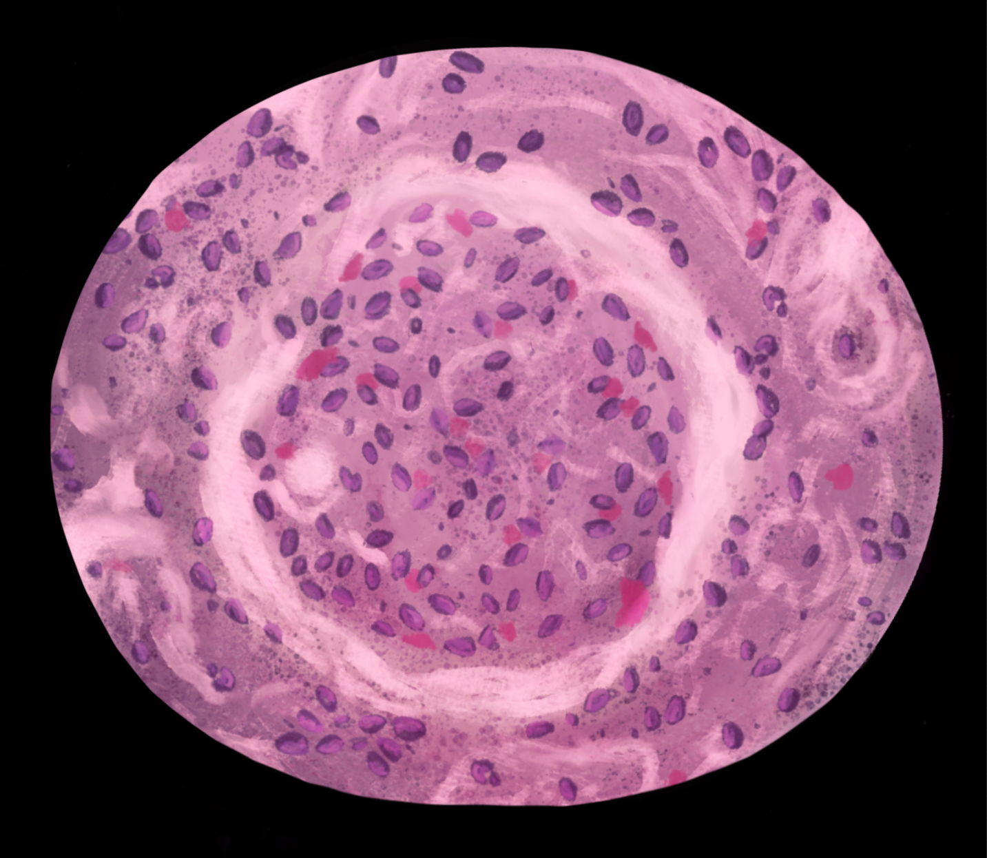
  • The diffuse proliferation of mesangial cells and matrix
  • Hypercellular or normal glomeruli with diffuse necrotizing crescentic glomerulonephritis
  • Mesangial involvement resembling focal and segmental glomerulosclerosis
  • Immunofluorescence will reveal a diffuse granular pattern of IgA deposits in the mesangium

Light Microscopy

The most commonly seen light microscopy findings are focal or diffuse mesangial proliferation and expansion of the extracellular matrix.[31] Morphology shows intracapillary and extracapillary proliferative lesions. Occasionally, focal glomerular sclerosis is seen as indistinguishable from focal segmental glomerulosclerosis. In advanced diseases, interstitial fibrosis, vascular sclerosis, and tubular atrophy can be seen. A few patients show segmental necrotizing areas with crescent formation because of extensive disruption of the capillaries.

Electron Microscopy

Electron microscopy reveals mesangial hypercellularity and excess mesangial matrix. An important finding is the presence of mesangial electron-dense deposits of IgA; however, subepithelial and subendothelial deposits of the glomerular capillary wall are seen in a minority of patients, particularly those with the more severe form of the disease.[32]

Immunofluorescence

Immunofluorescence reveals mesangial IgA deposits in a diffuse granular pattern. These deposits are majorly polymeric IgA of the IgA1 subclass. Additionally, IgG is found in 43% of patients and IgM in 54%.[15] C3 is also often present. The presence of C4d imparts a worse prognosis.

History and Physical

In most patients with IgA nephropathy, history and examination would be unremarkable. The most common complaint is gross hematuria. Acute renal failure may cause ankle edema, facial puffiness, and hypertension. Frothy urine may be present. A history of upper respiratory tract infections, such as pharyngitis, may be present just before hematuria.[33] The history of previous episodes of hematuria and proteinuria should be verified.

Physical examination should include checking for blood pressure and looking for signs of reduced renal function, such as edema, ascites, and lung basal crepitations. In addition, IgA nephropathy may coexist with cirrhosis, liver diseases, and celiac disease. Therefore, relevant general examination and abdominal examination should be done to exclude these clinically.

The range of clinical features of IgA nephropathy is broad, from asymptomatic hematuria to rapidly progressive glomerulonephritis. The mode of presentation differs according to age group and biopsy patterns. The two most common clinical phenotypes are:

  • Asymptomatic hematuria
  • Progressive kidney disease

Asymptomatic hematuria with mild proteinuria, such as 0.5 g/day, may be picked up on screening programs. A proportion of patients with isolated microscopic hematuria and mild proteinuria will ultimately develop significant proteinuria and hypertension, suggesting that long-term follow-up should be instituted.[34] Progressive chronic kidney disease is a common manifestation observed in many cohorts. Renal survival varies greatly according to biopsy timing and the introduction of lead-time bias. The actual renal 10-year survival is observed to be 57% to 91%.[35]

Less frequent manifestations include synpharyngitic macroscopic hematuria, which is a classic clinical syndrome seen as the first presentation of IgA nephropathy. Gross hematuria, in combination with pharyngitis or other infection, prompts patients to seek immediate medical attention. Recurrent macroscopic hematuria is also seen. Although nephrotic-range proteinuria is not uncommon in IgA nephropathy, they rarely coexist.[36][37]

Evaluation

Evaluation should start by establishing the diagnosis. The first investigation is urine analysis to look for microscopic hematuria. The presence of red cells and red cell casts indicate a glomerular injury. Protein to creatinine ratio in urine or 24-hour urinary protein excretion is done to look for proteinuria. Serum creatinine and eGFR are done to quantify renal function. Confirmation of the diagnosis relies on renal biopsy. Light microscopy, electron microscopy, and immunofluorescence are used to study renal histology. Immunofluorescence demonstrating the deposit of IgA in the glomerular basement membrane is the gold standard for diagnosis. IgA nephropathy should be classified using the Oxford classification, which can predict prognosis. The Oxford classification has its basis in a combination of histological, clinical, and biomarker criteria. 

The Oxford classification relies on the following criteria:

  • Mesangial cellularity
  • Endocapillary proliferation
  • Segmental glomerulosclerosis
  • Tubular atrophy
  • Crescents assessed by a renal biopsy[38]

Treatment / Management

The management of IgA nephropathy involves first confirming the diagnosis, followed by a renal biopsy. Secondary causes of IgA nephropathy should be ruled out. The amount of proteinuria, eGFR, blood pressure, and histological appearance is important in formulating the management plan. Treatment aims to induce remission and prevent the development of complications.[39]

Angiotensin-converting enzyme inhibitors or angiotensin receptor blockers are used to manage proteinuria and lower blood pressure. Salt intake is restricted to control blood pressure. The blood pressure target is 130/80 mmHg.[33]

Immunosuppression with corticosteroids or steroid-sparing agents is used to reduce the rate of progression.[40] Steroids have the most benefit if there is heavy proteinuria. Various regimens of oral prednisolone and methylprednisolone are available.[41] If there are contraindications for steroids or if the risks of therapy outweigh the anticipated benefits of steroid therapy, steroid-sparing agents may be an option. Cyclophosphamide, azathioprine, and cyclosporine are potential steroid-sparing agents.[42][43]

For the few who progress to develop end-stage renal disease (ESRD), renal transplantation is an option. There is still the risk of IgA nephropathy in the transplanted kidney. Treatment with an angiotensin-converting enzyme inhibitor or angiotensin receptor blocker may delay the progression of recurrent disease in allografts.[44]

Conservative Therapy

The role of conservative therapy in reducing proteinuria and slowing the rate of renal function decline in IgA nephropathy cannot be overemphasized. In the Supportive Versus Immunosuppressive Therapy of Progressive IgA Nephropathy (STOP-IgAN) study, around one-third of patients did not need immunosuppressive therapy after completing a 6-month run-in trial of optimization of conservative therapy.[45]

The role of renin-angiotensin system (RAS) blockade in IgA nephropathy is now well established. Dual RAS blockade should not be used due to the risks of hyperkalemia. Weight reduction may reduce proteinuria in IgA nephropathy.[46] Given the association between smoking and IgA nephropathy progression, smoking cessation should also be advised.[22]

Corticosteroids

There are many studies supporting that corticosteroids reduce proteinuria in IgA nephropathy. In an old study, patients received corticosteroid therapy or supportive therapy alone. The use of RAS blockade was random, and the run-in period of optimizing conservative treatment was not carried out. In this trial, 5-year renal survival was greater in the corticosteroid group.[47] This finding suggested that corticosteroids for a period of 6 months have a “legacy effect” with a sustained risk reduction in progressive renal dysfunction. In another similar study of patients with biopsy-proven IgA nephropathy, corticosteroids were reported to have reduced proteinuria.[48]

Rituximab

Although evidence for rituximab efficacy in other glomerular diseases is significant, early results in IgA nephropathy are not encouraging. A pilot study assessed the efficacy of rituximab versus conservative management in patients with proteinuria. No favorable effects on proteinuria or renal function were observed.[49]

Mycophenolate

Data around the efficacy of mycophenolate are equivocal, such that current guidelines recommend against using this agent in IgA nephropathy.[50] The majority of studies are limited by small sample size. There is also possible race-specific variation in response to mycophenolate, as in lupus nephritis.[51] However, systematic reviews and meta-analyses of the randomized trials of mycophenolate and systematic reviews suggest mixed results (64–66).[52][53]

Combination Therapy

Corticosteroids, in combination with an additional agent, are generally reserved for progressive disease. A single-center, randomized, prospective trial with “high-risk” IgA nephropathy demonstrated better renal survival in patients receiving prednisone in combination with cyclophosphamide/azathioprine as opposed to no immunotherapy.[54] In contrast, combination therapy does not always provide an advantage. A multicenter trial of 207 patients with IgA nephropathy demonstrated no difference between patients receiving corticosteroids alone and those taking combination therapy.[55]

Novel Agents

A novel potential therapy for IgA nephropathy is a formulation of oral budesonide. This agent can potentially act locally at the lymphoid tissue of the mucosa in the distal ileum and proximal large intestine to modulate IgA production. Higher first-pass metabolism theoretically minimizes systemic effects.

Differential Diagnosis

The diagnosis is usually apparent clinically and can be easily confirmed by investigations. The following is the list of differentials for IgA nephropathy:

  • Lupus nephritis
  • Membranoproliferative glomerulonephritis
  • Henoch-Schönlein purpura
  • Thin basement membrane nephropathy
  • Alport syndrome
  • Malignancies anywhere from the kidneys to the urethra
  • Local trauma
  • Urolithiasis
  • Urinary tract infections

Prognosis

Frequently IgA nephropathy patients have a benign course.[38] Infrequently, they may gradually progress to end-stage renal disease (ESRD), with the frequency of ESRD increasing with age.[3] The prognosis is predictable to some extent, based on the Oxford classification. Additionally, nephrotic range proteinuria, hypertension, high serum creatinine level, and widespread intestinal fibrosis of the kidneys on presentation indicate a poor prognosis.[9]

About 20% of patients will progress to ESRD within ten years. Currently, the MEST-C score is often used to predict the outcome. The score includes the following features:

  • M: Mesangial cellularity, defined as greater than 4 mesangial cells in any mesangial segment of the glomerulus
  • E: Endocapillary proliferation is the degree of hypercellularity
  • S: Segmental glomerulosclerosis is defined as sclerosis or adhesions in the glomerular tuft
  • T: Tubular atrophy or interstitial fibrosis
  • C: Presence or absence of crescents

In general, if any of the above features are seen, then the prognosis is poor. Other factors that determine outcomes include elevated creatinine, hypertension, need for antihypertensive treatment, proteinuria, decreased eGFR at diagnosis, significant interstitial fibrosis, and CD4 staining.[56][57] Isolated microscopic hematuria with mild proteinuria is considered favorable, particularly in Whites. Race may also be a significant determinant of outcome.[14]

Complications

Although only a small percentage of patients diagnosed with IgA nephropathy progress to ESRD, it remains a frequent cause of ESRD.[37] Complications of renal failure, such as hypertension, edema, anemia, heart failure, and pulmonary edema, may arise as the disease worsens. Side effects and complications of steroid and steroid-sparing therapy are common. Increased risk of infections, hypertension, fluid retention, weight gain, diabetes mellitus, osteoporosis, and iatrogenic Cushing syndrome is the most frequent side effects of steroid therapy.[41] Immunosuppression, anaphylaxis, renal, and hepatotoxicity are complications of steroid-sparing agents.

Postoperative and Rehabilitation Care

A low antigen diet with limitations in gluten, meat, and dairy products is recommended. Some studies indicate that low-protein diets may slow down the deterioration of renal function. The role of tonsillectomy remains controversial. Some experts believe that removing the tonsils may restrict the production of IgA. In the USA, tonsillectomy is only recommended for patients with tonsillar infections.

Consultations

Generally, the primary role in the management of patients with IgA nephropathy is of nephrologists. In establishing the diagnosis, histopathologists are critical in guiding physicians regarding biopsy results.

Deterrence and Patient Education

A minor upper respiratory tract infection can trigger the disease in genetically susceptible patients.[5] Routine screening to identify the persons at risk is not feasible. Prophylactic tonsillectomy has no proven benefit, nor does early antibiotic therapy.[33] Therefore, there are virtually no preventive measures. Patients with other autoimmune diseases and a strong family history of IgA nephropathy are more likely to have the disease. Screening for hematuria can detect patients early in the disease progression while asymptomatic.

Patients should receive instruction about the disease, the possibility of ESRD needing renal transplantation, the side effects of steroid therapy, and the importance of follow-up. Salt, protein, and saturated fat restriction are advised.

Enhancing Healthcare Team Outcomes

IgA nephropathy usually has a protracted course during which the patient may undergo management by an interprofessional team of healthcare providers. Ideally, a nephrologist (or pediatric nephrologist) plays a vital role in managing the patient. Coexisting autoimmune diseases may require management by a nurse practitioner, physician assistant, physician, endocrinologist, or gastroenterologist. The primary care team should be continuously updated about diagnosis and steroid therapy. A "steroid card" should be given to the patients.

The pharmacist should educate the patient regarding steroid therapy and promote compliance with communication as necessary to the clinical team. Laboratory staff should ensure the correct sample collection to avoid false positives for proteinuria. A dietitian should plan a low-salt, low-protein, and low-fat diet for the patient. Nurses are essential at every step of the way, assisting in patient assessment, offering patient counsel, and coordinating the activities of the various specialties on the case. All interprofessional team members must maintain accurate patient records and communicate with the rest of the team if they have any concerns regarding the patient's progress.

Fluid intake must be adjusted considering the fluid balance and renal function. If progression to ESRD is anticipated, the patient should be informed early in the course and be prepared for renal replacement therapy. A social worker would be a helpful team member at this stage. Due to the complexity of this disease, an interprofessional team of clinicians, pharmacists, nurses, and allied health professionals educating and assisting the patient and family will result in the best outcomes. [Level 5]


(Click Image to Enlarge)
<p>Illustration of IgA Nephropathy</p>

Illustration of IgA Nephropathy


Illustrated by Callahan Pels, MLS

Details

Author

Faten Limaiem

Updated:

7/7/2023 9:49:55 AM

References


[1]

Pattrapornpisut P, Avila-Casado C, Reich HN. IgA Nephropathy: Core Curriculum 2021. American journal of kidney diseases : the official journal of the National Kidney Foundation. 2021 Sep:78(3):429-441. doi: 10.1053/j.ajkd.2021.01.024. Epub 2021 Jul 9     [PubMed PMID: 34247883]


[2]

Working Group of the International IgA Nephropathy Network and the Renal Pathology Society, Coppo R, Troyanov S, Camilla R, Hogg RJ, Cattran DC, Cook HT, Feehally J, Roberts IS, Amore A, Alpers CE, Barratt J, Berthoux F, Bonsib S, Bruijn JA, D'Agati V, D'Amico G, Emancipator SN, Emma F, Ferrario F, Fervenza FC, Florquin S, Fogo AB, Geddes CC, Groene HJ, Haas M, Herzenberg AM, Hill PA, Hsu SI, Jennette JC, Joh K, Julian BA, Kawamura T, Lai FM, Li LS, Li PK, Liu ZH, Mezzano S, Schena FP, Tomino Y, Walker PD, Wang H, Weening JJ, Yoshikawa N, Zhang H. The Oxford IgA nephropathy clinicopathological classification is valid for children as well as adults. Kidney international. 2010 May:77(10):921-7. doi: 10.1038/ki.2010.43. Epub 2010 Mar 3     [PubMed PMID: 20200498]


[3]

Shen PC, He LQ, Tang Y, Wang Q, Wang W, Li J. Clinicopathological characteristics and prognostic factors of asymptomatic IgA nephropathy. Journal of investigative medicine : the official publication of the American Federation for Clinical Research. 2010 Mar:58(3):560-5. doi: 10.231/JIM.0b013e3181d20aa1. Epub     [PubMed PMID: 20215916]


[4]

Rodrigues JC, Haas M, Reich HN. IgA Nephropathy. Clinical journal of the American Society of Nephrology : CJASN. 2017 Apr 3:12(4):677-686. doi: 10.2215/CJN.07420716. Epub 2017 Feb 3     [PubMed PMID: 28159829]


[5]

Rollino C, Vischini G, Coppo R. IgA nephropathy and infections. Journal of nephrology. 2016 Aug:29(4):463-8. doi: 10.1007/s40620-016-0265-x. Epub 2016 Jan 22     [PubMed PMID: 26800970]


[6]

Woo KT, Lau YK, Choong HL, Tan HK, Foo MW, Lee EJ, Anantharaman V, Lee GS, Yap HK, Yi Z, Fook-Chong S, Wong KS, Chan CM. Genomics and disease progression in IgA nephritis. Annals of the Academy of Medicine, Singapore. 2013 Dec:42(12):674-80     [PubMed PMID: 24463829]


[7]

Lai KN, Tang SC, Schena FP, Novak J, Tomino Y, Fogo AB, Glassock RJ. IgA nephropathy. Nature reviews. Disease primers. 2016 Feb 11:2():16001. doi: 10.1038/nrdp.2016.1. Epub 2016 Feb 11     [PubMed PMID: 27189177]


[8]

Han SH, Kang EW, Kie JH, Yoo TH, Choi KH, Han DS, Kang SW. Spontaneous remission of IgA nephropathy associated with resolution of hepatitis A. American journal of kidney diseases : the official journal of the National Kidney Foundation. 2010 Dec:56(6):1163-7. doi: 10.1053/j.ajkd.2010.08.018. Epub 2010 Oct 8     [PubMed PMID: 20932622]


[9]

Soares MF, Roberts IS. IgA nephropathy: an update. Current opinion in nephrology and hypertension. 2017 May:26(3):165-171. doi: 10.1097/MNH.0000000000000312. Epub     [PubMed PMID: 28221174]

Level 3 (low-level) evidence

[10]

McGrogan A, Franssen CF, de Vries CS. The incidence of primary glomerulonephritis worldwide: a systematic review of the literature. Nephrology, dialysis, transplantation : official publication of the European Dialysis and Transplant Association - European Renal Association. 2011 Feb:26(2):414-30. doi: 10.1093/ndt/gfq665. Epub 2010 Nov 10     [PubMed PMID: 21068142]

Level 1 (high-level) evidence

[11]

Utsunomiya Y, Koda T, Kado T, Okada S, Hayashi A, Kanzaki S, Kasagi T, Hayashibara H, Okasora T. Incidence of pediatric IgA nephropathy. Pediatric nephrology (Berlin, Germany). 2003 Jun:18(6):511-5     [PubMed PMID: 12720079]


[12]

Schena FP, Nistor I. Epidemiology of IgA Nephropathy: A Global Perspective. Seminars in nephrology. 2018 Sep:38(5):435-442. doi: 10.1016/j.semnephrol.2018.05.013. Epub     [PubMed PMID: 30177015]

Level 3 (low-level) evidence

[13]

Kiryluk K, Li Y, Sanna-Cherchi S, Rohanizadegan M, Suzuki H, Eitner F, Snyder HJ, Choi M, Hou P, Scolari F, Izzi C, Gigante M, Gesualdo L, Savoldi S, Amoroso A, Cusi D, Zamboli P, Julian BA, Novak J, Wyatt RJ, Mucha K, Perola M, Kristiansson K, Viktorin A, Magnusson PK, Thorleifsson G, Thorsteinsdottir U, Stefansson K, Boland A, Metzger M, Thibaudin L, Wanner C, Jager KJ, Goto S, Maixnerova D, Karnib HH, Nagy J, Panzer U, Xie J, Chen N, Tesar V, Narita I, Berthoux F, Floege J, Stengel B, Zhang H, Lifton RP, Gharavi AG. Geographic differences in genetic susceptibility to IgA nephropathy: GWAS replication study and geospatial risk analysis. PLoS genetics. 2012:8(6):e1002765. doi: 10.1371/journal.pgen.1002765. Epub 2012 Jun 21     [PubMed PMID: 22737082]


[14]

Barbour SJ, Cattran DC, Kim SJ, Levin A, Wald R, Hladunewich MA, Reich HN. Individuals of Pacific Asian origin with IgA nephropathy have an increased risk of progression to end-stage renal disease. Kidney international. 2013 Nov:84(5):1017-24. doi: 10.1038/ki.2013.210. Epub 2013 Jun 5     [PubMed PMID: 23739233]


[15]

Magistroni R, D'Agati VD, Appel GB, Kiryluk K. New developments in the genetics, pathogenesis, and therapy of IgA nephropathy. Kidney international. 2015 Nov:88(5):974-89. doi: 10.1038/ki.2015.252. Epub 2015 Sep 16     [PubMed PMID: 26376134]


[16]

Suzuki H, Kiryluk K, Novak J, Moldoveanu Z, Herr AB, Renfrow MB, Wyatt RJ, Scolari F, Mestecky J, Gharavi AG, Julian BA. The pathophysiology of IgA nephropathy. Journal of the American Society of Nephrology : JASN. 2011 Oct:22(10):1795-803. doi: 10.1681/ASN.2011050464. Epub 2011 Sep 23     [PubMed PMID: 21949093]


[17]

Kiryluk K, Moldoveanu Z, Sanders JT, Eison TM, Suzuki H, Julian BA, Novak J, Gharavi AG, Wyatt RJ. Aberrant glycosylation of IgA1 is inherited in both pediatric IgA nephropathy and Henoch-Schönlein purpura nephritis. Kidney international. 2011 Jul:80(1):79-87. doi: 10.1038/ki.2011.16. Epub 2011 Feb 16     [PubMed PMID: 21326171]


[18]

Gharavi AG, Moldoveanu Z, Wyatt RJ, Barker CV, Woodford SY, Lifton RP, Mestecky J, Novak J, Julian BA. Aberrant IgA1 glycosylation is inherited in familial and sporadic IgA nephropathy. Journal of the American Society of Nephrology : JASN. 2008 May:19(5):1008-14. doi: 10.1681/ASN.2007091052. Epub 2008 Feb 13     [PubMed PMID: 18272841]


[19]

Knoppova B, Reily C, Maillard N, Rizk DV, Moldoveanu Z, Mestecky J, Raska M, Renfrow MB, Julian BA, Novak J. The Origin and Activities of IgA1-Containing Immune Complexes in IgA Nephropathy. Frontiers in immunology. 2016:7():117. doi: 10.3389/fimmu.2016.00117. Epub 2016 Apr 12     [PubMed PMID: 27148252]


[20]

Lamm ME, Emancipator SN, Robinson JK, Yamashita M, Fujioka H, Qiu J, Plaut AG. Microbial IgA protease removes IgA immune complexes from mouse glomeruli in vivo: potential therapy for IgA nephropathy. The American journal of pathology. 2008 Jan:172(1):31-6. doi: 10.2353/ajpath.2008.070131. Epub 2007 Dec 28     [PubMed PMID: 18165266]

Level 2 (mid-level) evidence

[21]

Huang ZQ, Raska M, Stewart TJ, Reily C, King RG, Crossman DK, Crowley MR, Hargett A, Zhang Z, Suzuki H, Hall S, Wyatt RJ, Julian BA, Renfrow MB, Gharavi AG, Novak J. Somatic Mutations Modulate Autoantibodies against Galactose-Deficient IgA1 in IgA Nephropathy. Journal of the American Society of Nephrology : JASN. 2016 Nov:27(11):3278-3284     [PubMed PMID: 26966014]


[22]

Yamamoto R, Nagasawa Y, Shoji T, Iwatani H, Hamano T, Kawada N, Inoue K, Uehata T, Kaneko T, Okada N, Moriyama T, Horio M, Yamauchi A, Tsubakihara Y, Imai E, Rakugi H, Isaka Y. Cigarette smoking and progression of IgA nephropathy. American journal of kidney diseases : the official journal of the National Kidney Foundation. 2010 Aug:56(2):313-24. doi: 10.1053/j.ajkd.2010.02.351. Epub 2010 May 14     [PubMed PMID: 20471735]


[23]

Kataoka H, Ohara M, Honda K, Mochizuki T, Nitta K. Maximal glomerular diameter as a 10-year prognostic indicator for IgA nephropathy. Nephrology, dialysis, transplantation : official publication of the European Dialysis and Transplant Association - European Renal Association. 2011 Dec:26(12):3937-43. doi: 10.1093/ndt/gfr139. Epub 2011 Mar 22     [PubMed PMID: 21427079]


[24]

Zhai YL, Zhu L, Shi SF, Liu LJ, Lv JC, Zhang H. Increased APRIL Expression Induces IgA1 Aberrant Glycosylation in IgA Nephropathy. Medicine. 2016 Mar:95(11):e3099. doi: 10.1097/MD.0000000000003099. Epub     [PubMed PMID: 26986150]


[25]

Kiryluk K, Li Y, Scolari F, Sanna-Cherchi S, Choi M, Verbitsky M, Fasel D, Lata S, Prakash S, Shapiro S, Fischman C, Snyder HJ, Appel G, Izzi C, Viola BF, Dallera N, Del Vecchio L, Barlassina C, Salvi E, Bertinetto FE, Amoroso A, Savoldi S, Rocchietti M, Amore A, Peruzzi L, Coppo R, Salvadori M, Ravani P, Magistroni R, Ghiggeri GM, Caridi G, Bodria M, Lugani F, Allegri L, Delsante M, Maiorana M, Magnano A, Frasca G, Boer E, Boscutti G, Ponticelli C, Mignani R, Marcantoni C, Di Landro D, Santoro D, Pani A, Polci R, Feriozzi S, Chicca S, Galliani M, Gigante M, Gesualdo L, Zamboli P, Battaglia GG, Garozzo M, Maixnerová D, Tesar V, Eitner F, Rauen T, Floege J, Kovacs T, Nagy J, Mucha K, Pączek L, Zaniew M, Mizerska-Wasiak M, Roszkowska-Blaim M, Pawlaczyk K, Gale D, Barratt J, Thibaudin L, Berthoux F, Canaud G, Boland A, Metzger M, Panzer U, Suzuki H, Goto S, Narita I, Caliskan Y, Xie J, Hou P, Chen N, Zhang H, Wyatt RJ, Novak J, Julian BA, Feehally J, Stengel B, Cusi D, Lifton RP, Gharavi AG. Discovery of new risk loci for IgA nephropathy implicates genes involved in immunity against intestinal pathogens. Nature genetics. 2014 Nov:46(11):1187-96. doi: 10.1038/ng.3118. Epub 2014 Oct 12     [PubMed PMID: 25305756]


[26]

Roos A, Rastaldi MP, Calvaresi N, Oortwijn BD, Schlagwein N, van Gijlswijk-Janssen DJ, Stahl GL, Matsushita M, Fujita T, van Kooten C, Daha MR. Glomerular activation of the lectin pathway of complement in IgA nephropathy is associated with more severe renal disease. Journal of the American Society of Nephrology : JASN. 2006 Jun:17(6):1724-34     [PubMed PMID: 16687629]


[27]

Roos A, Bouwman LH, van Gijlswijk-Janssen DJ, Faber-Krol MC, Stahl GL, Daha MR. Human IgA activates the complement system via the mannan-binding lectin pathway. Journal of immunology (Baltimore, Md. : 1950). 2001 Sep 1:167(5):2861-8     [PubMed PMID: 11509633]


[28]

Maillard N, Wyatt RJ, Julian BA, Kiryluk K, Gharavi A, Fremeaux-Bacchi V, Novak J. Current Understanding of the Role of Complement in IgA Nephropathy. Journal of the American Society of Nephrology : JASN. 2015 Jul:26(7):1503-12. doi: 10.1681/ASN.2014101000. Epub 2015 Feb 18     [PubMed PMID: 25694468]

Level 3 (low-level) evidence

[29]

Gharavi AG, Kiryluk K, Choi M, Li Y, Hou P, Xie J, Sanna-Cherchi S, Men CJ, Julian BA, Wyatt RJ, Novak J, He JC, Wang H, Lv J, Zhu L, Wang W, Wang Z, Yasuno K, Gunel M, Mane S, Umlauf S, Tikhonova I, Beerman I, Savoldi S, Magistroni R, Ghiggeri GM, Bodria M, Lugani F, Ravani P, Ponticelli C, Allegri L, Boscutti G, Frasca G, Amore A, Peruzzi L, Coppo R, Izzi C, Viola BF, Prati E, Salvadori M, Mignani R, Gesualdo L, Bertinetto F, Mesiano P, Amoroso A, Scolari F, Chen N, Zhang H, Lifton RP. Genome-wide association study identifies susceptibility loci for IgA nephropathy. Nature genetics. 2011 Mar 13:43(4):321-7. doi: 10.1038/ng.787. Epub 2011 Mar 13     [PubMed PMID: 21399633]


[30]

Zhu L, Zhai YL, Wang FM, Hou P, Lv JC, Xu DM, Shi SF, Liu LJ, Yu F, Zhao MH, Novak J, Gharavi AG, Zhang H. Variants in Complement Factor H and Complement Factor H-Related Protein Genes, CFHR3 and CFHR1, Affect Complement Activation in IgA Nephropathy. Journal of the American Society of Nephrology : JASN. 2015 May:26(5):1195-204. doi: 10.1681/ASN.2014010096. Epub 2014 Sep 9     [PubMed PMID: 25205734]


[31]

Hassler JR. IgA nephropathy: A brief review. Seminars in diagnostic pathology. 2020 May:37(3):143-147. doi: 10.1053/j.semdp.2020.03.001. Epub 2020 Mar 16     [PubMed PMID: 32241578]


[32]

Kusaba G, Ohsawa I, Ishii M, Inoshita H, Takagi M, Tanifuji C, Takahashi K, Nakamoto J, Yoshida M, Ohi H, Horikoshi S, Kurihara H, Tomino Y. Significance of broad distribution of electron-dense deposits in patients with IgA nephropathy. Medical molecular morphology. 2012 Dec:45(1):29-34. doi: 10.1007/s00795-011-0538-3. Epub 2012 Mar 20     [PubMed PMID: 22431181]


[33]

Appel GB, Waldman M. The IgA nephropathy treatment dilemma. Kidney international. 2006 Jun:69(11):1939-44     [PubMed PMID: 16641925]


[34]

Shen P, He L, Huang D. Clinical course and prognostic factors of clinical early IgA nephropathy. The Netherlands journal of medicine. 2008 Jun:66(6):242-7     [PubMed PMID: 18689907]


[35]

D'Amico G. Natural history of idiopathic IgA nephropathy and factors predictive of disease outcome. Seminars in nephrology. 2004 May:24(3):179-96     [PubMed PMID: 15156525]


[36]

Lai KN, Lai FM, Chan KW, Ho CP, Leung AC, Vallance-Owen J. An overlapping syndrome of IgA nephropathy and lipoid nephrosis. American journal of clinical pathology. 1986 Dec:86(6):716-23     [PubMed PMID: 3538845]


[37]

Herlitz LC, Bomback AS, Stokes MB, Radhakrishnan J, D'Agati VD, Markowitz GS. IgA nephropathy with minimal change disease. Clinical journal of the American Society of Nephrology : CJASN. 2014 Jun 6:9(6):1033-9. doi: 10.2215/CJN.11951113. Epub 2014 Apr 10     [PubMed PMID: 24721885]


[38]

Walsh M, Sar A, Lee D, Yilmaz S, Benediktsson H, Manns B, Hemmelgarn B. Histopathologic features aid in predicting risk for progression of IgA nephropathy. Clinical journal of the American Society of Nephrology : CJASN. 2010 Mar:5(3):425-30. doi: 10.2215/CJN.06530909. Epub 2010 Jan 14     [PubMed PMID: 20089495]


[39]

Pozzi C. Treatment of IgA nephropathy. Journal of nephrology. 2016 Feb:29(1):21-5. doi: 10.1007/s40620-015-0248-3. Epub 2015 Nov 17     [PubMed PMID: 26577268]


[40]

Gharavi AG, Yan Y, Scolari F, Schena FP, Frasca GM, Ghiggeri GM, Cooper K, Amoroso A, Viola BF, Battini G, Caridi G, Canova C, Farhi A, Subramanian V, Nelson-Williams C, Woodford S, Julian BA, Wyatt RJ, Lifton RP. IgA nephropathy, the most common cause of glomerulonephritis, is linked to 6q22-23. Nature genetics. 2000 Nov:26(3):354-7     [PubMed PMID: 11062479]


[41]

Lv J, Xu D, Perkovic V, Ma X, Johnson DW, Woodward M, Levin A, Zhang H, Wang H, TESTING Study Group. Corticosteroid therapy in IgA nephropathy. Journal of the American Society of Nephrology : JASN. 2012 Jun:23(6):1108-16. doi: 10.1681/ASN.2011111112. Epub 2012 Apr 26     [PubMed PMID: 22539830]


[42]

Coppo R. Treatment of IgA nephropathy: Recent advances and prospects. Nephrologie & therapeutique. 2018 Apr:14 Suppl 1():S13-S21. doi: 10.1016/j.nephro.2018.02.010. Epub     [PubMed PMID: 29606258]

Level 3 (low-level) evidence

[43]

Lai KN, Leung JC, Tang SC. The Treatment of IgA Nephropathy. Kidney diseases (Basel, Switzerland). 2015 May:1(1):19-26. doi: 10.1159/000381508. Epub 2015 Apr 15     [PubMed PMID: 27536661]


[44]

Courtney AE, McNamee PT, Nelson WE, Maxwell AP. Does angiotensin blockade influence graft outcome in renal transplant recipients with IgA nephropathy? Nephrology, dialysis, transplantation : official publication of the European Dialysis and Transplant Association - European Renal Association. 2006 Dec:21(12):3550-4     [PubMed PMID: 16968729]


[45]

Rauen T, Eitner F, Fitzner C, Sommerer C, Zeier M, Otte B, Panzer U, Peters H, Benck U, Mertens PR, Kuhlmann U, Witzke O, Gross O, Vielhauer V, Mann JF, Hilgers RD, Floege J, STOP-IgAN Investigators. Intensive Supportive Care plus Immunosuppression in IgA Nephropathy. The New England journal of medicine. 2015 Dec 3:373(23):2225-36. doi: 10.1056/NEJMoa1415463. Epub     [PubMed PMID: 26630142]


[46]

Kittiskulnam P, Kanjanabuch T, Tangmanjitjaroen K, Chancharoenthana W, Praditpornsilpa K, Eiam-Ong S. The beneficial effects of weight reduction in overweight patients with chronic proteinuric immunoglobulin a nephropathy: a randomized controlled trial. Journal of renal nutrition : the official journal of the Council on Renal Nutrition of the National Kidney Foundation. 2014 May:24(3):200-7. doi: 10.1053/j.jrn.2014.01.016. Epub     [PubMed PMID: 24759301]

Level 1 (high-level) evidence

[47]

Pozzi C, Bolasco PG, Fogazzi GB, Andrulli S, Altieri P, Ponticelli C, Locatelli F. Corticosteroids in IgA nephropathy: a randomised controlled trial. Lancet (London, England). 1999 Mar 13:353(9156):883-7     [PubMed PMID: 10093981]

Level 1 (high-level) evidence

[48]

Manno C, Torres DD, Rossini M, Pesce F, Schena FP. Randomized controlled clinical trial of corticosteroids plus ACE-inhibitors with long-term follow-up in proteinuric IgA nephropathy. Nephrology, dialysis, transplantation : official publication of the European Dialysis and Transplant Association - European Renal Association. 2009 Dec:24(12):3694-701. doi: 10.1093/ndt/gfp356. Epub 2009 Jul 23     [PubMed PMID: 19628647]

Level 1 (high-level) evidence

[49]

Lafayette RA, Canetta PA, Rovin BH, Appel GB, Novak J, Nath KA, Sethi S, Tumlin JA, Mehta K, Hogan M, Erickson S, Julian BA, Leung N, Enders FT, Brown R, Knoppova B, Hall S, Fervenza FC. A Randomized, Controlled Trial of Rituximab in IgA Nephropathy with Proteinuria and Renal Dysfunction. Journal of the American Society of Nephrology : JASN. 2017 Apr:28(4):1306-1313. doi: 10.1681/ASN.2016060640. Epub 2016 Nov 7     [PubMed PMID: 27821627]

Level 1 (high-level) evidence

[50]

Beck L, Bomback AS, Choi MJ, Holzman LB, Langford C, Mariani LH, Somers MJ, Trachtman H, Waldman M. KDOQI US commentary on the 2012 KDIGO clinical practice guideline for glomerulonephritis. American journal of kidney diseases : the official journal of the National Kidney Foundation. 2013 Sep:62(3):403-41. doi: 10.1053/j.ajkd.2013.06.002. Epub 2013 Jul 18     [PubMed PMID: 23871408]

Level 2 (mid-level) evidence

[51]

Appel GB, Contreras G, Dooley MA, Ginzler EM, Isenberg D, Jayne D, Li LS, Mysler E, Sánchez-Guerrero J, Solomons N, Wofsy D, Aspreva Lupus Management Study Group. Mycophenolate mofetil versus cyclophosphamide for induction treatment of lupus nephritis. Journal of the American Society of Nephrology : JASN. 2009 May:20(5):1103-12. doi: 10.1681/ASN.2008101028. Epub 2009 Apr 15     [PubMed PMID: 19369404]


[52]

Chen Y, Li Y, Yang S, Li Y, Liang M. Efficacy and safety of mycophenolate mofetil treatment in IgA nephropathy: a systematic review. BMC nephrology. 2014 Dec 5:15():193. doi: 10.1186/1471-2369-15-193. Epub 2014 Dec 5     [PubMed PMID: 25475967]

Level 1 (high-level) evidence

[53]

Tian L, Shao X, Xie Y, Wang L, Wang Q, Che X, Ni Z, Mou S. The long-term efficacy and safety of immunosuppressive therapy on the progression of IgA nephropathy: a meta-analysis of controlled clinical trials with more than 5-year follow-up. Expert opinion on pharmacotherapy. 2015 Jun:16(8):1137-47. doi: 10.1517/14656566.2015.1038238. Epub 2015 Apr 20     [PubMed PMID: 25892092]

Level 3 (low-level) evidence

[54]

Ballardie FW, Roberts ISD. Controlled prospective trial of prednisolone and cytotoxics in progressive IgA nephropathy. Journal of the American Society of Nephrology : JASN. 2002 Jan:13(1):142-148. doi: 10.1681/ASN.V131142. Epub     [PubMed PMID: 11752031]


[55]

Pozzi C, Andrulli S, Pani A, Scaini P, Roccatello D, Fogazzi G, Pecchini P, Rustichelli R, Finocchiaro P, Del Vecchio L, Locatelli F. IgA nephropathy with severe chronic renal failure: a randomized controlled trial of corticosteroids and azathioprine. Journal of nephrology. 2013 Jan-Feb:26(1):86-93. doi: 10.5301/jn.5000110. Epub     [PubMed PMID: 22460183]

Level 1 (high-level) evidence

[56]

Barbour SJ, Reich HN. Risk stratification of patients with IgA nephropathy. American journal of kidney diseases : the official journal of the National Kidney Foundation. 2012 Jun:59(6):865-73. doi: 10.1053/j.ajkd.2012.02.326. Epub 2012 Apr 11     [PubMed PMID: 22497792]


[57]

D'Amico G. Natural history of idiopathic IgA nephropathy: role of clinical and histological prognostic factors. American journal of kidney diseases : the official journal of the National Kidney Foundation. 2000 Aug:36(2):227-37     [PubMed PMID: 10922300]