Continuing Education Activity
Xeroderma, also known as dry skin, xerosis cutis, or asteatosis, is a prevalent condition resulting from inadequate hydrolipids in the skin. This deficiency can manifest as roughness, tightness, flaking, and scaling of the skin, resulting from various factors such as age, underlying medical conditions, medications, or environmental changes. This activity provides learners with insights into the evaluation and holistic management of xeroderma, including strategies to alleviate pruritus, minimize the risk of skin infections, and ultimately improve patient outcomes. Furthermore, this activity underscores the essential contribution of the interprofessional healthcare team in efficiently addressing the complexities associated with xeroderma, thereby fostering enhanced patient care and satisfaction.
Objectives:
Identify the clinical signs and symptoms of xeroderma, including skin roughness, tightness, flaking, and scaling, to diagnose the condition accurately.
Screen patients for the risk factors and underlying causes of xeroderma, such as comorbidities, medication use, systemic diseases, and environmental exposures.
Select suitable active therapeutic ingredients and products for xeroderma based on individual symptoms and preferences, the severity of xeroderma, and potential interactions.
Communicate effectively with patients to provide clear explanations about the condition, its management, and the importance of proper moisturization, thereby ensuring informed decision-making.
Introduction
Xeroderma, also known as dry skin, xerosis cutis, or asteatosis, is a prevalent condition resulting from inadequate hydrolipids in the skin. This deficiency can manifest as roughness, tightness, flaking, and scaling of the skin.[1][2] The condition may lead to pruritus, resulting in excoriations and an elevated susceptibility to skin infections. Xeroderma has a multifactorial etiology, often triggered by environmental changes, underlying diseases, medications, or advanced age.[2]
Although xeroderma can impact all body parts, it tends to occur more frequently in areas with fewer sebaceous glands, such as the lower legs, forearms, hands, and feet.[3] The primary treatment approach for xeroderma involves using moisturizers, which effectively repair the epidermal skin barrier and restore proper hydration.[4]
Etiology
Many people worldwide will encounter xeroderma at some point in their lives, primarily due to the depletion of lipids in the skin.[2][5] Xeroderma can present as either acute or chronic, and its onset can be attributed to diverse factors, which are elaborated upon below.
Exogenous Factors
- Skin cleansing: Repeated and lengthy hot showers and the use of harsh, alkaline soaps.
- Environmental factors: Exposure to cold weather, low humidity, dry indoor heating, and intense sunlight.
- Occupational factors: Contact with irritant agents, such as chemicals used in hairdressing or housekeeping.
Endogenous Factors
Skin diseases:
- Inflammatory skin disorders: Atopic dermatitis, allergic contact dermatitis, irritant contact dermatitis, dyshidrotic eczema, seborrheic dermatitis, and psoriasis.
- Chronic-phase infectious skin conditions: Scabies, bacterial, or fungal infections.
- Genodermatoses: Xeroderma pigmentosum and ichthyoses.
- Neoplasms: Cutaneous T-cell lymphoma.
Internal or systemic diseases:
- Endocrine or metabolic: Diabetes mellitus, hypothyroidism, hyperthyroidism, primary biliary cholangitis, cholestasis, hyperparathyroidism, and renal failure.
- Inflammatory: Crohn disease and ulcerative colitis.
- Infections: HIV and hepatitis B or C virus.
- Hormonal: Pregnancy and menopause.
- Hematologic: Myeloproliferative disorders, multiple myeloma, and Hodgkin and non-Hodgkin lymphomas.
Psychiatric diseases:
- Obsessive-compulsive disorders: Excessive skin washing.
- Eating disorders: Anorexia.
- Addictions: Alcohol or drug abuse.
Dietary factors:
- Dehydration: Often resulting from excessive perspiration and inadequate water intake.
- Malnutrition: Associated with deficiencies in vitamins A and D, zinc, or iron.
Medication-related effects:
- Adverse drug effects: Linked to diuretics, beta-blockers, contraceptives, retinoids, prolonged use of topical steroids, lipid-lowering agents, and radiation therapy.
Epidemiology
Xeroderma can manifest as an isolated condition, co-occur with other dermatological conditions such as atopic or irritant contact dermatitis, or be present in individuals with a family history of dry skin. Although the precise incidence of xeroderma remains unknown, xeroderma is a common condition affecting individuals of all age groups—both males and females.
Research has shown a greater prevalence of xeroderma in older patients, especially in individuals 60 or older. Furthermore, xeroderma is frequently observed in individuals with underlying medical conditions, including diabetes mellitus, renal failure, and hypothyroidism, or those taking specific medications.[6]
Pathophysiology
Xerotic skin develops as a result of impaired skin barrier function. This natural skin barrier consists of 15 to 20 layers of corneocytes embedded in a lipid intercellular substance, organized in a regular columnar pattern within the stratum corneum of the skin. These corneocytes originate from keratinocytes, which eventually undergo differentiation into nucleus- and organelle-free cells with a rigid horn protein coat as they progress toward the skin surface.
In the lower stratum corneum, profilaggrin within keratinocytes undergoes conversion into filaggrin. Filaggrins play a crucial role in strengthening the skin barrier by facilitating the crosslinking of keratin filaments through disulfide bridges.[3] To maintain skin health, the moisture level in the stratum corneum should ideally range from 10% to a maximum of 30%. Water loss from the stratum corneum to the environment happens primarily in low-humidity conditions, which requires replenishment from the lower epidermal layers and the dermis. A reduction in the water content of this layer results in abnormal corneocyte desquamation, thereby causing damage to the skin barrier and an elevation in transepidermal water loss.[7]
Sphingolipids, free sterols, and free fatty acids are essential for maintaining the skin barrier. Among these components, ceramides play a significant role as a major lipid component in the stratum corneum, constituting a substantial portion of its weight. Ceramides undergo glycosylation and transform into sphingolipids, which are pivotal for preserving the integrity of the skin barrier. An escalation in transepidermal water loss serves as a signal for skin barrier repair, triggering a cascade of cytokine changes that ultimately lead to ceramide production.[7]
Natural moisturizing factors (NMFs), including lactic acid, sugars, amino acids, and urea, are essential for the skin's ability to retain water. Approximately 10% of the dry weight of the stratum corneum consists of NMFs derived from the breakdown of filaggrin. Alterations in the structure or composition of any of these components can lead to reduced stratum corneum hydration, resulting in dry skin with clinical symptoms like dullness, roughness, scaliness, tightness, fissuring, itching, and, in severe cases, even bleeding.[8][9][10]
Histopathology
Usually, xeroderma can be diagnosed clinically through a comprehensive history and physical examination. However, a skin biopsy can be performed in cases where uncertainty arises or there is a need to distinguish xeroderma from other conditions with similar presentations.[9]
History and Physical
A comprehensive history and focused physical examination are adequate for an accurate diagnosis of xeroderma.
When gathering information about a patient's medical history, it is crucial to have a detailed discussion about the criteria mentioned below.[2]
- Atopic history: Allergies, atopic dermatitis, and asthma
- External factors: Occupational exposure, environment, diet, and skincare regimen
- Age: Higher likelihood with older age
- Pregnancy or menopause: Hormonal influences
- Comorbidities: Renal failure, chronic kidney disease, thyroid disorders, diabetes mellitus, and inflammatory bowel disease
- Medications: Diuretics and lipid-lowering agents
- Prior history of xeroderma and any previous attempted treatments
- Symptoms: Itching, burning sensations, pain, and skin tightness
- Duration: Acute or chronic conditions
- Timing: Symptoms occurring intermittently, persisting constantly, or coinciding with specific triggers
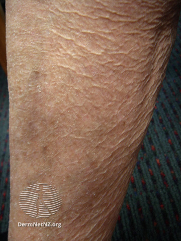
Individuals with xeroderma may encounter subjective symptoms, including pain, a burning sensation, tightness, and pruritus (see Image. Xerosis Cutis). When xeroderma is associated with pruritus, it can significantly diminish the patient's quality of life. The most frequently affected body areas include the lower legs, forearms, hands, and feet. A physical examination typically reveals dry, rough, and scaly skin with a somewhat grayish hue. Moreover, reduced skin elasticity, wrinkling, erythema, and fissures may also be observed.[2]
Evaluation
The diagnosis of xeroderma primarily relies on clinical assessment, with a comprehensive history and physical examination adequate to identify this prevalent condition. Healthcare providers may conduct laboratory testing to evaluate the potential underlying causes of the condition, including assessments of thyroid hormone and vitamin levels. In rare instances, a skin biopsy may be warranted to differentiate xeroderma from conditions that mimic its presentation. However, the assessment of xeroderma usually does not include techniques such as "teewametry" for measuring transepidermal water loss or "corneometry" for hydration measurement. These methods are not typically recommended or used for diagnosing xerosis and are more relevant in clinical trials or research.[2]
Dermoscopy enables the assessment of subtle morphological changes in the skin that may not be apparent to the naked eye. To categorize the degree of xerosis, a grading system based on specific criteria has been established, as mentioned below.
- Mild: Scaling confined to skin furrows.
- Moderate: Scaling extending beyond skin furrows with distinct skin markings.
- Severe: Plate-like scaling extending beyond skin furrows and accompanied by the development of deep skin fissures.
This dermoscopic grading system enables clinicians to accurately assess variations in the severity of xerosis attributed to various exacerbating factors and to evaluate the clinical response to treatment.[11]
Treatment / Management
The treatment of xeroderma should prioritize restoring physiological lipids in the epidermis, improving skin hydration, optimizing skin barrier function, and promoting epidermal differentiation.[9][12] Proper skincare practices are crucial in managing xeroderma, and a few recommended strategies are listed below.
Infrequent bathing and using lukewarm water: To effectively manage dry skin, it is advisable that individuals adopt infrequent bathing practices and opt for lukewarm water, steering clear of aggressive skin washing and hot water. These measures help prevent abrasion and the depletion of the skin's protective oils.
Gentle cleansers: Gentle and synthetic detergent cleansers are recommended because of their acidic pH, which closely matches the skin's natural pH. Syndet cleansers, in particular, are less irritating than traditional soaps and have been observed to reduce pruritus due to their lower pH levels.[13][14] Traditional soaps should be avoided as they raise the skin's pH, exacerbating dry skin and itching.
Skin moisturizers: Routine application of oil-based creams is advisable due to their thicker consistency, which is more effective at moisturizing the skin than water-based lotions. With their greasier texture, ointments are especially valuable for preventing transepidermal water loss.[10] For optimal results, moisturizers can be applied to damp skin after bathing, which will help reduce evaporation.
Room humidifiers: Room humidifiers are particularly beneficial during winter as they help retain the skin's moisture.
Hydration: Individuals dealing with xeroderma should stay hydrated by consuming adequate fluids.
Skin moisturization can be achieved through various methods, each offering distinct advantages. One approach involves occlusion, where a water-impermeable barrier is applied to the skin's surface, creating an environment that supports barrier repair. Petrolatum is renowned as the most occlusive moisturizer.
Humectancy is an alternative method for skin moisturization, where humectants function like sponges, drawing and retaining water within the skin. Within the dermis, glycosaminoglycans, including hyaluronic acid, serve as effective humectants. Additional humectants comprise glycerin, honey, sodium lactate, urea, and propylene glycol. Among these humectants, glycerin is the most potent one, which can draw water from the deeper epidermis and dermis layers of the skin, thereby filling gaps in the stratum corneum. Notably, unopposed humectants have the potential to draw moisture from the skin into the drier surrounding environment. Consequently, an ideal moisturizer should incorporate both humectants and occlusives for adequate skin hydration.
Hydrophilic matrices are less commonly used for skin moisturization, as they create a physical protective coating to prevent water loss. Colloidal oatmeal baths and other high-molecular-weight substances, such as growth factors and collagen fragments, can serve as hydrophilic matrices for skin moisturization. However, it is noteworthy that occlusives and humectants are generally more effective than hydrophilic matrices in this context.[7]
Various active ingredients with unique benefits are used in skincare products to improve skin texture and hydration, some of which are mentioned below.[2][7]
Petrolatum: This is recognized as the most effective moisturizing ingredient that acts as an occlusive by forming an oily barrier that prevents water from evaporating, thereby preserving the skin's moisture content while facilitating barrier repair.
Silicone: This is a non-greasy occlusive agent capable of filling gaps between desquamating corneocytes, resulting in smoother skin surfaces. The 2 most frequently utilized derivatives of silicone are dimethicone and cyclomethicone.
Ceramides: They act to enhance intercellular lipids, thereby maintaining the natural skin barrier. Moisturizer formulations often include 9 distinct ceramides, each characterized by its polar head group.
NMFs: They are synthetically formulated blends of amino acids that attempt to moisturize the skin by mimicking naturally occurring substances.
Urea and lactic acid: These ingredients reveal water-binding sites on corneocytes, thereby enhancing skin hydration. Due to their keratolytic properties, they hold particular significance, especially in addressing foot calluses.
Licochalcone A: This ingredient obtained from the roots of the Chinese licorice plant (Glycyrrhiza inflata) possesses anti-inflammatory properties that can soothe irritated skin and reduce erythema.
Dexpanthenol: This is the precursor of vitamin B5 and supports wound healing by stimulating epithelization and fibroblast proliferation. As a result, it finds utility in conditions such as atopic dermatitis, diabetic foot care, diaper dermatitis, burn injuries, and skin grafts.
Polidocanol: This ingredient is a nonionic surfactant with local anesthetic properties that aid in alleviating pruritus linked to xerosis.
The selection of ingredients should correspond to the patient's symptoms. Urea can be advantageous for addressing scaling, licochalcone A may be considered for erythema, and polidocanol can help alleviate pruritus. Urea or dexpanthenol can offer relief for fissures. Furthermore, clinicians may consider including topical corticosteroids or calcineurin inhibitors in the treatment regimen if dermatitis accompanies xeroderma.[15]
Differential Diagnosis
A differential diagnosis for xerosis may encompass the following conditions:
- Ichthyosis Vulgaris
- Atopic dermatitis
- Stasis dermatitis
- Irritant contact dermatitis
- Allergic contact dermatitis
- Nummular dermatitis
- Scabies
- Tinea corporis
- Psoriasis
- Cutaneous T-cell lymphoma
Prognosis
Xeroderma is typically a benign condition that may persist for years. The prognosis is favorable when triggers are avoided and a skincare regimen, which prioritizes gentle cleansing and adequate moisturization, is followed. In some instances, dry skin may be associated with a genetic disorder, such as xeroderma pigmentosum, resulting in more severe consequences, including an increased risk of developing skin cancer.
Complications
Xeroderma can lead to pruritus, which increases the susceptibility to skin infections, as microorganisms infiltrate the skin's surface through breaks. Asteatotic eczema, which resembles cracked porcelain in severe cases, can exacerbate and coexist with xerosis during winter and cause bleeding from damaged dermal capillaries. This distinctive appearance, akin to cracked porcelain or a pattern resembling crazy paving, is called eczema craquelé. Other potential complications include food allergies, which are linked to filaggrin mutations and atopy, and allergic contact dermatitis, which is attributed to the impaired function of the skin barrier.
Deterrence and Patient Education
Xeroderma can present as either an acute or chronic condition; for many individuals, it becomes a persistent, lifelong issue characterized by dry skin. In most instances, conservative management through gentle cleansing and proper moisturization of the skin effectively alleviates symptoms. Patients should recognize and avoid triggers, which can include using harsh soaps and detergents, extreme weather conditions, rough and tight clothing, stress, excessive consumption of alcohol, and eating spicy foods and citrus fruits.
Enhancing Healthcare Team Outcomes
Xeroderma is a prevalent condition that can be identified by various members of healthcare professionals, including nurses, physician assistants, nurse practitioners, podiatrists, primary care providers, and dermatologists. Every healthcare provider plays an essential role in diagnosing dry skin in patients and providing valuable education to them regarding practical treatment approaches. For instance, pharmacists are adept at identifying medication-related factors contributing to xeroderma, which include adverse effects or improper dosing. Pharmacists are also proficient in formulating creams with active ingredients to enhance emollient and humectant properties.
Nursing professionals are well-equipped to educate patients on proper skin cleansing and moisturization techniques, which may involve the application of occlusive dressings. Primary care providers and specialists can collaborate effectively to address patients' comorbidities and systemic diseases. Interprofessional communication and coordinated care are crucial for delivering high-quality healthcare and ensuring patient satisfaction in cases of xeroderma, which is typically a benign condition.