Continuing Education Activity
Tinea barbae is a superficial fungal infection of the skin, hair, and hair follicles caused by dermatophytes. Though dermatophyte infections are one of the most common skin infections in humans, tinea barbae is relatively rare. Lack of awareness about the disease in the medical community often leads to missed diagnosis and mismanagement. To avoid the morbidity and healthcare costs associated with this condition, it must be promptly diagnosed and treated. This activity reviews the etiology, clinical presentations, diagnosis, and treatment of tinea barbae and highlights the role of the interprofessional team in evaluating and treating patients with this condition.
Objectives:
- Identify the etiology of tinea barbae.
- Summarize the appropriate evaluation of tinea barbae.
- Outline the management options available for tinea barbae.
- Review the importance of collaboration amongst the interprofessional team to enhance the care of patients with tinea barbae.
Introduction
Dermatophytes are fungi that invade keratinized tissues such as skin, hair, and nails.[1] Dermatophytosis is an infection caused by dermatophytes; these infections are commonly referred to as "tinea" infections.[2] Dermatophytes need keratin for their growth (keratinophilic); hence they do not infect mucosal surfaces.[3] Dermatophyte infections can affect any superficial keratinized surface from the scalp to the toes. Tinea infections are classified according to the affected body site.
Tinea barbae is a rare dermatophyte infection affecting the skin, hair, and hair follicles of the beard and mustache.[1] Tinea barbae was first described by Gruby in 1842 as a fungal infection of the beard area, where the fungal elements formed a continuous sheath around the hair. Gruby named the fungus "mentagrophyte," which means "plant of the chin."[4] Tinea barbae is also known as tinea sycosis, as one of the clinical manifestations is inflammation of the hair follicles.[1][5] Tinea barbae in the past was commonly attributed to being transmitted by unsanitary razors used by the barbers. Hence, it was commonly referred to as barber's itch and beard ringworm.[4][6]
Etiology
Transmission of dermatophyte infections to humans happens through direct contact with infected humans (anthropophilic), soil (geophilic), or infected animals (zoophilic).[7] Literature has shown that tinea barbae is exclusively caused by zoophilic and anthropophilic dermatophytes.[1] Trichophyton verrucosum, Trichophyton mentagrophytes, and Trichophyton rubrum are the most commonly reported causative organisms in the literature.[1][7][8][9][10][11] Other causative organisms that were documented are Trichophyton violaceum, Trichophyton megninii, Trichophyton schoenleinii, Trichophyton tonsurans, Trichophyton interdigitale, Trichophyton ernacei, Microsporum canis, Microsporum nanum, Mycroscporum gypseum, and Epidermophyton floccosum.[1]
Until 2000, most of the tinea barbae cases that were documented in the literature were caused by zoophilic dermatophytes from infected farm animals and domestic animals, especially dairy cows (T. verrucosum), sheep, pigs (T. ernacei), horses, dogs, and cats (M. canis).[1][12] More recently, infections with anthropophilic T. rubrum have been a frequently reported causative organism, as noted in multiple individual case reports worldwide and case series published from Portugal and Mexico.[1][8][9][10][11] Human to human transmission of tinea barbae is rare but possible. Individual case reports in the recent past have documented autoinoculations with T. rubrum in patients with tinea pedis.[8][9][10]
Epidemiology
Though superficial fungal infections are very common worldwide, tinea barbae is relatively uncommon. Tinea barbae was first reported in 1842 by Gruby,[4] and since then, only a little over 150 cases have been reported in the literature until 1990.[1] Most of the data since 1990 on tinea barbae in the literature are either isolated case reports or small case series.[1]
Due to the rarity of the disease, predicting a true incidence or lifetime risk is difficult. The distribution of dermatophytes is worldwide, and so are the cases of tinea barbae that were reported in the literature. Since tinea barbae is an infection of hair and hair follicles of the beard and mustache area, it is exclusively seen in adolescent boys and men. In an extreme rarity, a case of tinea barbae has been reported in a hirsute woman in a longitudinal study from Portugal.[11]
Pathophysiology
Dermatophytes are keratinophilic fungi that cause superficial and deep infections of the keratinized tissues. The dryness of the skin’s outer layer, shedding of the outer layers of the dead skin, and the innate peptides and medium chain-fatty acids secreted on to the skin generally prevent the colonization of the microorganisms.[3][13] However, certain factors like age, immune status, steroid use, diabetes mellitus, trauma, and occupational exposures increase the likelihood of dermatophyte infections.[3][13]
Dermatophytes, along with the inflammatory response by the host to the pathogen combined, are responsible for the pathology.[3] Both fungal-specific and host-specific factors play a role in the inflammatory process. The fungal-specific factors that are responsible for pathogenesis are an adaptation to the specific host, release of enzymes, production of inflammatory factors and toxins, and release of immunomodulating agents. The host-specific factors that play a role in the inflammation process are the site of entry, non-specific defense mechanisms, and the immune response.
The infection process begins with the inoculation of the arthroconidium (spores) to the keratinized tissue. Once inoculated, the carbohydrate microfibrils present on the fungal spores anchor to the keratinocytes. Once anchored, the spores germinate and produce hyphae, which spread centrifugally into the deeper layers of the stratum corneum.[3][14]
Invading fungal hyphae produce keratinases, proteases, elastases, and other pathogenic factors into the stratum corneum. These enzymes help in digestion and utilization of the keratin and other proteins needed for the survival and growth of the dermatophytes.[3] Dermatophytes develop a specialized structure called the penetrating organ to invade the hair shafts.[3].Keratinases activate the keratinocytes to release inflammatory mediators like cytokines and interleukins. The interleukins and cytokines released by the keratinocytes will recruit both humoral and cell-mediated immune response depending on the specific pathogen.[14]
History and Physical
As tinea barbae is a relatively rare dermatophyte infection, knowledge about the disease, and taking a good history will help to make a prompt diagnosis of the condition. Zoophilic fungi predominantly cause tinea barbae; hence occupational history, contact with pets, and domestic animals will help in narrowing the differential diagnosis. Factors like diabetes mellitus, local trauma, steroid use, and immunosuppressive therapy can suppress local defense mechanisms and predispose them to infections with dermatophytes.[1] Considering that there have been multiple cases of anthropophilic dermatophytoses via autoinoculation and transmission from person to person, it is important to know the history of other dermatophyte infections like tinea capitis, tinea pedis, etc. in the patient or their close contacts.[15]
Clinically tinea barbae presents as two different morphologies, inflammatory and noninflammatory.[1][6][14] Inflammatory presentation is seen with zoophilic dermatophytosis and noninflammatory presentation with anthropophilic dermatophytosis.[7][8]
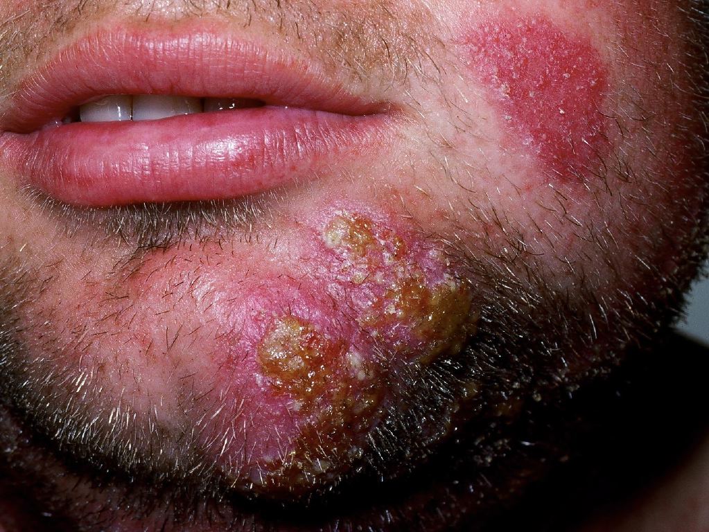
The classic inflammatory form of tinea barbae develops a characteristic lesion called kerion. Kerion is an erythematous, boggy, tender, often sterile, weeping nodule or plaque with pustules and draining sinuses.[6][7] Hair in these areas appears lusterless, brittle, and is easily plucked. Kerion can be solitary or multiple but is usually unilateral.[6][7] Patients with inflammatory tinea barbae may have constitutional symptoms like fever and malaise.[6] Kerion is a coalesced sterile pustule, but occasionally they can get superinfected with cutaneous bacteria and develop regional lymphadenopathy.[7]
Noninflammatory tinea barbae is a pruriginous superficial dermatophytosis, which presents as a diffusely erythematous squamous plaque with perifollicular pustules and papules.[7] Occasionally, superficial tinea barbae lesions can be indistinguishable from the tinea of the face with flat scaly plaques with central clearing and a ring-shaped active border consisting of vesicles and pustules.[6]
Evaluation
The diagnosis of tinea barbae is often clinical. Knowledge about the disease and its clinical presentation, along with a good history and physical examination, can help in early diagnosis. Since the mainstay of treatment for tinea barbae is systemic antifungal therapy for an extended period with the potential for side effects, it is important to verify the diagnosis. The diagnosis can be established by simple, inexpensive in-office testing by direct microscopic examination of the skin scrapings from the lesion or by direct visualization of the affected area under a Wood's lamp. Visualization of the affected area under Wood's lamp is of limited use, as most dermatophytes that cause tinea barbae do not fluoresce except for Microsporum species.[1]
Direct Microscopic Examination
Obtain scales and skin scrapings from the active border of the lesion by using a scalpel or moist cotton tip along with epilated hairs. Transfer the collected sample onto a slide and add a few drops of 10%-20% solution of potassium hydroxide (KOH). To speed up the emulsification of the skin cells, gently warm the slide or add dimethyl sulfoxide.[7][15] Fungal stains such as chlorazol black E or Parker's blue-black ink may be added to the sample to highlight the fungal elements.[15] Examining the wet-mount preparation under 20-times and 40-times magnification will show branching, rod-shaped fungal filaments with uniform width called hyphae and minute spores called arthroconidia, which is diagnostic.[7][13] Depending on the causative organism, fungal elements can be observed either covering the hair (ectothrix) or invading the hair shaft (endothrix).[7]
Fungal Culture
Innoculate the collected skin scrapings and hair samples into the specialized dermatophyte test medium. Mycocel and mycobiotic agar are some of the commercially available test media for dermatophytes. These specialized media have cycloheximide and chloramphenicol to inhibit the growth of contaminating fungi and bacteria.[7] These test media also have phenol red as an indicator, which turns from yellow to pink/red in the presence of alkaline metabolites of dermatophytes.[7][13][15] It takes about 7 to 14 days for the fungus to grow on this medium. The inoculated medium should show no growth for 21 days before declaring the culture to be negative.[13] Based on the colony and microscopic morphology, individual species responsible for dermatophytoses can be differentiated. Fungal cultures are time taking and expensive, but they have better sensitivity and specificity compared to direct microscopy. A fungal culture is not mandatory before initiating antifungal therapy, but it helps in determining the appropriateness of treatment.
Skin Biopsy and Histopathological Examination
The majority of the tinea barbae infections can be diagnosed either by direct microscopic examination or by fungal cultures. Skin biopsy and histopathological examination of the tissue is reserved for cases refractory to treatment or where a definitive diagnosis can not be established by other methods.[15] Periodic acid-Schiff (PAS) stain is the recommended staining as fungal elements are often not differentiated with hematoxylin and eosin staining.[6] Microscopy under PAS stain shows fungal hyphae and arthroconidia along with inflammatory changes in the epidermis and dermis. Inflammatory patterns seen in the biopsy sections are consistent with folliculitis, perifolliculitis, and microabscesses.
Conventional diagnostic methods mentioned above are adequate for diagnosing tinea barbae, but may not be specific to identify the exact causative pathogen by species. Newer diagnostic modalities like polymerase chain reaction-restriction fragment length polymorphism (PCR-RFLP) on the ribosomal DNA internal transcribed spacer (ITS) region will help in the identification of the specific pathogen. Currently, these tests are expensive and are limited to specialized centers, mostly participating in studying epidemiology, transmission patterns, and drug development.[2]
Treatment / Management
The mainstay of treatment for tinea barbae is oral antifungal therapy. Topical agents can be used, but only as an adjunct therapy.[15] Oral antifungal agents that have proven effective in the management of tinea barbae are terbinafine, azoles, and griseofulvin.[15] Griseofulvin used to be the preferred drug, but it is rarely used now due to its side effects, drug resistance, need for prolonged therapy, and increased relapse rates due to the rapid clearance of the drug from the skin.[7][13] In the past, tinea barbae was treated for 12 weeks with griseofulvin. Recent case reports have demonstrated complete resolution of the tinea barbae with 4-6 weeks of therapy with both terbinafine and azoles.[5][8][9][10][11] Even though four weeks of treatment is sufficient, some authors have suggested continuing treatment for 2 to 3 weeks after the resolution of the lesions. Recommended dosages for the available antifungal agents for tinea barbae are:
- Terbinafine 125 mg to 250 mg once a day
- Ketoconazole 200 to 400 mg a day
- Fluconazole 200 mg once a day
- Itraconozole 100 mg once a day
Common side effects for terbinafine and azoles include nausea, abdominal pain, diarrhea, rash, elevated transaminases, and visual disturbances.[7] When on systemic antifungal therapy, periodic monitoring of hepatic, renal, and hematopoietic function is recommended. Significant drug interactions can happen when hepatically metabolized drugs are taken along with azoles.[7] The role of treatment with steroids and other topical agents like selenium sulfide is not well defined, and it is the prescriber’s discretion on a case to case basis. Oral or systemic antibiotics are warranted when a secondary bacterial infection is suspected. Surgery is rarely indicated, and incision and drainage of kerion are not recommended.[15]
Differential Diagnosis
Tinea barbae is a rare disease and is frequently misdiagnosed as more common conditions that affect the beard and mustache area like bacterial folliculitis, perioral dermatitis, pseudofolliculitis barbae, contact dermatitis, herpes simplex, sporotrichosis, acne vulgaris, acne rosacea, and rarely neoplasm.[3] It is important to know the common characteristics that can differentiate tinea barbae from other conditions clinically to aid with prompt diagnosis.
Bacterial folliculitis or sycosis barbae caused by Staphylococcus species or other bacteria can be differentiated from tinea barbae by the presence of systemic symptoms like fever and malaise. Regional lymphadenopathy is more common with bacterial folliculitis, and hair removal is painless in tinea barbae in contrast to sycosis barbae. Pseudofolliculitis barbae is a form of irritant folliculitis caused secondary to shaving, and its presentation is bilateral and diffuse in comparison to tinea barbae. Candidal infections can be differentiated from tinea barbae by the involvement of adjacent mucosal surfaces.
Prognosis
Tinea barbae is a rare dermatophytosis infection with only a few hundred cases reported in the literature. Most of the reported cases of tinea barbae in the literature were either single case reports or small case series. Though the data available is limited, it has been very reassuring, as almost all cases responded well to oral antifungal therapy with complete resolution.
Complications
Other than morbidity and healthcare costs associated with the therapy, no significant complications have been reported in the literature.
Deterrence and Patient Education
Tinea barbae is a contagious disease where an individual can be infected from either an animal or human source via direct or indirect contact. Once the diagnosis of tinea barbae is made, the emphasis should be placed on identifying the source of the infection. Patients and family members should be screened for other dermatophyte infections like tinea capitis, tinea pedis, onychomycosis, etc. Domestic and farm animals should be checked for zoonotic dermatophytoses and treated appropriately with the help of a veterinarian to prevent reinfections.[7] Avoid sharing combs, brushes, and hats to reduce transmission between family members.[15]
Enhancing Healthcare Team Outcomes
Though dermatophyte infections are the most common fungal infections in humans, tinea barbae is a rare dermatophyte infection. Due to its rarity, no large randomized clinical trials are reported in the literature. More detailed, multi-centered randomized control studies are needed before concluding incidence, prevalence, and mortality.
Based on the available information from the literature, the majority of the tinea barbae cases can be handled by primary clinicians in the office setting. However, in special situations involving an interprofessional team, including a dermatologist, pharmacist, veterinarian, and microbiologist will help in attaining the best possible outcomes and reducing healthcare costs. [Level 5] Oral antifungal agents are commonly associated with drug interactions and side effects; hence involving a pharmacist can help in increasing drug safety by choosing the right medication based on the patient's comorbidities and current medications. As discussed, zoophilic dermatophytes are the predominant causative agents for tinea barbae in humans. Once contact with animals is established, the evaluation of domestic pets and farm animals by a veterinarian will help in identifying the source and preventing reinfection. In cases refractory to management with oral antifungal therapy consulting a dermatologist and microbiologist can be helpful in considering alternate diagnosis/causative organism/therapy.