Introduction
The inferior hypogastric plexus is also known as the pelvic ganglion. Classic anatomical studies have provided few details of the inferior hypogastric plexus morphology or the location and nature of the associated nerves. The fusion of the pelvic splanchnic nerves, sacral splanchnic nerves, and superior hypogastric plexus along with visceral afferent fibers forms the inferior hypogastric plexus. There have also been studies showing the contribution of spinal nerves S2-S5 to the inferior hypogastric plexus.[1]
Dissection of the inferior hypogastric plexus is difficult due to its location, the multiplicity of its sympathetic and parasympathetic roots, and the complex distribution of its terminal branches. The density of surrounding connective tissue also precludes the pelvic-perineal anatomy; therefore, it is difficult to determine the exact relationship between the nerves and pelvic connective tissue. The inferior hypogastric plexus lends its clinical relevance to various urogenital pain syndromes, including endometriosis, prostatitis, and chronic pain of the sacral region, postherpetic neuralgia, and rectal pain, among others.[2] Therefore, any surgical procedure in this anatomical region must be met with due diligence, with respect to the underlying plexus that may be inadvertently damaged, thus causing a slew of related symptoms of the urogenital tract as well compromise of the pelvic autonomic innervation.[3][4]
Structure and Function
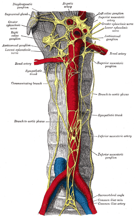
The hypogastric nerve starts at the fork of the superior hypogastric plexus: right and left plexus, continuing inferiorly bilaterally on the sides of the body, descending into the pelvis forming the inferior hypogastric plexus. The inferior hypogastric plexus forms a network of nerves that supply the viscera of the pelvic cavity. The plexus lies in the extraperitoneal connective tissue on the pelvic sidewall; the plexus lies on the rectum in males and the rectum and vagina in females. It is located anterolaterally to the mesorectum.[2]
The inferior hypogastric plexus is commonly described as a triangle. The inferior edge of the triangle constantly stretches from the fourth sacral root; its posterior (dorsal) side the point of contact with the sacral roots, from which it receives its afferents; its cranial edge lays parallel to the posterior edge of the hypogastric artery.[1]
Contributions to the inferior hypogastric plexus include:
- superior hypogastric plexus in the form of the hypogastric nerve
- sacral splanchnic nerves from the sympathetic trunk at vertebral levels T10-L2
- pelvic splanchnic nerves
The pelvic splanchnic nerves contribute parasympathetic efferent fibers to the plexus.
Embryology
The 'pelvic plexus,' more specifically known as the inferior hypogastric plexus, consists of a complex fibro-nervous structural network of afferent and efferent fibers providing function for the pelvic and perineal organs. Knowing this important relationship is extremely important from a surgical point of view. Understanding this anatomy is crucial to preventing errors that can potentially be fatal to patients undergoing surgery or abdominopelvic procedures.[5]
The bladder is innervated by the superior and inferior levels of the efferent fibers of the pelvic plexus. The superior level arose from the hypogastric nerves at the ureterovesical junction. The inferior level fibers arose from the anterior plexus edge of the bladder neck at 5 and 7 o'clock. Proximal seminal vesicle fibers follow the posterior (between the seminal vesicles and rectal fascia) and lateral (runs along the lateral wall of the seminal vesicles and the prostate) courses.
It is important to note that the posterior courses are intimately connected with the seminal vesicles and provide fibers to the vas deferens and ejaculatory canal, reaching the colliculus seminalis via the prostate gland.
Efferent Fibers from the Pelvic Plexus
The efferent fibers innervate the bladder and are situated superior and inferior. Fibers of the superior level arise from the hypogastric nerves at the ureterovesical junction, unlike the inferior level fibers that arise from the anterior plexus edge and approach the bladder neck at 5 and 7 o'clock positions.
Proximal seminal vesicle fibers follow two courses: lateral and posterior, the latter which lays between the rectal fascia and the seminal vesicles and connected intimately with the seminal vesicles. This intimate posterior fiber connection provides fibers to the vas deferens and ejaculatory canal, eventually reaching the colliculus seminalis via the prostate. The lateral fibers run along the lateral wall of the prostate and seminal vesicles, and some even penetrate the prostate from its lateral side, unlike others that run anterolateral, eventually reaching the prostatic apex and external urethral sphincter. The two different courses of fibers contain a cholinergic and adrenergic function. The fibers involved in innervating erectile bodies are postero-lateral to the prostate (distant from the prostatic capsule at the base level) and are mainly cholinergic with few adrenergic fibers.
Blood Supply and Lymphatics
At the level of the area between the ureter and the posterior portion of the uterine artery, one finds the inferior hypogastric plexuses, which create a plexiform structure (similar to a folding fan) at the end of both hypogastric nerves; as another anatomical landmark, we find the plexus dorsally to the parametric vessels in the area of the uterine vein. We can find a small number of lymph nodes adjacent to the inferior hypogastric plexus.
Nerves
The plexus divides into numerous branches and includes:
- postganglionic sympathetic fibers
- visceral afferent fibers
- preganglionic and postganglionic parasympathetic fibers
The branches are not given off directly, but they instead pass through the viscera via small subsidiary plexuses such as the middle rectal plexus, the prostatic plexus, and the uterovaginal plexus.[6] As the focal part of all autonomic control within the pelvis, the inferior hypogastric plexus pairs a meshwork of nerves located bilaterally alongside the rectum, medial to the internal iliac arteries. There are two sources of sympathetic contribution in the inferior hypogastric plexus; the largest contribution comes from the superior hypogastric plexus, purely containing sympathetic fibers and receives upper lumbar communication from the intermesenteric plexus and the L3 and L4 splanchnic nerves.
fThe superior hypogastric plexus is located within the abdomen at the bifurcation of the aorta, eventually leading to the left and right hypogastric nerves, forming a vast meshwork of solid nerve trunks. The nerve meshwork resides lateral to the rectum, bilaterally, and curve down and out as they make their way into the pelvis, eventually interconnecting the superior and inferior hypogastric plexuses. There are no ganglia within this nerve plexus. The majority of the sympathetic contribution to the hypogastric nerves are derived from the superior into the inferior hypogastric plexuses.
Muscles
The inferior hypogastric nerve supplies innervation to the urinary bladder, the urethra, and the corpora cavernosa.[2]
The inferior hypogastric plexus fibers innervate the smooth muscle fibers of the visceral portion of the levator ani muscle.
Physiologic Variants
There is not much data on the physiological variations of this plexus. Still, one must remember that anatomy is usually variable, and we cannot exclude the presence of anatomical variations.
Surgical Considerations
Because the inferior hypogastric plexus is a paired structure, injury is a commonly known complication of pelvic surgeries. Damage to the plexus can lead to urinary dysfunction, specifically urinary incontinence. The bladder will be non-compliant with a fixed external sphincter tone. The integrity of the somatic and autonomic pelvic nerves is essential to maintaining normal sexual function and a normal micturition-continence cycle.[5] The main difficulty in preserving these nerves is due to their small architecture in combination with the narrowness of the pelvis.
Clinical Significance
The inferior hypogastric plexus mediates sensation, such as pain, through the sympathetic chain for the lower abdominal and pelvic viscera. It is thought to be a major structure involved in numerous pelvic and perineal pain syndromes and conditions. Multiple studies have investigated possible nerve blocks treating chronic and more acute pain in this area. Hypogastric plexus blocks relieve pelvic pain that potentially refers to the colon, lower intestines, bladder, uterus and ovaries, prostate or testicles, and other parts of the pelvis.[7][8][9]
Surgeons performing hysterectomy must keep in mind the inferior hypogastric plexus as disruption of nerves may cause sexual, genitourinary, and colonic motility dysfunction. Radical hysterectomies may result in disruption of pelvic autonomic nerves.[10]
Other Issues
The inferior hypogastric plexus sends small filaments to innervate the rectum through the pre-hypogastric fascia and the anterior portion of the Denonvilliers' fascia.[11]
We can identify three small plexuses from the inferior hypogastric plexus, such as the vesical plexus, vaginal-rectal plexus, and inferior rectal plexus.[12]