Graham-Little-Piccardi-Lasseur Syndrome

Earn CME/CE in your profession:


Continuing Education Activity

Graham-Little-Piccardi-Lasseur syndrome (GLPLS) is a type of lichen planopilaris. Lichen planopilaris is the follicular type of lichen planus. There are 3 variants of lichen planopilaris, including GLPLS, frontal fibrosing alopecia, and classic lichen planopilaris. The onset of symptoms in GLPLS can occur in any order, though the condition often begins with developing the hyperkeratotic papules on the trunk and extremities before the development of alopecia. This activity reviews the evaluation and treatment of Graham-Little-Piccardi-Lasseur Syndrome and highlights the role of the interprofessional team in evaluating and treating this condition.

Objectives:

  • Identify the etiology of Graham-Little-Piccardi-Lasseur syndrome.

  • Evaluate the presentation of Graham-Little-Piccardi-Lasseur syndrome.

  • Apply the treatment of Graham-Little-Piccardi-Lasseur syndrome.

  • Communicate modalities to improve care coordination among interprofessional team members to improve outcomes for patients affected by Graham-Little-Piccardi-Lasseur syndrome.

Introduction

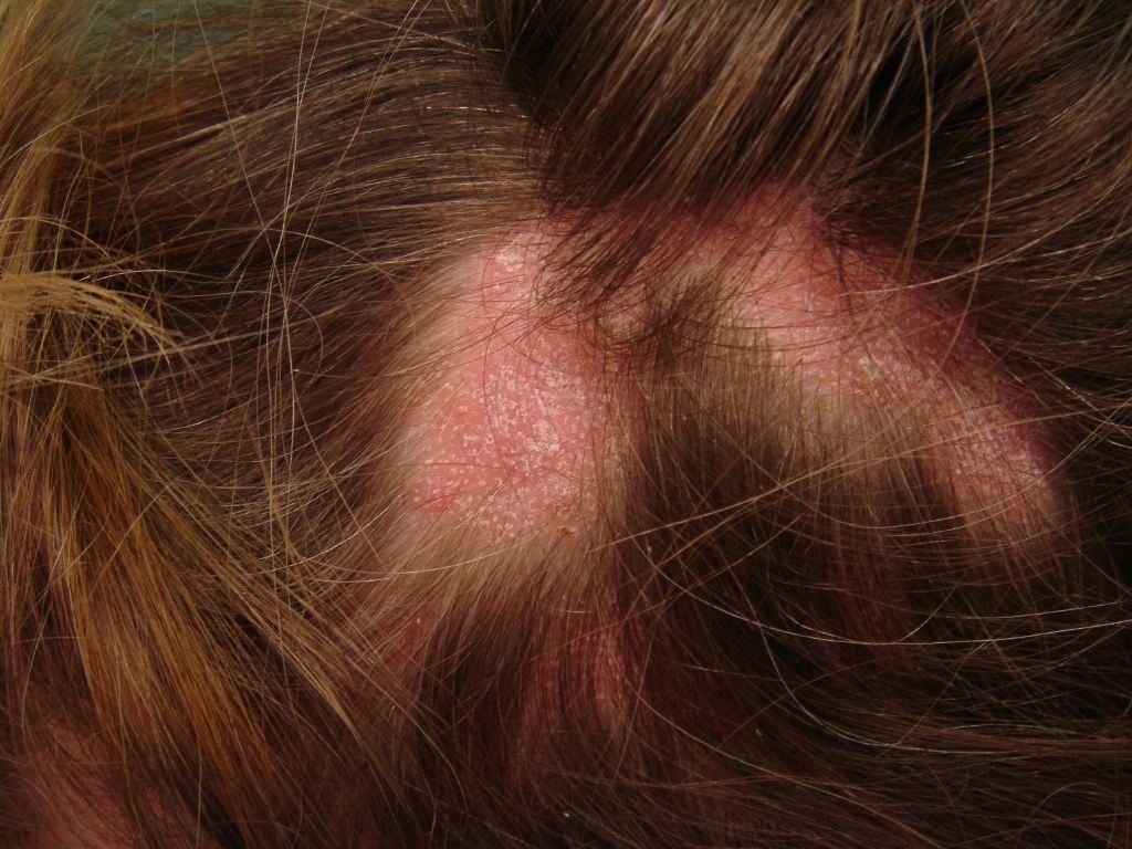
Graham-Little-Piccardi-Lasseur Syndrome (GLPLS) is a sub-type of lichen planopilaris, first discovered by Piccardi in 1913 and 2 years later by Ernst Graham Little in a mutual patient with Lasseur.[1] Lichen planopilaris is the follicular iteration of lichen planus. There are 3 variants of lichen planopilaris, including GLPLS, frontal fibrosing alopecia, and classic lichen planopilaris.[2] Classic lichen planopilaris is a chronic inflammatory process that results in patchy progressive cicatricial alopecia, most commonly found on the scalp. Frontal fibrosing alopecia has band-like cicatricial alopecia of the frontal and temporal zones of the scalp.[3] GLPLS has the specific characteristics of multifocal, patchy, cicatricial alopecia present on the scalp, non-cicatricial alopecia of the axillae, non-cicatricial alopecia of the perineum, and follicular hyperkeratosis of the trunk and extremities.[4] The onset of these symptoms in the outbreak of GLPLS can occur in any order, with a predilection for the development of the hyperkeratotic papules on the trunk and extremities before alopecia occurs in any location (See Image. Lichen Planopilaris).

Etiology

GLPLS etiology is an enigma. However, most schools of thought believe the symptomatology found in GLPLS is via an autoimmune mechanism that is T-cell mediated.[5] Many mechanisms of lichen planopilaris are present in GLPLS, including altered integrin expression and interferon-gamma dysregulation. Inflammation of the hair follicles and altered integrin expression may contribute to the cicatricial alopecia in GLPLS. The altered integrin expression creates the follicular root's gelatinous consistency, resulting in a positive anagen hair pull test of active affected sites.[6][7] A case report by Rodriguez-Bayona et al found that there is a strong auto-immune reactivity to a centromere-specific protein INCENP, which leads to the development of mitotic cellular deficits. While further investigation is necessary to determine INCENP auto-antibody prevalence in GLPLS, this marks the first GLPLS-linked antibody.[8]

Epidemiology

GLPLS is an illness that mainly affects middle-aged Caucasian women with an average age of onset varying from 30 to 70 years old.[9] This variant of lichen planopilaris comprises a small portion of the overall cases. In a case report of 80 lichen planopilaris cases, only 1 report was consistent with GLPLS.[2] The correlation between GLPLS and multiple other entities exists, including hepatitis B vaccination, androgen insensitivity syndrome, HLA DR-1 positivity in a mother and daughter, vitamin A deficiency, hormonal dysfunction, and neuropsychological issues.[5][10]

Pathophysiology

Damage to the pilosebaceous unit is a crucial factor in the pathogenesis of GLPLS, a variant of lichen planopilaris. Lesional skin shows increased interferon-gamma-induced chemokines, which recruit the cytotoxic T-cells to the follicular infundibulum.[11] Dysregulation of the interferon-gamma signaling pathway in lichen planopilaris also causes increased MHC class 1 and 2 expression, which leads to increased antigenic presentation to T-cells. Besides interferon-gamma dysregulation, damage to the chromosomal passenger protein INCENP inhibits binding to microtubules, which causes improper localization and function of chromosome passenger complexes.[12] This inability to direct chromosome passenger complexes to the correct locality causes impairment of chromosomal segregation, central spindle formation, and cytoplasmic allocation.[13] These alterations create metaphase abnormalities which lead to a decrease in overall mitotic function. 

Histopathology

Histopathologic examination within cicatricial alopecia areas of GLPLS shows a dilated follicular lumen superior to the sebaceous gland with a dense hyperkeratotic plug in the lower portion of the infundibulum. Epithelium in the nearby periphery of the follicular infundibulum shows hypergranulosis, vacuolar degradation of keratinocytes, and a lymphocytic infiltrate that obscures the dermal-epidermal junction with increased dense bundles of collagen at the follicular base. Examination of the hyperkeratotic papules on the trunk and extremities shows an expanded keratin-filled follicular opening vacuolar degradation of adjacent keratinocytes and a lymphohistiocytic infiltrate at the dermal-epidermal junction.[14]

History and Physical

A thorough history and physical exam in patients with GLPLS is a core component of a satisfactory diagnosis. Determining if a patient has experienced significant hair loss, psychosocial stress, or endocrinological abnormalities can lead you to other potential causes of alopecia that may exclude GLPLS as a final diagnosis. Physical examination should include a full body skin check with additional attention on the perineum and axillae for signs of non-cicatricial alopecia. Examination of the trunk and extremities for signs of hyperkeratotic papules with surrounding perifollicular erythema is also a key component of GLPLS. Investigation of the scalp in GLPLS should reveal a multifocal, patchy, and cicatricial alopecia of the scalp. In addition, an anagen hair pull is often positive in GLPLS because of the relatively weak stabilization of the hair follicles of affected individuals.

Evaluation

Skin biopsy, in conjunction with physical exam findings, is the primary method of diagnosis in GLPLS. Histology of the cicatricial areas and the hyperkeratotic papules reveals features of lichen planopilaris. Biopsy shows areas of perifollicular inflammation leading to pilosebaceous destruction, with late-stage GLPLS causing corruption of arrector pili muscles. Clinical evaluation of GLPLS shows erythematous follicular papules varying in size from 1 to 4 mm, non-cicatricial alopecia of the perineum and axillae, and patchy cicatricial alopecia on the scalp. Hyperkeratotic papules are typically present on the trunk, thighs, inner arms, outer arms, and wrists with or without pruritus. The initial onset of the hyperkeratotic papules can precede the development of alopecia on the scalp. However, there is no set chronological order of symptoms.[15]

Treatment / Management

The management of GLPLS remains elusive, with no current therapeutic regimen highlighted to be dominant. Treatment focuses on reducing the immunological response before the development of follicular scarring. Novel research has shown that JAK inhibition with tofacitinib leads to the normalization of interferon-mediated T-cell chemotaxis, thus preventing infundibular inflammation. A case series of 8 lichen planopilaris patients treated with a twice-a-day 5mg dose of tofacitinib reported a 30-94% improvement in disease severity.[11] Zegarska et al reported a case of a prednisone 30 mg dosage for 2 weeks, then a subsequent reduction to 20 mg of prednisone with psoralen and ultraviolet light A at a dosage of 12 joules. This treatment regimen led to the moderate improvement of the follicular hyperkeratosis of the trunk and extremities. While the follicular papules were reduced, there was no reduction in the cicatricial alopecia of the scalp or the non-cicatricial alopecia in the axillae and perineum.[16] Besides these modalities, there have been reports of the following therapies: intralesional glucocorticoids, topical glucocorticoids, systemic glucocorticoids, cyclosporine, tacrolimus ointment, and systemic retinoids. Most treatment methods reduce perifollicular erythema and inflammation without affecting the cicatricial alopecia or perineal and axillary non-cicatricial alopecia.[4][9][16]

Differential Diagnosis

Differential diagnosis in GLPLS includes classical lichen planopilaris, pseudopelade of Brocq, frontal fibrosing alopecia, lichen spinulosus, androgen insensitivity syndrome, discoid lupus erythematosus, secondary syphilis, and Addison disease.[17][18][19][20]

Prognosis

The course of GLPLS is often drawn out and may improve with treatment. However, full resolution of alopecic symptoms with current modalities of treatment is rare. Limited data is present to determine the average disease duration, with some cases extending for over 20 years. However, Zegarska et al report a range of 6 months to 10 years.[16] The prognosis of GLPLS may see a significant improvement in the reduction of hyperkeratotic papules on the trunk and extremities with adequate medical management.

Complications

While GLPLS can bring about significant psycho-social stress from the superficial alteration to appearance, there are no known systemic manifestations of the disease. Further study of the INCENP protein and its role in overall mitotic function may lead to the discovery of concomitant morbidities in GLPLS, yet no such comorbidity is present. The primary lasting complication in GLPLS is the existence of follicular scarring that occurs after the infundibular inflammation. This cicatricial portion of the disease manifests on the scalp and permanently damages the pilosebaceous unit.

Deterrence and Patient Education

Patients suffering from GLPLS require a thorough explanation of prognosis and treatment options to lead them towards realistic expectations for the management of their disease. With current therapy often not affecting the alopecic portion of the disease, many patients forego treatment. While treatment modalities cannot cause improvement towards the prior cicatricial damage, early initiation of a therapeutic agent may lead to a reduction in further progression of the non-cicatricial alopecia, cicatricial alopecia of the scalp, and hyperkeratotic papule formation. Giving patients a clear insight into the goals of GLPLS therapy can improve compliance and lead to better patient-physician relationships.

Enhancing Healthcare Team Outcomes

An interprofessional approach to the initial evaluation of patients with GLPLS is the key to providing optimal care and improved outcomes. Multiple medical specialties can assist in the diagnostic and long-term care processes including dermatologists, dermatopathologists, pharmacists, hair-loss support groups, and psychologists. Satisfactory diagnosis is of utmost importance, followed by psycho-social assistance from trained professionals and alopecia support groups. Support groups of patients with alopecia can lead to increased feelings of belonging and reduce psycho-social stress associated with the diagnosis of GLPLS.[21]


(Click Image to Enlarge)
<p>Lichen Planopilaris

Lichen Planopilaris. Classic lichen planopilaris is a chronic inflammatory process that results in patchy progressive cicatricial alopecia, most commonly found on the scalp. Frontal fibrosing alopecia has band-like cicatricial alopecia of the frontal and temporal zones of the scalp.


DermNet New Zealand

Details

Editor:

Cory Maughan

Updated:

9/12/2022 9:14:34 PM

References


[1]

László FG. Graham-Little-Piccardi-Lasseur syndrome: case report and review of the syndrome in men. International journal of dermatology. 2014 Aug:53(8):1019-22. doi: 10.1111/j.1365-4632.2012.05672.x. Epub 2013 Mar 14     [PubMed PMID: 23489018]

Level 3 (low-level) evidence

[2]

Soares VC, Mulinari-Brenner F, Souza TE. Lichen planopilaris epidemiology: a retrospective study of 80 cases. Anais brasileiros de dermatologia. 2015 Sep-Oct:90(5):666-70. doi: 10.1590/abd1806-4841.20153923. Epub     [PubMed PMID: 26560212]

Level 2 (mid-level) evidence

[3]

Valesky EM, Maier MD, Kaufmann R, Zöller N, Meissner M. Single-center analysis of patients with frontal fibrosing alopecia: evidence for hypothyroidism and a good quality of life. The Journal of international medical research. 2019 Feb:47(2):653-661. doi: 10.1177/0300060518807335. Epub 2018 Oct 31     [PubMed PMID: 30378444]

Level 2 (mid-level) evidence

[4]

Griffiths AAC, Sancho MI, Plaza AI. Piccardi-Lassueur-Graham-Little syndrome associated with frontal fibrosing alopecia. Anais brasileiros de dermatologia. 2017 Nov-Dec:92(6):867-869. doi: 10.1590/abd1806-4841.20176741. Epub     [PubMed PMID: 29364452]


[5]

Yorulmaz A, Artuz F, Er O, Guresci S. A case of Graham-Little-Piccardi-Lasseur syndrome. Dermatology online journal. 2015 Jun 16:21(6):. pii: 13030/qt7gj157xg. Epub 2015 Jun 16     [PubMed PMID: 26158366]

Level 3 (low-level) evidence

[6]

d'Ovidio R, Sgarra C, Conserva A, Angelotti UF, Erriquez R, Foti C. Alterated integrin expression in lichen planopilaris. Head & face medicine. 2007 Feb 8:3():11     [PubMed PMID: 17288588]


[7]

Assouly P, Reygagne P. Lichen planopilaris: update on diagnosis and treatment. Seminars in cutaneous medicine and surgery. 2009 Mar:28(1):3-10. doi: 10.1016/j.sder.2008.12.006. Epub     [PubMed PMID: 19341936]


[8]

Rodríguez-Bayona B, Ruchaud S, Rodríguez C, Linares M, Astola A, Ortiz M, Earnshaw WC, Valdivia MM. Autoantibodies against the chromosomal passenger protein INCENP found in a patient with Graham Little-Piccardi-Lassueur syndrome. Journal of autoimmune diseases. 2007 Jan 12:4():1. doi: 10.1186/1740-2557-4-1. Epub 2007 Jan 12     [PubMed PMID: 17222351]


[9]

Pai VV, Kikkeri NN, Sori T, Dinesh U. Graham-little piccardi lassueur syndrome: an unusual variant of follicular lichen planus. International journal of trichology. 2011 Jan:3(1):28-30. doi: 10.4103/0974-7753.82129. Epub     [PubMed PMID: 21769233]


[10]

Viglizzo G, Verrini A, Rongioletti F. Familial Lassueur-Graham-Little-Piccardi syndrome. Dermatology (Basel, Switzerland). 2004:208(2):142-4     [PubMed PMID: 15057005]


[11]

Yang CC, Khanna T, Sallee B, Christiano AM, Bordone LA. Tofacitinib for the treatment of lichen planopilaris: A case series. Dermatologic therapy. 2018 Nov:31(6):e12656. doi: 10.1111/dth.12656. Epub 2018 Sep 27     [PubMed PMID: 30264512]

Level 2 (mid-level) evidence

[12]

Samejima K,Platani M,Wolny M,Ogawa H,Vargiu G,Knight PJ,Peckham M,Earnshaw WC, The Inner Centromere Protein (INCENP) Coil Is a Single α-Helix (SAH) Domain That Binds Directly to Microtubules and Is Important for Chromosome Passenger Complex (CPC) Localization and Function in Mitosis. The Journal of biological chemistry. 2015 Aug 28     [PubMed PMID: 26175154]


[13]

Terada Y. Role of chromosomal passenger complex in chromosome segregation and cytokinesis. Cell structure and function. 2001 Dec:26(6):653-7     [PubMed PMID: 11942622]


[14]

Brar B, Khanna E, Mahajan BB. Graham little piccardi lasseur syndrome: a rare case report with concomitant hypertrophic lichen planus. International journal of trichology. 2013 Oct:5(4):199-200. doi: 10.4103/0974-7753.130403. Epub     [PubMed PMID: 24778531]

Level 3 (low-level) evidence

[15]

Errichetti E, Figini M, Croatto M, Stinco G. Therapeutic management of classic lichen planopilaris: a systematic review. Clinical, cosmetic and investigational dermatology. 2018:11():91-102. doi: 10.2147/CCID.S137870. Epub 2018 Feb 27     [PubMed PMID: 29520159]

Level 1 (high-level) evidence

[16]

Zegarska B, Kallas D, Schwartz RA, Czajkowski R, Uchanska G, Placek W. Graham-Little syndrome. Acta dermatovenerologica Alpina, Pannonica, et Adriatica. 2010 Oct:19(3):39-42     [PubMed PMID: 20976421]


[17]

Diwan N, Gohil S, Nair PA. Primary idiopathic pseudopelade of brocq: five case reports. International journal of trichology. 2014 Jan:6(1):27-30. doi: 10.4103/0974-7753.136759. Epub     [PubMed PMID: 25114452]

Level 3 (low-level) evidence

[18]

McDaniel B, Sukumaran S, Koritala T, Tanner LS. Discoid Lupus Erythematosus. StatPearls. 2024 Jan:():     [PubMed PMID: 29630197]


[19]

Qiao J, Fang H. Moth-eaten alopecia: a sign of secondary syphilis. CMAJ : Canadian Medical Association journal = journal de l'Association medicale canadienne. 2013 Jan 8:185(1):61. doi: 10.1503/cmaj.120229. Epub 2012 Jul 3     [PubMed PMID: 22761476]

Level 2 (mid-level) evidence

[20]

Lause M, Kamboj A, Fernandez Faith E. Dermatologic manifestations of endocrine disorders. Translational pediatrics. 2017 Oct:6(4):300-312. doi: 10.21037/tp.2017.09.08. Epub     [PubMed PMID: 29184811]


[21]

Aschenbeck KA, McFarland SL, Hordinsky MK, Lindgren BR, Farah RS. Importance of Group Therapeutic Support for Family Members of Children with Alopecia Areata: A Cross-Sectional Survey Study. Pediatric dermatology. 2017 Jul:34(4):427-432. doi: 10.1111/pde.13176. Epub 2017 May 16     [PubMed PMID: 28512762]

Level 2 (mid-level) evidence