Caput Succedaneum

Earn CME/CE in your profession:


Continuing Education Activity

Caput succedaneum is a condition in which edema is observed on an infant's scalp shortly after delivery. It is a benign condition associated with birth-related trauma to the scalp during delivery. This activity describes the evaluation and management of caput succedaneum and explains the role of the healthcare team in evaluating and treating patients with this condition.

Objectives:

  • Describe the various etiologies of the development of caput succedaneum.
  • Review the risk factors of developing caput succedaneum.
  • Outline the typical presentation of a patient with caput succedaneum.
  • Summarize the management for patients with caput succedaneum.

Introduction

Caput succedaneum refers to common benign edema that crosses cranial suture lines and midline that appears on an infant’s scalp shortly after birth. This swelling is associated with multiple etiologies related to birth trauma, including strain to the newborn’s head during vertex (head down) and vacuum-assisted delivery. It is important to differentiate caput succedaneum from other, more concerning, etiologies of fetal head trauma that require closer monitoring.[1]

Etiology

The etiology of caput succedaneum is birth-related trauma to the fetal head during vaginal birth. It is more often associated with an extended labor course in vertex delivery. The fetal head is subjected to pressure by both the uterine and vaginal walls as it passes through the cervix. Additionally, the condition has been associated with both forceps and vacuum-assisted delivery when the vacuum cup contacts the head.[2] Operative vaginal delivery represents roughly five percent of deliveries in the United States.[3]

Epidemiology

Risk factors for caput succedaneum included a protracted active labor course, operative vaginal delivery, oligohydramnios, primigravida pregnancy, Braxton-Hicks contractions, and premature rupture of membranes. Vacuum-assisted vaginal delivery is the most significant risk factor for developing caput succedaneum and scalp injury, even in comparison to forceps-assisted vaginal delivery. Indications for vacuum-assisted vaginal delivery include but are not limited to a prolonged second stage of labor, nonreassuring fetal heart patterns, and maternal exhaustion.[3] 

Protracted active labor course: This diagnosis is made when there are less than 1.2 centimeters of cervical dilation per hour in nulliparous women and less than 1.5 centimeters of cervical dilation per hour for multiparous women. 

The prolonged second stage of labor: This differs for nulliparous and multiparous mothers. A diagnosis of "prolonged second stage of labor" is made in a multiparous mother when there is no dilation after three hours using anesthetics or no dilation after two hours without the use of anesthetics. This diagnosis is made in a nulliparous mother when there is no dilation after two hours with the use of anesthetics or no dilation after one hour without the use of anesthetics.

  • Operative vaginal delivery: vacuum-assisted and forceps-assisted delivery.
  • Primigravida pregnancy: Mothers who are pregnant for the first time. 
  • Braxton-Hicks contractions: irregular contractions also known as false labor pains.
  • Premature rupture of membranes: Rupture of fluids before regular contractions that result in cervical dilation (onset of labor).

Pathophysiology

The serosanguinous and/or hemorrhagic fluid collection associated with caput succedaneum notably lies superior to the cranial suture lines and thus will also cross the midline. This important distinguishing characteristic is due to the site of fluid accumulation superior to the periosteum and epicranial aponeurosis and inferior to the subcutis and cutis layer of the scalp. The cranial suture lines are collagenous sites for future intramembranous bone growth that lay inferior to the periosteum. The fluid accumulation in caput succedaneum is superior to the cranial sutures leading to an edematous, fluctuant, pitting mass that crosses the cranial sutures lines on an exam. This is an important distinguishing feature from cephalohematoma (birth trauma resulting in edema that does not cross cranial suture lines) and other related injuries.[4]

The initial location of the edema depends on the etiology of the trauma. For example, during protracted labor, the fetal head is compressed by the uterus. The resulting edema moves to the side opposite of contact while following the path of least resistance. Contrasted with instrument-assisted delivery, the resulting edema presents quickly at the site that the instrument makes contact with the scalp. 

History and Physical

History

The clinician should have high clinical suspicion for caput succedaneum when evaluating swelling present on a newborns’ scalp when the following risk factors are present:

  1. Protracted labor
  2. Maternal nulliparity
  3. Vacuum-assisted delivery
  4. Forceps assisted delivery
  5. Oligohydramnios
  6. Premature rupture of membranes 
  7. Macrosomia 

Physical Exam

Caput succedaneum can be reliably assessed as early as the first stage of labor with a digital vaginal examination of the mass on the scalp and ultrasound.[5] When evaluating the edema on a newborn's scalp, the clinician needs to note whether the edema crosses the midline (pathognomonic finding for caput succedaneum). Bruising, petechiae, and ecchymosis may be present around the area of swelling as well. When palpating the edematous area, the mass should feel soft, boggy, and fluctuant. Diagnosis of caput succedaneum is most often made on a physical exam alone. Chiefly, it is most important for the clinician should observe the distribution and palpate the swelling. Evaluating and palpating the scalp is an important component of the initial newborn assessment.

Evaluation

During prepartum visits, it is important to elicit risk factors for developing caput succedaneum or any other birth-related trauma and counsel parents on the possibility of complication and prognosis. Caput succedaneum is a clinical diagnosis that typically does not warrant further imaging or laboratory testing. Both intrapartum and postpartum ultrasound of the scalp will show an echogenic hemorrhage above the skull. However, an ultrasound is not required to make a diagnosis of caput succedaneum.[6][7]

Treatment / Management

Treatment and management of caput succedaneum are observational. A majority of cases will self-resolve within forty-eight hours, and management includes observation and reassurance only. Infants should continue to receive a standard of care monitoring for neonatal jaundice. Rarely, caput succedaneum can result in new or worsening hyperbilirubinemia as the bilirubin is resorbed back into the systemic circulation.[2]

Differential Diagnosis

A thorough history and physical should be completed on every neonate that presents with scalp swelling to differentiate caput succedaneum, a benign self-resolving condition, from more life-threatening etiologies of scalp edema that require closer monitoring and further management. Other common etiologies of neonatal scalp swelling that present similarly include but are not limited to:

  1. Cephalohematoma refers to hematoma associated with birth trauma (instrument-assisted delivery, prolonged delivery) that results in rupture of capillaries located inferior to the periosteum. The resulting hematoma is a firm fluctuant mass that does not cross cranial suture lines or midline. This mass size grows during the first day of life and is rarely associated with intracranial hemorrhage and infection, requiring monitoring for hyperbilirubinemia in the newborn. The swelling typically resolves itself between two weeks to six months. Cephalohematoma can sometimes present bilaterally as two separate masses that should be differentiated from a caput succedaneum, a singular mass that crosses the midline.[2][8]
  2. Subgaleal hemorrhage refers to hemorrhage associated with birth trauma located inferior to the epicranial aponeurosis that can present as edema on the scalp that crosses suture lines and then spreads diffusely. This condition is associated with a high mortality rate and is associated with seizures, skull fractures, and hypotonia. Subgaleal hemorrhage can result in hypovolemia, coagulopathy, and hyperbilirubinemia. Subgaleal hemorrhage is also associated with vacuum and forceps-assisted delivery. The subgaleal space is large, and the fluid collection has the potential to extend behind the orbits and back on the neck as well. In comparison to the edema found in caput succedaneum, both will cross midline as the fluid collection is above the cranial suture lines in both cases. However, subgaleal hemorrhage will be much more diffuse.[9][10]
  3. Subdural hemorrhage refers to hemorrhage associated with birth trauma that occurs between the dura and arachnoid space.[11][12]

Prognosis

Most cases of caput succedaneum self-resolve within forty-eight hours without complication. The condition is considered benign and has an excellent clinical prognosis with observation.

Complications

Complications associated with caput succedaneum are rare. The following are noted in the literature[13][14]:

  1. Scarring 
  2. Jaundice 
  3. Halo scalp ring alopecia

Deterrence and Patient Education

Overall, caput succedaneum is a clinically benign condition. Clinicians involved in neonatal care may frequently encounter the condition. However, parents and family members of newborns may find the swelling quite distressing. They should be reassured and provided with education regarding the condition and favorable short and long-term prognosis. It is important, when possible, to set expectations and provide patient education during prenatal visits about maternal and fetal complications associated with delivery.

Pearls and Other Issues

Pearls

  1. Caput succedaneum is benign edema associated with birth trauma that crosses cranial suture lines. On palpation, it is described as boggy, soft, and fluctuant. Parents should provide reassurance that the condition typically resolves within forty-eight hours and is not associated with significant complications.
  2. The most common risk factors for caput succedaneum include an extended labor course in primigravida pregnancy and instrument-assisted vaginal delivery.
  3. Monitoring any edema on a newborn's scalp is important to rule out more concerning etiologies of hemorrhage that require more aggressive management associated with birth trauma. 
  4. Evaluating and palpating the scalp is an important component of the initial newborn assessment. 

Additional Information 

Vacuum-assisted delivery is associated with the development of caput succedaneum, cephalohematoma, subgaleal hemorrhage, and other scalp injuries. However, healthcare practitioners and patients must be aware that it is a largely safe procedure, and it has been shown not to impact long-term development adversely.[3]

Enhancing Healthcare Team Outcomes

Clinicians involved in the care of newborns should be familiar with the history and physical exam findings of caput succedaneum and be able to differentiate and exclude etiologies of birth-trauma-associated edema. Clinical decision-making and exam findings consistent with a diagnosis of caput succedaneum should be documented in the electronic health record. Parents and caregivers must be educated on the condition. The patient’s primary care provider should be made aware of the finding through the documentation so that they can ensure resolution of the swelling during early well-child checks. [Level 1]


(Click Image to Enlarge)
Caput Succedaneum vs. Cephalohematoma fluid collection
Contributed by Rian Kabir, MD.
Details

Author

Kevin Jacob

Updated:

8/14/2023 10:13:41 PM

References


[1]

Ojumah N, Ramdhan RC, Wilson C, Loukas M, Oskouian RJ, Tubbs RS. Neurological Neonatal Birth Injuries: A Literature Review. Cureus. 2017 Dec 12:9(12):e1938. doi: 10.7759/cureus.1938. Epub 2017 Dec 12     [PubMed PMID: 29464145]


[2]

Abbas RA, Qadi YH, Bukhari R, Shams T. Maternal and Neonatal Complications Resulting From Vacuum-Assisted and Normal Vaginal Deliveries. Cureus. 2021 May 11:13(5):e14962. doi: 10.7759/cureus.14962. Epub 2021 May 11     [PubMed PMID: 34123659]


[3]

Ali UA, Norwitz ER. Vacuum-assisted vaginal delivery. Reviews in obstetrics & gynecology. 2009 Winter:2(1):5-17     [PubMed PMID: 19399290]


[4]

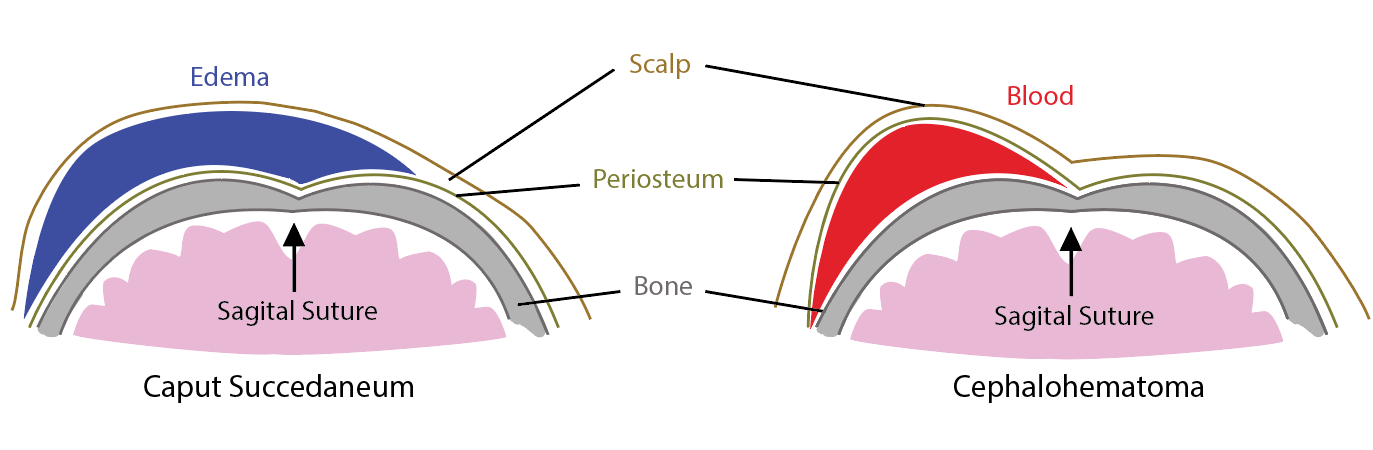
Nicholson L. Caput succedaneum and cephalohematoma: the cs that leave bumps on the head. Neonatal network : NN. 2007 Sep-Oct:26(5):277-81     [PubMed PMID: 17926657]


[5]

Gilboa Y, Kivilevitch Z, Kedem A, Spira M, Borkowski T, Moran O, Katorza E, Achiron R. Caput succedaneum thickness in prolonged second stage of labour: a clinical evaluation. The Australian & New Zealand journal of obstetrics & gynaecology. 2013 Oct:53(5):459-63. doi: 10.1111/ajo.12104. Epub 2013 Jun 26     [PubMed PMID: 23802621]


[6]

Usman S, Wilkinson M, Barton H, Lees CC. The feasibility and accuracy of ultrasound assessment in the labor room. The journal of maternal-fetal & neonatal medicine : the official journal of the European Association of Perinatal Medicine, the Federation of Asia and Oceania Perinatal Societies, the International Society of Perinatal Obstetricians. 2019 Oct:32(20):3442-3451. doi: 10.1080/14767058.2018.1465553. Epub 2018 Apr 30     [PubMed PMID: 29712501]

Level 2 (mid-level) evidence

[7]

Hassan WA, Eggebo TM, Salvesen KA, Lindtjorn E, Lees C. Intrapartum assessment of caput succedaneum by transperineal ultrasound: a two-centre pilot study. The Australian & New Zealand journal of obstetrics & gynaecology. 2015 Aug:55(4):401-3. doi: 10.1111/ajo.12342. Epub 2015 Jul 23     [PubMed PMID: 26201530]

Level 3 (low-level) evidence

[8]

Shah NA, Wusthoff CJ. Intracranial Hemorrhage in the Neonate. Neonatal network : NN. 2016:35(2):67-71. doi: 10.1891/0730-0832.35.2.67. Epub     [PubMed PMID: 27052980]


[9]

Levin G, Mankuta D, Eventov-Friedman S, Ezra Y, Koren A, Yagel S, Rottenstreich A. Factors associated with the severity of neonatal subgaleal haemorrhage following vacuum assisted delivery. European journal of obstetrics, gynecology, and reproductive biology. 2020 Feb:245():205-209. doi: 10.1016/j.ejogrb.2019.12.012. Epub 2019 Dec 28     [PubMed PMID: 31902544]


[10]

Colditz MJ, Lai MM, Cartwright DW, Colditz PB. Subgaleal haemorrhage in the newborn: A call for early diagnosis and aggressive management. Journal of paediatrics and child health. 2015 Feb:51(2):140-6. doi: 10.1111/jpc.12698. Epub 2014 Aug 11     [PubMed PMID: 25109786]


[11]

Shekhar S, Rana N, Jaswal RS. A prospective randomized study comparing maternal and fetal effects of forceps delivery and vacuum extraction. Journal of obstetrics and gynaecology of India. 2013 Apr:63(2):116-9. doi: 10.1007/s13224-012-0282-1. Epub 2012 Aug 28     [PubMed PMID: 24431617]

Level 1 (high-level) evidence

[12]

Jayawant S, Parr J. Outcome following subdural haemorrhages in infancy. Archives of disease in childhood. 2007 Apr:92(4):343-7     [PubMed PMID: 17376941]


[13]

Lykoudis EG, Spyropoulou GA, Lavasidis LG, Paschopoulos ME, Paraskevaidis EA. Alopecia associated with birth injury. Obstetrics and gynecology. 2007 Aug:110(2 Pt 2):487-90     [PubMed PMID: 17666638]


[14]

Martín JM, Jordá E, Alonso V, Villalón G, Montesinos E. Halo scalp ring in a premature newborn and review of the literature. Pediatric dermatology. 2009 Nov-Dec:26(6):706-8. doi: 10.1111/j.1525-1470.2009.01017.x. Epub     [PubMed PMID: 20199445]