Pediatric Cataract

Earn CME/CE in your profession:


Continuing Education Activity

A pediatric cataract is an important cause of treatable childhood blindness, provided it is recognized and managed early. Leukocoria, strabismus, and nystagmus are common presenting signs. Various other differentials of leukocoria must be ruled out before managing a case of pediatric cataract. This activity reviews the etiology, pathophysiology, evaluation, management, and postoperative rehabilitation of pediatric cataracts and highlights the role of the interprofessional team in evaluating and treating children with this condition.

Objectives:

  • Identify the etiology of pediatric cataracts.
  • Review the appropriate history and systemic and ocular evaluation of pediatric cataracts.
  • Outline the management options available for pediatric cataracts.
  • Explain interprofessional team strategies for improving final visual outcomes in children with pediatric cataracts.

Introduction

Pediatric cataracts are one of the leading causes of treatable childhood blindness. If left untreated, it can have a significant impact socially, economically, and emotionally on the child, family, and society at large. This remains a challenging entity in ophthalmological practice because of the need to identify, diagnose and manage the condition as early as possible in order to prevent amblyopia.

Routine screening and awareness among parents about leukocoria and strabismus lead to early diagnosis and management. A good pre-op evaluation, intraocular lens (IOL) power calculation, meticulous surgery, and equally efficient postoperative care and visual rehabilitation are important for a favorable outcome. An interdepartmental effort involving pediatrics, anesthesia, ophthalmology, and optometry help in the proper and effective management of pediatric cataract.[1][2][3][4]

Etiology

Causes of pediatric cataracts are diverse and can range from being idiopathic to being associated with systemic disorders. They can be unilateral or bilateral based on etiology. A majority of unilateral and some bilateral cases are idiopathic.[3]

  • Idiopathic
  • Intrauterine infection (TORCH infections- toxoplasma, CMV retinitis, rubella, HSV)[5]
  • Drug-induced
    • Corticosteroids
  • Metabolic disorders[6]
    • Galactosemia
    • Galactokinase deficiency
    • Hypocalcemia
    • Hypoglycemia
  • Trauma
    • Accidental
    • Non-accidental
    • Radiation
    • Laser photocoagulation
  • Other associated ocular diseases
    • Microphthalmia, Microcornea
    • Aniridia
    • Persistent hyperplastic primary vitreous (PHPV)
    • Peter anomaly
    • Corneal guttae
    • Coloboma
  • Inherited without systemic abnormalities[7]
    • Autosomal dominant (most common)
    • Autosomal recessive (mostly seen in families with a history of consanguinity)
    • X-linked
  • Associated with systemic abnormalities[6]
    • Chromosomal abnormalities
      • Trisomy 21
      • Turner syndrome
      • Trisomy 13
      • Trisomy 18
      • Cri-du-chat syndrome
    • Cerebro-oculofacial-skeletal syndrome (COFS)
    • Mitochondrial abnormalities
      • Complex I deficiency
    • Renal disease
      • Lowe syndrome
    • Skeletal disease
      • Smith-Lemli-Opitz
      • Conradi syndrome
      • Weill-Marchesani syndrome
    • Syndactyly, polydactyly, or digital abnormalities
      • Bardet-Biedl syndrome
      • Rubenstein-Taybi syndrome
    • Central nervous system abnormalities
      • Zellweger syndrome
      • Meckel-Gruber syndrome
    • Cardiac disease
      • Hypertrophic cardiomyopathy
    • Dermatological
      • Cockayne syndrome
      • Rothmund-Thomson
      • Atopic dermatitis
      • Incontinentia pigmenti
      • Progeria
      • Ichthyosis
      • Ectodermal dysplasia
    • Dental Anomalies
      • Nance-Horan syndrome
      • Lenz syndrome

Causes of bilateral congenital cataracts:

  • Idiopathic
  • Intrauterine infection (TORCH infections- toxoplasma, CMV retinitis, rubella, HSV)[5]
  • Multisystem genetic disorder
  • Inborn errors of metabolism
  • Endocrinopathies
  • Trauma
  • Uveitic cataract

Epidemiology

Various epidemiological studies have been performed to evaluate the causes of childhood blindness, of which pediatric cataract accounts for about 7.4% to 15.3% if not diagnosed and treated early.[2] Incidence and prevalence of pediatric cataracts differ from region to region, being lower in high socioeconomic countries and higher in low socioeconomic countries. The incidence ranges between 1.8 to 3.6/10000 per year, while prevalence varies between 0.63 to 13.6 per 10000 (in low-income economies like Bangladesh, Pakistan, India) to 0.42-2.05/10000 (in high-income economies like the US, UK).[8] There is no sex-based or laterality-based difference in the prevalence of pediatric cataracts.[9]

Pathophysiology

Pediatric cataracts can have a diverse etiology ranging from heritable genetic defects to any insult during lenticular development in the fetal period or the growing age to associated systemic syndromes. The autosomal dominant type of inheritance is commonly seen in hereditary cataracts.[7] As many as 51 genes and loci have been identified in a genetic screening study in Australia.[10] Mutations in genes coding for transcription proteins like PAX6, FoxE3, C-MAF, PITX3, MIP, CRYAA, etc., are frequent. Crystallin and connexin mutations are also seen in the majority of cases.[11][12]

History and Physical

History, ocular examination, and systemic examination are essential when evaluating a patient with a pediatric cataract. Various ocular and syndromic associations must be ruled out.

History

  • Age of onset, duration of symptoms
  • Antenatal and perinatal history
  • Developmental history, milestones
  • History of any change in behavior, inability to catch hold of objects thrown at them, frequent falls, squeezing eyes in bright light
  • Any history suggestive of any associated systemic abnormality
  • Trauma, Previous treatment, or surgery
  • Family history of congenital or developmental cataract or history of consanguinity

General Physical Examination:

  • Pediatric consultation – to rule out any systemic association or genetically transmitted disorder
  • Head circumference. Congenital cataracts associated with dysmorphic syndromes where head circumference measurement holds importance: Trisomy 21, Hallermann-Streiff-Francois syndrome, Lowe's Oculo-Cerebro-renal syndrome, Cri-du-chat syndrome (5p deletion), Nance-Horan syndrome, Edward syndrome, etc.
  • If the attending physician or the parents are not sure about when they noticed the white reflex in the eye or the onset of cataract, the family album can be reviewed. This is important because cataracts developing during the critical period of ocular and visual development have a poorer prognosis.
  • Examining the family members: an ocular examination of family members, especially parents and other siblings, may reveal undiagnosed lenticular changes in them suggestive of an inherited cause of pediatric cataract. Autosomal dominant forms are the commonest. The X-linked recessive pattern may also present in children with Lowe syndrome and Nance-Horan syndrome.[3][13][6][14][6]

Ocular Examination

  1. Visual acuity: 
    1. Preverbal Children
      • Bruckner test: both eyes are visualized simultaneously with the direct ophthalmoscope. If there is a deviation of an eye, the deviated eye will have a lighter red reflex. In the presence of anisometropia, one of the reflexes may also be brighter. 
      • Cover test
      • Resistance to occlusion of the better eye
      • Monocular fixation
      • Fixation preference
      • Vertical Prism Test
      • Forced choice preferential looking
      • Teller acuity cards
      • Optokinetic nystagmus
      • Pattern VEP
    2. Verbal Children:
      1. Detection acuity test:
        • Catford drum test
        • STYCAR graded ball test
      2. Recognition acuity test
        1. Direction identification test: Landolt C test, Snellen E test.
        2. Letter identification test: Snellen charts, Lippman HOTV tests.
        3. Picture identification charts: Allen picture cards, Domino cards tests.
        4. Picture identification on the behavioral pattern: Cardiff acuity test, Optokinetic drum test
  2. Slit-lamp examination: any associated anterior segment findings like microcornea, anterior segment dysgenesis, Iris coloboma, the morphology of cataract, any microspherophakia, ectopia lentis or lens subluxation, pre-existing posterior capsular defect. Morphological classification of pediatric cataract is as follows:[15][3]
    1. Anterior cataracts- anterior polar, anterior pyramidal, anterior subcapsular, anterior lenticonus (Alport syndrome)
    2. Cataract involving whole lens – Total cataract, membranous cataract
    3. Posterior cataracts- Mittendrofs dot, posterior lenticonus, posterior subcapsular cataract
    4. Central cataract- Lamellar (zonular), pulverulent, oil drop, coronary, blue dot cataract
    5. Sutural cataract
    6. Wedge-shaped cataract
  3. Ocular motility evaluation: Strabismus and nystagmus should be specifically looked for in these children as sometimes these may be the presenting signs. Strabismus is usually seen in children with unilateral cataracts and develops when an irreparable visual loss has already occurred. Congenital sensory nystagmus is usually associated with pediatric cataracts. The presence of either strabismus or nystagmus indicates that cataract is visually significant.[16][17][18]
  4. Intraocular pressure: To rule out glaucoma( as associated with congenital rubella syndrome) with the help of a tonometer.[19]
  5. Pupillary reactions: This can give a rough idea about the health of the optic nerve head.
  6. Direct and indirect ophthalmoscopy: To evaluate for associated vitreous or posterior segment abnormality such as vitreous hemorrhage, remnants of primary vitreous, fundal coloboma, optic or macular hypoplasia, etc.

Visually significant Cataract[20][21]

  • Visual acuity ≤ 20/60
  • Decrease in contrast sensitivity
  • Increased glare
  • Loss of stereo acuity
  • Centrally or posteriorly located
  • Size more than 3 mm
  • associated strabismus or nystagmus

Examination of The Red Reflex[22][23][24][25]

It is vital that every medical person looking after children, especially newborns and neonates, be familiar with performing the red reflex test. According to the American Academy of Pediatrics, red reflex testing should be performed in all patients in the neonatal period and all routine well-child examinations and health care appointments. 

The red reflex is seen commonly when photographs are taken with a flash: it is essentially a reflective phenomenon. The light that enters the eye through the pupil is reflected by the cornea creating a red glow. The presence of a red reflex confirms transparency in the optical structures of the eye, including the tear film, the cornea, the aqueous humor, the lens, the vitreous gel, and the retina. 

The red reflex is performed with emphasis on the following:

  • Presence of absence of the red reflex
  • Color of the reflex
  • Brightness of the reflex
  • Symmetry of the reflex between the eyes

Requirements to Perform a Red Reflex Test

  • Dim or turn off the room lights
  • Have the child on the parent's lap so that the height of the child's eyes are aligned with those of the examiner
  • The direct ophthalmoscope diopter power is set to that of the examiner's eye reading or to "0."
  • After being turned on, the ophthalmoscope is held close to the examiner's eye and 12 to 18 inches away from those of the child.
  • The child should be encouraged to look at the light, preferably with the aid of an assistant and toys as needed.
  • The light is directed at one eye and then the other to detect and compare the light reflexes.
  • Examine the symmetry by signing the light over both eyes (Bruckner test)

Evaluation

Laboratory Investigations

  1. Blood and Routine Investigations
    • Complete hemogram
    • Blood sugar
    • Urine routine microscopy
  2. Other Tests (case to case basis depending upon the history and examination)
    • Serum calcium for hyper or hypoparathyroidism.[26]
    • VDRL for syphilis[27]
    • Antibody titer for TORCH.[28][5]
    • Red cell galactokinase or uridyl transferase for galactosemia[29]
    • Urine protein for Alport syndrome[30]
    • Urine amino acid for Lowe syndrome[14]
    • Urine sodium nitroprusside/ plasma homocysteine for homocystinuria[31]
    • Urine/serum copper for Wilson disease[32]
    • Karyotyping for a genetic defect[7]

Ocular Investigations

  1. Ultrasound B scan: B scan ultrasonography has to be performed to rule out other posterior segment pathologies that mimic congenital cataract (other causes of leukocoria include retinoblastoma, persistent hyperplastic primary vitreous, coats disease, ROP with retrolental fibroplasia, organized vitreous hemorrhage, congenital falciform fold, ocular toxocariasis, and retinal hamartomas.[33][34]
  2. Ultrasound biometry can also help to obtain optical parameters such as axial length (AL), anterior chamber depth (ACD), and lens thickness (LT), which is crucial for IOL power calculation.[35][36]
  3. Keratometry: Using handheld keratometers. A child’s cooperation is the key to accurate keratometry readings. In the case of an uncooperative child, standard K values of 43.00 D can be used.[37]

Treatment / Management

Managing pediatric cataracts requires a team effort consisting of ophthalmologists, pediatricians, anesthesiologists, counselors, and parents/family members. Before surgery, the family members are counseled regarding the visual prognosis, the different available treatment options, and the need for regular follow-up, post-operative visual rehabilitation, and compliance to the amblyopia therapy, if required.

Treatment Options

Mydriasis: Use 2.5% phenylephrine as we need only mydriatic action. This method is used when the partial or nonamblyogenic cataract is present to permit vision through the non opacified area.

Optical iridectomy: no longer used. The same purpose as mydriatics; that is, it permits vision through the non-opacified area.

Surgery

  1. Indications:
    • Presence of any visually significant opacity
    • Cataract with vision ≤ 20/60 or disc not visible with an indirect ophthalmoscope
    • Central cataract ≥ 3 mm
    • Posterior subcapsular cataract
    • Nuclear cataract
    • Bilateral cataract
    • Cataract associated with strabismus, nystagmus
  2. Anesthesia: general anesthesia, along with constant monitoring of vital parameters, is preferred for cataract surgery. A trained anesthesiologist team and interdepartmental support are required in dealing with children.
  3. When to operate: surgery should be performed as soon as possible for visually significant cataracts, ideally within a few weeks after birth to prevent amblyopia. For nonamblyogenic cataract, surgery is better planned after 4years of age as ocular development will be complete and post-operative complications are less, but close follow-up and visual monitoring is required to prevent amblyopia and treat as indicated. The consensus is to operate a unilateral cataract as early as 4-6 weeks after birth. Bilateral cataracts should be operated on by 6-8 weeks, and the second eye should be operated on within 2 weeks of the first surgery.[21]
  4. Type of surgery and the surgical challenges: there has been a paradigm shift and advancement in cataract surgeries. Discission has been replaced by modern-day phacoaspiration and IOL implantation. Choyce and Binkhorst first performed the implantation of a monocular intraocular lens in a child's eye in 1959. Pediatric cataract surgery is different from adult cataract surgery and poses various intraoperative challenges: low scleral rigidity causes difficulty in incision construction and wound closure, the smaller size of the eyeball, shallow anterior chamber depth and small pupil size causing decreased maneuverability, elastic capsule, high positive intravitreal pressure and risk of vitreous loss and expulsion of intraocular contents. Advantages of primary IOL implantation are immediate post-operative refractive correction, minimal or no optical aberration, full visual field, less chance of development and progression of amblyopia, strabismus, nystagmus, and minimal dependence on patients' compliance. IOL implantation in below 2 years of age is still a controversial topic. Lack of long-term data to predict the success rate, associated other ocular abnormalities along with cataract, frequent occurrence of deprivational amblyopia, and increased postoperative complications are the main reason for limited uses of IOL implantation in children below 2 years of age. Infant Aphakia Treatment Study (IATS) showed higher intra-op and post-op complications and the need for additional surgeries in children with IOL implantation in less than 1 yr of age.[38] A consensus regarding surgery performed, and IOL implantation is as follows:[39][20][40][3]
    1. Less than 18 months to 2 years of age - Lens aspiration without IOL with posterior curvilinear capsulorhexis (PCCC) with limited anterior vitrectomy (LAV), leaving the child aphakic and giving post-op aphakic correction followed by secondary IOL implantation at a later age.
    2. 2 years to 5 years- Lens aspiration with IOL implantation with PCCC with LAV.
    3. More than 8 years - Lens aspiration with IOL implantation with PCCC without LAV.
    4. More than 8 years - Phacoaspiration with IOL implantation like an adult cataract surgery.
    5. PCCC (in < 8 years) and LAV (in < 5 years)  are important in younger children because of the higher risk of VAO post-surgery.
  5. IOL POWER CALCULATION IN CHILDREN: Accurate IOL power calculation is essential for post-surgical refractive outcomes in children. It is challenging both in terms of calculating it and adjusting it for the age of the child and/or axial length of the eye. Biometry and keratometry measurements differ according to the type of instruments and method used to measure them. Studies have shown that SRK/T and Holladay 2 formulae had the least predictive error (PE) among the various IOL formulas available. Newer IOL formulas are being investigated to optimize the variability of IOL power in children. Emmetropisation is the change of three major variables, namely axial length, corneal curvature, and lenticular power from its birth values to its adult values as the child grows. As the size of the eyeball grows, axial length increases, and there is a myopic shift. Thus, the initial desired refractive outcome after IOL implantation is hypermetropia. Various studies have shown that under correction of the required IOL power depending upon the child's age improves the refractive outcome in these children post-surgery. Dahan and Drusedau suggested an under correction of 20% in <2 years; 10% between 2 and 8 years. A consensus is to have residual refraction of +6D (1 to 2 years), +5 (2 to 4 years), +4 (4 to 5 years), +3 (5 to 6 years), +2 (6 to 7 years), and plano for >14 years.[41][42][43][44]

Differential Diagnosis

Pediatric cataracts usually present with the whitish opacity in the eye first noticed by the attending parents. Various causes of the white reflex (leukocoria) must be ruled out before diagnosing a pediatric cataract alone. Causes of leukocoria include:

  • Abnormalities in the cornea (corneal opacity)
  • Lens abnormalities (cataract)
  • Vitreous causes (vitreous hemorrhage, persistent hyperplastic primary vitreous)
  • Retinal diseases (Coat disease, retinoblastoma, familial exudative vitreoretinopathy, retinal detachment, retinopathy of prematurity, coloboma, etc.).
  • Tumors such as medulloepithelioma, retinal astrocytoma, etc.

Cataracts can occur in isolation or association with other causes of leukocoria mentioned above. The differential diagnosis can be narrowed down based on history, family history, and a thorough ophthalmic examination.[45][46]

Prognosis

Numerous factors affect the final visual outcome in a child with a pediatric cataract. Visually significant cataract produces blurred images on the retina and thus affect the development of visual pathways and connections in the occipital cortex. Today, with a better understanding of the consequences of cataracts in the early age group and the advancement of the surgical techniques to deal with the same, it is recommended to remove a visually significant cataract at the earliest to prevent sensory deprivation amblyopia. Unilateral cataracts are by far more amblyogenic than bilateral cataracts and thus to be operated on within the first few weeks to months.

The time of diagnosis of a pediatric cataract also plays a crucial role in prognosticating the final visual outcome—the earlier the diagnosis, the earlier the treatment, the better the prognosis, and vice versa. Associated ocular diseases include microcornea, corneal opacities, glaucoma, intraocular inflammation, posterior segment abnormalities, and ocular movement disorders such as unsteady fixation, strabismus, nystagmus, or nystagmoid movements denote poorer prognosis post-surgery.[3][47]

Complications

As with any other intraocular surgery, pediatric cataract surgery has its own set of associated complications that may require additional treatment. 

  1. Visual axis opacification (VAO): It is a potential cause of amblyopia after cataract surgery.[48] It is inevitable if the posterior capsule is left intact. It occurs as a result of proliferation, migration, and metaplasia of lens epithelial cells from the equator to the posterior capsule. Even after adequate posterior capsulotomy, secondary membranes and media opacity can occur. An intact hyaloid face can serve as a scaffold for the migration of lens epithelial cells and their subsequent proliferation and transformation. One of the crucial factors to influence is the age at which a child is being operated. It also depends upon the type of cataract and associated ocular abnormalities (e.g., PHPV, uveitis), aphakic or pseudophakic, type of IOL, size of the capsular opening, and completeness of cortical cleanup. Adequate posterior capsulotomy with or without vitrectomy is essential to prevent VAO. Implantation of IOL in the bag also reduces the chance of VAO. A new technique of bag in the IOL seems promising in decreasing its occurrence. Nd Yag laser capsulotomy can be done if the child is cooperative. A dense and thick VAO may also require additional surgery like membranectomy to clear the visual axis.[49]
  2. Glaucoma: The incidence of glaucoma is 10% to 25%.[50] Open-angle glaucoma is more common, but pupillary block glaucoma can also occur. In most cases, medical management is sufficient, but if uncontrolled, then surgical management is advised. Surgical procedures like trabeculectomy, implantation of glaucoma drainage devices, lastly, cyclodestructive procedures can be resorted to.[51]
  3. Post-operative anterior uveitis: It is more common in children than adults due to increased tissue reactivity. No-touch surgery and in-bag fixation reduce it. Post-operative frequent application of topical steroids and/or posterior sub tenon injection of triamcinolone acetonide may be needed. Sometimes, dense fibrinous exudates and post-op miosis are seen. In such cases, mydriatics such as atropine or even subconjunctival injected mydricaine can be used to break synechiae formation and achieve mydriasis.
  4. IOL-related complications- IOL deposits, synechiae, pupillary capture, IOL opacification, decentration can be seen in the post-op period.
  5. Other complications include cystoid macular edema, retinal detachment, endophthalmitis, heterochromia, corneal decompensation, bullous keratopathy, astigmatism, post-op refractive surprise, etc. may also be seen.[52]

Postoperative and Rehabilitation Care

Early visual rehabilitation of children with pediatric cataracts is as crucial as early diagnosis and treatment. This reduces the incidence of amblyopia, strabismus, and poor fusion.

There are some options available for visual rehabilitation in these children, namely spectacles, Contact lenses, intraocular lenses, epikeratophakia, low vision aids, and amblyopia therapy. Parents should be counseled about the need for regular follow-up and routine refraction due to frequent refractive changes as the eyeball grows with age.

Children less than 2 years need aphakic glasses or contact lens for visual rehabilitation after cataract surgery till the secondary IOL implantation is planned. Aphakic glasses are usually used for bilateral aphakic patients. Due to improved surgical technique and increased number of primary IOL implantation, the use of aphakic glasses has reduced. Aphakia treatment aims to achieve the correct optical correction as soon as cataract extraction is done to permit the early symmetrical stimulation of the visual system in the critical period of development.[53]

  • Spectacles are the earliest form of aphakia treatment. Aphakic glasses with bifocal addition remain the primary mode of visual rehabilitation in bilateral aphakic children owing to their low cost and easy availability, no ocular discomfort, or direct contact with the ocular surface. There are a few notable disadvantages, too, like the weight, large size, restriction of the field of vision, increasing nystagmus, jack in the box phenomenon, pin cushion phenomenon, and ring scotoma. In cases of unilateral cataract, anisometropia, and anisokenia lead to diplopia and amblyopia, which further causes permanent suppression and anomalous retinal correspondence (ARC).
  • Contact lenses (CL) are the best optical device to correct postoperative aphakia. These lenses should be worn during the daytime and removed during the night. It is superior to spectacles in both unilateral and bilateral cases. It offers a larger visual field, decreases glare, minimizes spherical aberrations, better binocularity, and cosmetic appearance. Intolerability, cost, and noncompliance pose significant problems among the children and their parents. A few notable contraindications to its use are chronic eye disease and ocular surface disorders. Three types of CL are available for pediatric patients, namely PMMA or Rigid gas permeable lenses, Silicon elastomer lenses, and Hydrogel lenses.[38] Ideal contact lens should be extended type, but because of its associated complications like giant papillary conjunctivitis, neovascularisation, abrasions, and infective keratitis, daily used lenses are more preferred.

 Children more than 2 years of age with primary IOL implantation also require adequate visual rehabilitation post-surgery. Spectacles or contact lenses may be prescribed after refraction.

Amblyopia Treatment

As mentioned earlier, deprivational amblyopia is more common in pediatric cataracts. Amblyopia developed due to unequal visual input in two eyes during the critical period of development i,e within 2 months. Therefore a unilateral cataract leads to denser amblyopia than a bilateral cataract, hence need earlier surgical intervention. Amblyopia therapy remains the most important part of visual rehabilitation. Treatment should commence as soon as possible. Occlusion therapy remains the mainstay of amblyopia therapy. However, compliance remains the main drawback of amblyopia therapy. Therefore proper counseling of parents is essential for successful visual outcomes.[54]

Patients are reviewed every 2 weeks for visual improvement or reversal of amblyopia.

Low Vision Aids

These devices enable the children to improve their visual performance and helped them to reach their full capacity. There are many devices available according to patient age and affordability of parents. Convex lenses are used in various forms as primary optical assistance. Other devices are standard additional bifocals, hand magnifier, stand magnifier, electronic magnifier, telescopes, and computer adaptation closed-circuit televisions.

Consultations

A pediatric cataract can be a manifestation of or an association of a systemic disease involving other organ systems such as cardiac, renal, or central nervous system, etc. A pediatric consultation to rule out any other systemic comorbidities and gross congenital anomalies is a must. Moreover, since general anesthesia is required for children to be operated upon, a thorough pre-anesthetic evaluation is required to avoid untoward anesthetic complications on the table.

Deterrence and Patient Education

It is essential to counsel parents about their child's condition, etiology, treatment options, and visual prognosis. They should also be informed about the possible complications of the surgery and following surgery together with the need for regular, timely follow-up with proper visual rehabilitation to give the best possible visual outcome to their child.

Enhancing Healthcare Team Outcomes

Pediatric cataract generally presents in primary care settings with apprehensive parents bringing their child after noticing a white reflex (leukocoria), squinting, or nystagmus in their eyes. Primary healthcare professionals play a vital role in the timely referral of such cases to centers with the expertise and capability to deal with the condition. Careful systemic and ocular examination to rule out other causes of leukocoria and any syndromic associations is essential before undertaking surgery. Timely diagnosis, management, and visual rehabilitation of a child with a pediatric cataract are crucial determinants of a favorable prognosis.

An interprofessional team approach involving pediatric ophthalmology specialty-trained ophthalmic surgeons, pediatricians, anesthetists, optometrists, nurses along with informed and motivated parents helps in bringing about the best possible visual outcome. [Level 5]


(Click Image to Enlarge)
Pediatric Zonular cataract, diffuse and oblique illumination on slit lamp examination
Contributed by Dr. Priyadarshi Gupta, MBBS, (MS) ophthalmology

(Click Image to Enlarge)
Pediatric Cataract. This slit lamp photograph depicts zonular type of pediatric cataract with blue dot cataract component.
Contributed by Dr. Priyadarshi Gupta, MBBS, (MS) Ophthalmology

(Click Image to Enlarge)
Pediatric cataract
Pediatric cataract. Posterior lenticonus. This slit lamp photograph shows posterior lenticonus variety of pediatric cataract. These, being close to the nodal point of eye affect visual acuity more than its anterior counterpart. Lowe syndrome is frequently associated with posterior lenticonus.
Contributed by Dr. Priyadarshi Gupta, MBBS, (MS) Ophthalmology

(Click Image to Enlarge)
Congenital Cataracts: peripheral lenticular opacities of the type often seen in patients with galactosemia or galactokinase d
Congenital Cataracts: peripheral lenticular opacities of the type often seen in patients with galactosemia or galactokinase deficiency
Contributed by Prof. Bhupendra C. K. Patel MD, FRCS

(Click Image to Enlarge)
The red reflex: a normal red reflex in each eye with equal clarity and brightness
The red reflex: a normal red reflex in each eye with equal clarity and brightness. This indicates the absence of opacification of the globe structures: cornea, aqueous, lens, vitreous and a normal retina
Contributed by Prof. Bhupendra C. K. Patel MD, FRCS
Details

Updated:

5/29/2023 5:05:42 PM

References


[1]

. Childhood cataract: magnitude, management, economics and impact. Community eye health. 2004:17(50):17-8     [PubMed PMID: 17491800]


[2]

Gilbert C, Foster A. Childhood blindness in the context of VISION 2020--the right to sight. Bulletin of the World Health Organization. 2001:79(3):227-32     [PubMed PMID: 11285667]


[3]

Khokhar SK, Pillay G, Dhull C, Agarwal E, Mahabir M, Aggarwal P. Pediatric cataract. Indian journal of ophthalmology. 2017 Dec:65(12):1340-1349. doi: 10.4103/ijo.IJO_1023_17. Epub     [PubMed PMID: 29208814]


[4]

Solebo AL, Teoh L, Rahi J. Epidemiology of blindness in children. Archives of disease in childhood. 2017 Sep:102(9):853-857. doi: 10.1136/archdischild-2016-310532. Epub 2017 May 2     [PubMed PMID: 28465303]


[5]

Mets MB. Eye manifestations of intrauterine infections. Ophthalmology clinics of North America. 2001 Sep:14(3):521-31     [PubMed PMID: 11705152]


[6]

Trumler AA. Evaluation of pediatric cataracts and systemic disorders. Current opinion in ophthalmology. 2011 Sep:22(5):365-79. doi: 10.1097/ICU.0b013e32834994dc. Epub     [PubMed PMID: 21832913]

Level 3 (low-level) evidence

[7]

Santana A, Waiswo M. The genetic and molecular basis of congenital cataract. Arquivos brasileiros de oftalmologia. 2011 Mar-Apr:74(2):136-42     [PubMed PMID: 21779674]


[8]

Wadhwani M, Vashist P, Singh SS, Gupta V, Gupta N, Saxena R. Prevalence and causes of childhood blindness in India: A systematic review. Indian journal of ophthalmology. 2020 Feb:68(2):311-315. doi: 10.4103/ijo.IJO_2076_18. Epub     [PubMed PMID: 31957718]

Level 1 (high-level) evidence

[9]

Sheeladevi S, Lawrenson JG, Fielder AR, Suttle CM. Global prevalence of childhood cataract: a systematic review. Eye (London, England). 2016 Sep:30(9):1160-9. doi: 10.1038/eye.2016.156. Epub 2016 Aug 12     [PubMed PMID: 27518543]

Level 1 (high-level) evidence

[10]

Javadiyan S, Craig JE, Souzeau E, Sharma S, Lower KM, Mackey DA, Staffieri SE, Elder JE, Taranath D, Straga T, Black J, Pater J, Casey T, Hewitt AW, Burdon KP. High-Throughput Genetic Screening of 51 Pediatric Cataract Genes Identifies Causative Mutations in Inherited Pediatric Cataract in South Eastern Australia. G3 (Bethesda, Md.). 2017 Oct 5:7(10):3257-3268. doi: 10.1534/g3.117.300109. Epub 2017 Oct 5     [PubMed PMID: 28839118]

Level 3 (low-level) evidence

[11]

Cvekl A, Duncan MK. Genetic and epigenetic mechanisms of gene regulation during lens development. Progress in retinal and eye research. 2007 Nov:26(6):555-97     [PubMed PMID: 17905638]


[12]

Verma IC, Paliwal P, Singh K. Genetic Testing in Pediatric Ophthalmology. Indian journal of pediatrics. 2018 Mar:85(3):228-236. doi: 10.1007/s12098-017-2453-7. Epub 2017 Oct 2     [PubMed PMID: 28971364]


[13]

Patil-Chhablani P, Kekunnaya R, Nischal KK. Complex Cases in Pediatric Cataract. Developments in ophthalmology. 2016:57():85-106. doi: 10.1159/000442505. Epub 2016 Apr 1     [PubMed PMID: 27043394]

Level 3 (low-level) evidence

[14]

Bökenkamp A,Ludwig M, The oculocerebrorenal syndrome of Lowe: an update. Pediatric nephrology (Berlin, Germany). 2016 Dec;     [PubMed PMID: 27011217]


[15]

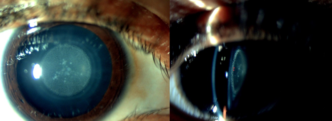
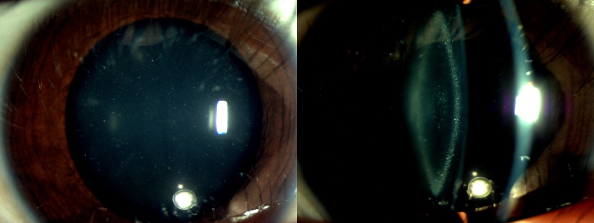
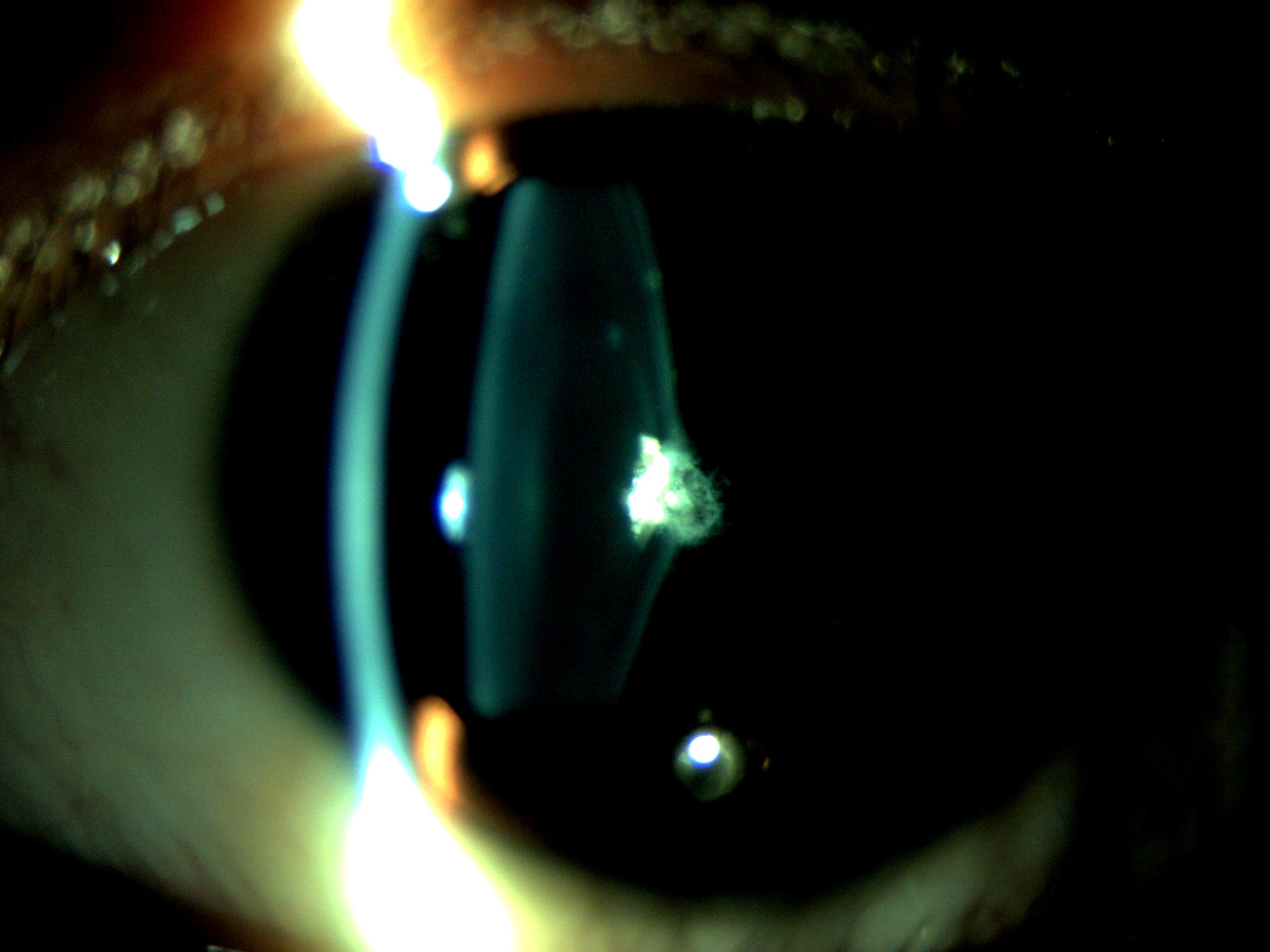
Amaya L, Taylor D, Russell-Eggitt I, Nischal KK, Lengyel D. The morphology and natural history of childhood cataracts. Survey of ophthalmology. 2003 Mar-Apr:48(2):125-44     [PubMed PMID: 12686301]

Level 3 (low-level) evidence

[16]

Zetterström C, Lundvall A, Kugelberg M. Cataracts in children. Journal of cataract and refractive surgery. 2005 Apr:31(4):824-40     [PubMed PMID: 15899463]


[17]

Braddick O, Atkinson J. Development of human visual function. Vision research. 2011 Jul 1:51(13):1588-609. doi: 10.1016/j.visres.2011.02.018. Epub 2011 Feb 26     [PubMed PMID: 21356229]


[18]

Papageorgiou E,McLean RJ,Gottlob I, Nystagmus in childhood. Pediatrics and neonatology. 2014 Oct;     [PubMed PMID: 25086850]


[19]

Giangiacomo A, Beck A. Pediatric glaucoma: review of recent literature. Current opinion in ophthalmology. 2017 Mar:28(2):199-203. doi: 10.1097/ICU.0000000000000349. Epub     [PubMed PMID: 27875350]

Level 3 (low-level) evidence

[20]

Vasavada AR, Nihalani BR. Pediatric cataract surgery. Current opinion in ophthalmology. 2006 Feb:17(1):54-61     [PubMed PMID: 16436925]

Level 3 (low-level) evidence

[21]

Vasavada V. Paradigms for Pediatric Cataract Surgery. Asia-Pacific journal of ophthalmology (Philadelphia, Pa.). 2018 Mar-Apr:7(2):123-127. doi: 10.22608/APO.2017202. Epub 2017 Sep 25     [PubMed PMID: 28971632]


[22]

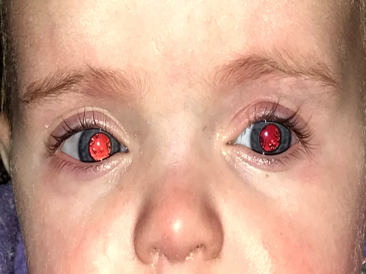
Adams GGW. The Enduring Value of Newborn Red Reflex Testing as a Screening Tool. JAMA ophthalmology. 2021 Jan 1:139(1):40-41. doi: 10.1001/jamaophthalmol.2020.4853. Epub     [PubMed PMID: 33180105]


[23]

Subhi Y, Schmidt DC, Al-Bakri M, Bach-Holm D, Kessel L. Diagnostic Test Accuracy of the Red Reflex Test for Ocular Pathology in Infants: A Meta-analysis. JAMA ophthalmology. 2021 Jan 1:139(1):33-40. doi: 10.1001/jamaophthalmol.2020.4854. Epub     [PubMed PMID: 33180103]

Level 1 (high-level) evidence

[24]

Sun M, Ma A, Li F, Cheng K, Zhang M, Yang H, Nie W, Zhao B. Sensitivity and Specificity of Red Reflex Test in Newborn Eye Screening. The Journal of pediatrics. 2016 Dec:179():192-196.e4. doi: 10.1016/j.jpeds.2016.08.048. Epub 2016 Sep 15     [PubMed PMID: 27640356]


[25]

Gurney SP, Makanjuola T, Kutubi M, Parulekar M, Abbott J. How to use…the direct ophthalmoscope. Archives of disease in childhood. Education and practice edition. 2018 Apr:103(2):102-109. doi: 10.1136/archdischild-2016-312378. Epub 2017 Sep 21     [PubMed PMID: 28935833]


[26]

Mannstadt M, Bilezikian JP, Thakker RV, Hannan FM, Clarke BL, Rejnmark L, Mitchell DM, Vokes TJ, Winer KK, Shoback DM. Hypoparathyroidism. Nature reviews. Disease primers. 2017 Aug 31:3():17055. doi: 10.1038/nrdp.2017.55. Epub 2017 Aug 31     [PubMed PMID: 28857066]


[27]

Drusin LM. Syphilis: clinical manifestations, diagnosis, and treatment. The Urologic clinics of North America. 1984 Feb:11(1):121-30     [PubMed PMID: 6369700]


[28]

Jyoti M,Shirke S,Matalia H, Congenital rubella syndrome: Global issue. Journal of cataract and refractive surgery. 2015 May;     [PubMed PMID: 26049858]


[29]

Anderson S. GALT Deficiency Galactosemia. MCN. The American journal of maternal child nursing. 2018 Jan/Feb:43(1):44-51. doi: 10.1097/NMC.0000000000000388. Epub     [PubMed PMID: 29215423]


[30]

Savige J, Sheth S, Leys A, Nicholson A, Mack HG, Colville D. Ocular features in Alport syndrome: pathogenesis and clinical significance. Clinical journal of the American Society of Nephrology : CJASN. 2015 Apr 7:10(4):703-9. doi: 10.2215/CJN.10581014. Epub 2015 Feb 3     [PubMed PMID: 25649157]


[31]

Carrillo-Carrasco N, Chandler RJ, Venditti CP. Combined methylmalonic acidemia and homocystinuria, cblC type. I. Clinical presentations, diagnosis and management. Journal of inherited metabolic disease. 2012 Jan:35(1):91-102. doi: 10.1007/s10545-011-9364-y. Epub 2011 Jul 12     [PubMed PMID: 21748409]


[32]

Ferenci P. Diagnosis of Wilson disease. Handbook of clinical neurology. 2017:142():171-180. doi: 10.1016/B978-0-444-63625-6.00014-8. Epub     [PubMed PMID: 28433100]


[33]

Kaya A. Preoperative usage of ultrasound biomicroscopy in pediatric cataract. Arquivos brasileiros de oftalmologia. 2016 Feb:79(1):62. doi: 10.5935/0004-2749.20160019. Epub     [PubMed PMID: 26840173]


[34]

Khokhar S, Tejwani LK, Kumar G, Kushmesh R. Approach to cataract with persistent hyperplastic primary vitreous. Journal of cataract and refractive surgery. 2011 Aug:37(8):1382-5. doi: 10.1016/j.jcrs.2011.06.012. Epub     [PubMed PMID: 21782083]


[35]

Capozzi P, Morini C, Piga S, Cuttini M, Vadalà P. Corneal curvature and axial length values in children with congenital/infantile cataract in the first 42 months of life. Investigative ophthalmology & visual science. 2008 Nov:49(11):4774-8. doi: 10.1167/iovs.07-1564. Epub 2008 May 23     [PubMed PMID: 18502997]


[36]

Moshirfar M, Buckner B, Ronquillo YC, Hofstedt D. Biometry in cataract surgery: a review of the current literature. Current opinion in ophthalmology. 2019 Jan:30(1):9-12. doi: 10.1097/ICU.0000000000000536. Epub     [PubMed PMID: 30394990]

Level 3 (low-level) evidence

[37]

Trivedi RH, Wilson ME. Keratometry in pediatric eyes with cataract. Archives of ophthalmology (Chicago, Ill. : 1960). 2008 Jan:126(1):38-42. doi: 10.1001/archophthalmol.2007.22. Epub     [PubMed PMID: 18195216]


[38]

Infant Aphakia Treatment Study Group, Lambert SR, Buckley EG, Drews-Botsch C, DuBois L, Hartmann E, Lynn MJ, Plager DA, Wilson ME. The infant aphakia treatment study: design and clinical measures at enrollment. Archives of ophthalmology (Chicago, Ill. : 1960). 2010 Jan:128(1):21-7. doi: 10.1001/archophthalmol.2009.350. Epub     [PubMed PMID: 20065212]


[39]

Lim ME, Buckley EG, Prakalapakorn SG. Update on congenital cataract surgery management. Current opinion in ophthalmology. 2017 Jan:28(1):87-92     [PubMed PMID: 27653605]

Level 3 (low-level) evidence

[40]

Lin AA, Buckley EG. Update on pediatric cataract surgery and intraocular lens implantation. Current opinion in ophthalmology. 2010 Jan:21(1):55-9. doi: 10.1097/ICU.0b013e32833383cb. Epub     [PubMed PMID: 19855277]

Level 3 (low-level) evidence

[41]

Eibschitz-Tsimhoni M, Archer SM, Del Monte MA. Intraocular lens power calculation in children. Survey of ophthalmology. 2007 Sep-Oct:52(5):474-82     [PubMed PMID: 17719370]

Level 3 (low-level) evidence

[42]

Vasavada V, Shah SK, Vasavada VA, Vasavada AR, Trivedi RH, Srivastava S, Vasavada SA. Comparison of IOL power calculation formulae for pediatric eyes. Eye (London, England). 2016 Sep:30(9):1242-50. doi: 10.1038/eye.2016.171. Epub 2016 Aug 5     [PubMed PMID: 27494083]


[43]

Dahan E, Drusedau MU. Choice of lens and dioptric power in pediatric pseudophakia. Journal of cataract and refractive surgery. 1997:23 Suppl 1():618-23     [PubMed PMID: 9278814]


[44]

Enyedi LB, Peterseim MW, Freedman SF, Buckley EG. Refractive changes after pediatric intraocular lens implantation. American journal of ophthalmology. 1998 Dec:126(6):772-81     [PubMed PMID: 9860000]


[45]

Cheng KP, Hiles DA, Biglan AW. The differential diagnosis of leukokoria. Pediatric annals. 1990 Jun:19(6):376-83, 386     [PubMed PMID: 2200999]


[46]

Kembhavi SA, Sable N, Vora T, Arora B. Leukokoria: All That's White Is Not Retinoblastoma. Journal of clinical oncology : official journal of the American Society of Clinical Oncology. 2011 Jul 1:29(19):e586-7. doi: 10.1200/JCO.2011.34.8334. Epub 2011 Apr 18     [PubMed PMID: 21502547]


[47]

Kalia A, Gandhi T, Chatterjee G, Swami P, Dhillon H, Bi S, Chauhan N, Gupta SD, Sharma P, Sood S, Ganesh S, Mathur U, Sinha P. Assessing the impact of a program for late surgical intervention in early-blind children. Public health. 2017 May:146():15-23. doi: 10.1016/j.puhe.2016.12.036. Epub 2017 Feb 1     [PubMed PMID: 28404468]


[48]

Shrestha UD, Shrestha MK. Visual Axis Opacification in Children Following Paediatric Cataract Surgery. JNMA; journal of the Nepal Medical Association. 2014 Oct-Dec:52(196):1024-30     [PubMed PMID: 26982905]


[49]

Khaja WA, Verma M, Shoss BL, Yen KG. Visual axis opacification in children. Ophthalmology. 2011 Jan:118(1):224-5. doi: 10.1016/j.ophtha.2010.08.030. Epub     [PubMed PMID: 21199720]


[50]

Baden C, Shija F, Lewallen S, Courtright P, Hall A. Glaucoma after pediatric cataract surgery in a population with limited access to care. Journal of AAPOS : the official publication of the American Association for Pediatric Ophthalmology and Strabismus. 2013 Apr:17(2):158-62. doi: 10.1016/j.jaapos.2012.11.009. Epub 2013 Mar 22     [PubMed PMID: 23528376]


[51]

Whitman MC, Vanderveen DK. Complications of pediatric cataract surgery. Seminars in ophthalmology. 2014 Sep-Nov:29(5-6):414-20. doi: 10.3109/08820538.2014.959192. Epub     [PubMed PMID: 25325868]


[52]

Gasper C, Trivedi RH, Wilson ME. Complications of Pediatric Cataract Surgery. Developments in ophthalmology. 2016:57():69-84. doi: 10.1159/000442502. Epub 2016 Apr 1     [PubMed PMID: 27043393]


[53]

Repka MX. Visual Rehabilitation in Pediatric Aphakia. Developments in ophthalmology. 2016:57():49-68. doi: 10.1159/000442501. Epub 2016 Apr 1     [PubMed PMID: 27043392]


[54]

Vagge A, Nelson LB. Amblyopia update: new treatments. Current opinion in ophthalmology. 2016 Sep:27(5):380-6. doi: 10.1097/ICU.0000000000000293. Epub     [PubMed PMID: 27152486]

Level 3 (low-level) evidence