Neurally Adjusted Ventilatory Assist (NAVA)

Earn CME/CE in your profession:


Continuing Education Activity

Neurally-adjusted ventilatory assist (NAVA) is a relatively newer mode of ventilation in which a ventilator utilizes the electrical activity of the diaphragm (Edi) to generate appropriate breaths and assist ventilated patients. This topic highlights and reviews the role of NAVA in the management of respiratory failure in both neonates and adults by an interprofessional team.

Objectives:

  • Review the difference between proportional assisted ventilation and NAVA ventilation.
  • Identify indications and contraindications to the NAVA ventilator.
  • Describe the clinical significance and possible benefits of NAVA ventilation.
  • Outline the equipment, personnel, preparation, and technique for initial set-up by the interprofessional team.

Introduction

Although the earliest reported use of mechanical ventilation is in the 16th century, its use has become widespread in the 20th century in patients with respiratory failure. Over the decades, optimal respiratory care and ventilator support have been supported by strong clinical evidence. Mechanical ventilation is triggered by either change in respiratory flow or pressure. Change in the respiratory flow (commonly termed as flow trigger) may cause false triggering or missed triggering. This produces patient-ventilator asynchrony.[1][2] 

Invasive mechanical ventilation causes alveolar overdistention, pulmonary air leaks, and small airway injury.[3] In 1999, the concept of neural control of mechanical ventilation was first described by Sinderby et al.[4] NAVA ventilator detects the electrical activity (electromyographic signal) of the diaphragm by a specially placed nasogastric catheter. In 2007, FDA approved the use of NAVA in all patients as small as 500 grams birth weight.

Anatomy and Physiology

Normal Respiration

The respiratory center in the brain activates spontaneous breathing via the transmission of impulses through the phrenic nerve. Lourenco et al. showed that diaphragmatic activity is proportional to phrenic nerve activity.[5] This leads to a muscle contraction and allows the inflow of air by creating a negative alveolar pressure. Phrenic nerve impulses precede the contraction of the diaphragm. Depth and cycling of the breathing movement are dependent on the respiratory center output. 

Physiology of Edi Signal

All muscles in the body stimulate muscle contraction by generating electrical activity. The nerve stimulus controls this electrical activity. The NAVA ventilator utilizes this electrical activity to assist the patient’s respiratory efforts in a synchronized manner. Edi is the primary requirement for NAVA to function and the main source for ventilator trigger. In theory, NAVA overcomes the limitation of proportional assist ventilation such as air leaks and asynchrony between patient and ventilator. The ventilator breath is triggered and terminated by changes in this electrical activity. The pressure is delivered in proportion to the Edi signal and ends when the Edi signal subsides. The ventilator displays two forms of Edi; Edi max (peak inspiration) and Edi min (tonic activity of diaphragm).

Figure 1: A process of inspiration and ventilator trigger[4]

NAVA Level

NAVA level converts the Edi signal into an appropriate pressure. NAVA level is expressed as cmHO/ µV. During ventilation, the ventilator will deliver pressure by multiplying each Edi by the NAVA level (see below). Increasing or decreasing the NAVA level will change the pressure delivery for the same Edi. However, pressure delivery varies with each breathing effort, and it depends on the measured electrical activity of the diaphragm. Hence, the patient controls both own and ventilator pressure and thus improves synchrony and comfort.

Calculation of Peak inspiratory pressure (PIP) = NAVA level x (Edi max – Edi min) + PEEP

Indications

There is no specific indication for the use of NAVA in adults, children, or neonates. It can be used in all conditions where conventional ventilators are indicated.

Adults[6]

  • Acute respiratory distress syndrome (ARDS)
  • Acute hypoxemic respiratory failure 

Neonates or Children

  • Respiratory distress syndrome (RDS)
  • Pulmonary hypertension (primary or secondary)
  • Assess breathing activity in the certain condition such as central hypoventilation syndrome[7]

Contraindications

Any condition that limits the respiratory drive or any diaphragmatic anatomical defect in cases of an intact respiratory drive may be contraindicated, including:

  1. Central: paralytics, suppressed respiratory drive due to heavy sedation or brain injury
  2. Peripheral: phrenic nerve injury or use of paralytic agent
  3. Structural: esophageal atresia and diaphragmatic hernia

Equipment

The Servo-I ventilator is a commonly used ventilator and is the only one compatible with the NAVA catheter.

Personnel

A skilled respiratory therapist must set up the machine, and a nurse will insert an Edi catheter.

Preparation

Personnel must insert and verify proper Edi catheter position and check the machine for proper functioning.

Technique or Treatment

An array of nine miniaturized electrodes are embedded within a catheter which is positioned in the lower esophagus at the level of the diaphragm. These electrodes continuously detect the electrical activity of the diaphragm and transmit this information to the ventilator.

There are three steps for the proper positioning of the Edi catheter.

  • Anatomical placement: The most common practice to place a catheter is to measure a distance from the nose to the ear lobe to the xiphoid process (NEX method).[8] The use of lubricants is not recommended as it may interfere with Edi signal measurement. Instead of lubricants, an Edi catheter can be dipped into the water.
  • Verification of the position of electrodes: ECG waveforms are visualized in the “Edi catheter positioning” window of the ventilator. P and QRS waves are present in the top electrodes (close to the right atrium). Loss of P wave and dampen QRS wave are present in the lower leads (close to the stomach)
  • Verification of the Edi signal: A weak or absent signal may suggest neural disorder, sedation, or muscle relaxant use.

Secure the catheter once the second and third leads are highlighted in blue and record the insertion.

Initial NAVA Settings[9]

  1. NAVA level
  2. Positive end-expiratory pressure (PEEP)
  3. Trigger Edi
  4. Backup ventilator settings
  5. fraction of inspired oxygen (FiO)
  6. Alarm settings

NAVA Level

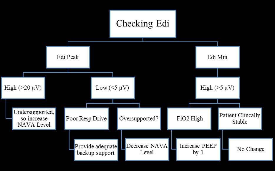
NAVA level should be chosen to achieve a goal Edi range of (5-20µV). Lower NAVA level if Edi max < 5 µV and increase level if Edi max > 20 µV. NAVA level typically adjusted by 0.1-0.2 cmHO/ µV increments.

PEEP

Oxygenation and lung expansion are mainly determined by PEEP. Set the same PEEP according to earlier ventilator settings or according to the age of the patients.

Trigger Edi

Trigger Edi should be 0.5 µV.

Backup Ventilator Settings

An appropriate ventilator setting should be chosen in terms of respiratory rate, PEEP, tidal volume, inspiratory time, and peak inspiratory pressure. It could be similar to the earlier ventilator settings. These are set to kick in if there is a lack of Edi signal triggering a NAVA breath.

FiO

Oxygen requirements should be adjusted depending on targeted oxygen saturations depends on the condition of the disease.

Alarm Settings

The maximum pressure and apnea time should be chosen according to gestation age and maturity. Apnea time should be 2-4 s.

Monitoring During NAVA Ventilation

  • Oxygen saturation
  • Transcutaneous carbon dioxide pressure
  • Edi signal (monitor trends on a ventilator)
  • Arterial/capillary blood gas
  • Trends
    1. The number of switches to back-up per minute: This indicates how often the patient goes into back-up mode. If the numbers are higher, then the current apnea time may be too short, and a patient can tolerate longer apnea time.
    2. Percentage of time on backup mode: if patients remain mostly in backup ventilation, then patients may not be ready to be weaned or may indicate malpositioning of the catheter.

Weaning From NAVA Ventilator

Unlike pressure-assist ventilation, weaning happens spontaneously in NAVA.[10] Arterial or capillary blood gas is an important tool to monitor ventilator requirements. NAVA ventilator weaning is achieved by adjusting the NAVA level. If blood gas is acceptable, wean NAVA level by 0.5-1 cmHO/ µV. Consider extubating the patient once the NAVA level reaches 1 cmHO/ µV. If the patient appears clinically stable, then the following things can be considered before extubation.

  • Increasing apnea time
  • Decreasing back-up settings
  • Lowering NAVA level

Troubleshooting During NAVA

  • The patient develops respiratory distress and oxygen requirement increasing
  1. Catheter malposition
  2. Upper airway obstruction
  3. Exceeding pressure limit
  4. High Edi (under-supported ventilation)
  5. Not enough Backup pressure
  • Edi monitoring (Figure 2)

Complications

There are no specific complications to NAVA ventilation mode use. However, it can cause the same complications as other mechanical ventilator modes, such as ventilator-induced lung injury, ventilator-induced diaphragmatic dysfunction, ventilator-induced pneumonia, and pneumothorax. The chances of ventilator-induced lung injury and ventilator-induced diaphragmatic dysfunction are less with NAVA as delivery of pressure is proportional to the patient’s efforts, and reports have shown the lower need for PIP when using NAVA.

Clinical Significance

As mentioned earlier, NAVA was aimed to overcome the limitation of limitations of proportional assisted ventilation. However, NAVA has several potential benefits.

  • Patient-ventilator synchrony: Mechanical ventilator synchrony depends on two factors- the timing of the mechanical breath delivery and the amount of pressure from a ventilator. There is a delay in diaphragm activity (signaled from the brain) and ventilator response in the flow trigger ventilator. However, NAVA delivers mechanical breath when the diaphragm receives a signal from the brain. Beck et al. and Breatnach et al. reported improved patent-ventilator synchrony during their NAVA trial in infants and children. It decreases the discomfort and agitation in patients while improving synchrony. Synchronized respiratory efforts have been associated with improved ventilation and oxygenation.[11][12] Improved synchronization will prevent further lung injury by limiting barotrauma and volutrauma. Improved synchrony also leads to decreased use of sedation (opioids)
  • Air leaks: Air leaks are very common in both invasive and non-invasive ventilation. Although, leaks can be minimized by choosing the proper size endotracheal tube (cuffed or uncuffed) and compensating circuit in ventilators. Air-leak can be a bigger problem in non-invasive ventilation. It has shown that NAVA continues to trigger effectively even in the presence of 75% air leaks.[10][13]
  • Benefits in preterm infants: In recent years, the use of NAVA has been increased and studied extensively in both preterm and term infants. Kallio et al. compared NAVA with conventional ventilators in preterm infants (28 to 36 weeks GA) and showed lower PIP needs in infants treated with NAVA, but the difference in the secondary outcomes such as bronchopulmonary dysplasia, time on mechanical ventilation, or pneumothorax.[14] In a multi-center retrospective review, NAVA was found to be successful as respiratory stability in 67% of the infants with severe BPD.[15] Thus, NAVA can also be used safely in premature infants. However, a well-designed randomized controlled trial is required to assess the long-term effectiveness of NAVA ventilation in terms of reducing the incidence of BPD.
  • Non-invasive mode: Non-invasive NAVA works exactly the same as invasive NAVA, but ventilation is delivered via nasal prongs or a mask. Non-invasive NAVA has shown to be effective even in the settings of high leaks.[13] (19218884). When infants extubated to non-invasive NAVA ventilation, infants remained extubated longer and required lower PIP as compared to non-invasive pressure ventilation.[16] Setting up non-invasive NAVA and other monitoring approaches remains the same as invasive NAVA.

Enhancing Healthcare Team Outcomes

Like any other ventilators, NAVA also requires close coordination of physicians, registered nurses, and respiratory therapists for better management. Proper teaching and guidance are required before its implementation in any intensive care unit. Although NAVA has shown some promising results in few studies, larger studies are required for its widespread use. Proper monitoring, documentation, and trending ventilator data are required for better outcomes in the given patients.


(Click Image to Enlarge)
Edi Monitoring
Edi Monitoring
Contributed by Sanket D Shah, MD
Details

Editor:

Fatima Anjum

Updated:

5/22/2023 9:55:21 PM

References


[1]

Keszler M. State of the art in conventional mechanical ventilation. Journal of perinatology : official journal of the California Perinatal Association. 2009 Apr:29(4):262-75. doi: 10.1038/jp.2009.11. Epub 2009 Feb 26     [PubMed PMID: 19242486]


[2]

Mellott KG,Grap MJ,Munro CL,Sessler CN,Wetzel PA, Patient-ventilator dyssynchrony: clinical significance and implications for practice. Critical care nurse. 2009 Dec;     [PubMed PMID: 19724065]


[3]

Stein H, Alosh H, Ethington P, White DB. Prospective crossover comparison between NAVA and pressure control ventilation in premature neonates less than 1500 grams. Journal of perinatology : official journal of the California Perinatal Association. 2013 Jun:33(6):452-6. doi: 10.1038/jp.2012.136. Epub 2012 Oct 25     [PubMed PMID: 23100042]


[4]

Sinderby C, Navalesi P, Beck J, Skrobik Y, Comtois N, Friberg S, Gottfried SB, Lindström L. Neural control of mechanical ventilation in respiratory failure. Nature medicine. 1999 Dec:5(12):1433-6     [PubMed PMID: 10581089]


[5]

Lourenço RV, Cherniack NS, Malm JR, Fishman AP. Nervous output from the respiratory center during obstructed breathing. Journal of applied physiology. 1966 Mar:21(2):527-33     [PubMed PMID: 5934459]


[6]

Navalesi P,Costa R, New modes of mechanical ventilation: proportional assist ventilation, neurally adjusted ventilatory assist, and fractal ventilation. Current opinion in critical care. 2003 Feb;     [PubMed PMID: 12548030]

Level 3 (low-level) evidence

[7]

Szczapa T, Beck J, Migdal M, Gadzinowski J. Monitoring diaphragm electrical activity and the detection of congenital central hypoventilation syndrome in a newborn. Journal of perinatology : official journal of the California Perinatal Association. 2013 Nov:33(11):905-7. doi: 10.1038/jp.2013.89. Epub     [PubMed PMID: 24169930]


[8]

Ellett ML, Beckstrand J, Flueckiger J, Perkins SM, Johnson CS. Predicting the insertion distance for placing gastric tubes. Clinical nursing research. 2005 Feb:14(1):11-27; discussion 28-31     [PubMed PMID: 15604226]


[9]

Sindelar R, McKinney RL, Wallström L, Keszler M. Proportional assist and neurally adjusted ventilation: Clinical knowledge and future trials in newborn infants. Pediatric pulmonology. 2021 Jul:56(7):1841-1849. doi: 10.1002/ppul.25354. Epub 2021 Mar 15     [PubMed PMID: 33721418]


[10]

Narchi H, Chedid F. Neurally adjusted ventilator assist in very low birth weight infants: Current status. World journal of methodology. 2015 Jun 26:5(2):62-7. doi: 10.5662/wjm.v5.i2.62. Epub 2015 Jun 26     [PubMed PMID: 26140273]


[11]

Breatnach C, Conlon NP, Stack M, Healy M, O'Hare BP. A prospective crossover comparison of neurally adjusted ventilatory assist and pressure-support ventilation in a pediatric and neonatal intensive care unit population. Pediatric critical care medicine : a journal of the Society of Critical Care Medicine and the World Federation of Pediatric Intensive and Critical Care Societies. 2010 Jan:11(1):7-11. doi: 10.1097/PCC.0b013e3181b0630f. Epub     [PubMed PMID: 19593246]


[12]

Donn SM, Sinha SK. Can mechanical ventilation strategies reduce chronic lung disease? Seminars in neonatology : SN. 2003 Dec:8(6):441-8     [PubMed PMID: 15001116]


[13]

Beck J, Reilly M, Grasselli G, Mirabella L, Slutsky AS, Dunn MS, Sinderby C. Patient-ventilator interaction during neurally adjusted ventilatory assist in low birth weight infants. Pediatric research. 2009 Jun:65(6):663-8. doi: 10.1203/PDR.0b013e31819e72ab. Epub     [PubMed PMID: 19218884]


[14]

Kallio M, Koskela U, Peltoniemi O, Kontiokari T, Pokka T, Suo-Palosaari M, Saarela T. Neurally adjusted ventilatory assist (NAVA) in preterm newborn infants with respiratory distress syndrome-a randomized controlled trial. European journal of pediatrics. 2016 Sep:175(9):1175-1183. doi: 10.1007/s00431-016-2758-y. Epub 2016 Aug 9     [PubMed PMID: 27502948]

Level 1 (high-level) evidence

[15]

McKinney RL, Keszler M, Truog WE, Norberg M, Sindelar R, Wallström L, Schulman B, Gien J, Abman SH, Bronchopulmonary Dysplasia Collaborative. Multicenter Experience with Neurally Adjusted Ventilatory Assist in Infants with Severe Bronchopulmonary Dysplasia. American journal of perinatology. 2021 Aug:38(S 01):e162-e166. doi: 10.1055/s-0040-1708559. Epub 2020 Mar 24     [PubMed PMID: 32208500]


[16]

Makker K, Cortez J, Jha K, Shah S, Nandula P, Lowrie D, Smotherman C, Gautam S, Hudak ML. Comparison of extubation success using noninvasive positive pressure ventilation (NIPPV) versus noninvasive neurally adjusted ventilatory assist (NI-NAVA). Journal of perinatology : official journal of the California Perinatal Association. 2020 Aug:40(8):1202-1210. doi: 10.1038/s41372-019-0578-4. Epub 2020 Jan 7     [PubMed PMID: 31911641]