Continuing Education Activity
White lesions discovered upon routine oral examination can range from benign processes to invasive malignancies. It is prudent for the clinician to collect a thorough medical history, particularly focusing on potential carcinogenic habits like smoking, to formulate a comprehensive differential diagnosis. The significance of the clinical examination is highlighted, emphasizing the need for appropriate and complete documentation of findings. This activity reviews the most common benign chronic white lesions on the oral mucosa and their etiologies. This activity also highlights the role of the healthcare team in evaluating, managing, and potentially treating patients with these described conditions.
Clinicians can expect to gain valuable insights into the diverse spectrum of white lesions discovered during routine oral examinations. Participants will gain a deeper understanding of the etiologies of these lesions and their typical locations within the oral cavity. By participating in this activity, clinicians can enhance their diagnostic skills, improve patient care, and stay informed about the latest developments in the evaluation and management of benign chronic white lesions of the oral mucosa.
Objectives:
Differentiate between benign chronic white lesions of the oral mucosa, recognizing key clinical and pathological features that distinguish various conditions.
Select the potential etiologies of the most common benign chronic white lesions of the oral mucosa.
Identify high-risk features in benign chronic white lesions that may warrant further evaluation, referral to specialists, or close monitoring to enhance patient safety and outcomes.
Collaborate amongst the interdisciplinary healthcare team to integrate diverse perspectives and develop comprehensive care plans for patients with benign chronic white lesions of the oral mucosa.
Introduction
Benign chronic white lesions of the oral mucosa encompass a diverse array of conditions that can present both diagnostic challenges and clinical significance in patient care. Discovered during routine oral examinations, these lesions vary from innocuous to potentially malignant, necessitating a comprehensive understanding of their etiologies, clinical presentations, and management strategies. Pseudomembranous candidiasis, erythema migrans, morsicatio buccarum, linea alba, leukoedema, and lichen planus are among the most common benign chronic white lesions, often manifesting on the dorsal aspect of the tongue or the buccal mucosa. This introductory exploration aims to unravel the complexities surrounding these lesions, providing clinicians with insights into effective diagnostic approaches, patient-centered care strategies, and interdisciplinary collaboration in oral pathology.
Etiology
Pseudomembranous Candidiasis
Pseudomembranous candidiasis is an opportunistic infection, with the primary etiology being an overgrowth of the fungal species Candia albicans. Pseudomembranous candidiasis results from a disturbance and shift of the normal oral microbiota, allowing Candida species to grow uninhibited.[1][2] Commonly, this lesion is identified by a thick, white plaque that can easily be wiped off, often to reveal erythematous tissue underneath.
Erythema Migrans
Erythema migrans is an inflammatory condition of unknown etiology but has been found more frequently in patients with psoriasis.[3] Erythema migrans may present with immunogenetic patterns similar to psoriasis.[4] These lesions can present with inflammatory infiltrates, resulting in the loss of normal papillae on the tongue. The inflammatory response is also responsible for the erythematous color of the tissue.
Morsicatio Buccarum
Morsicatio buccarum, commonly known as "cheek biting," can present on the buccal mucosa or the lateral surface of the tongue (morsicatio linguarum). This lesion is the result of repeated trauma from occlusion on the mucosa.[5]
Linea Alba
Linea alba can present either unilaterally or bilaterally and is attributed to the negative pressure associated with sucking on the cheeks. This pressure will lead the tissue to stretch and appear less vascular, giving the white color to the lesion. This lesion results from habit, typically induced subconsciously in times of stress.
Leukoedema
Leukoedma presents on either the buccal or labial mucosa. Its etiology is unknown, but it has been suggested to appear in areas of irritation.[6] These lesions arise due to intracellular edema, causing the tissue to appear blanched or white. Upon being stretched, this edema resolves, giving the tissue its normal appearance.
Oral Lichen Planus
Oral lichen planus is a chronic inflammatory condition impacting mucosal surfaces. This condition involves the epithelium and underlying lamina propria of the oral mucosa.[7] It is a T-cell-mediated disorder of unknown etiology.[8] Many stimuli, from stress, infections, or cell-mediated inflammatory autoimmune responses, can trigger oral lesions. This chronic inflammatory response is the link between the potential malignant transformation of untreated oral lichen planus lesions.[7]
Epidemiology
Pseudomembranous Candidiasis
Pseudomembranous candidiasis can be present in any patient but is most frequently found in immunocompromised patients.[9] There is no gender predilection.[10]
Erythema Migrans
Erythema migrans affects 1% to 2% of individuals, with more frequent cases in younger individuals.[3] This condition has a slightly higher propensity for females than males.[11]
Morsicatio Buccarum
Morsicatio buccarum can present in any patient, most likely patients undergoing stressful situations or with adverse habits. Amadori et al discovered cheek biting to be present in 4.7% of a cohort of adolescents aged 13 to 18 years. There is no gender predilection.[12]
Linea Alba
Linea alba can be present in any patient, especially those undergoing stressful situations. Amadori et al identified linea alba to be present in 5.3% of a cohort of teenagers aged 13 to 18 years, while Gonçalves Vieira-Andrade et al found it to be present in 33.9% of a cohort of adults, with a female predilection.[12][13]
Leukoedema
Leukoedema’s prevalence varies among different ethnic and racial groups, with Martin et al discovering it in 58% of black individuals with no predilection for gender.[14][15]
Oral Lichen Planus
Oral lichen planus affects 1% to 2% of patients, with the highest incidence in middle-aged females. Globally, its incidence is estimated at 0.5% to 1.5%. Oral involvement is found between 70% to 77% of patients with systemic lichen planus.[16]
Histopathology
Pseudomembranous Candidiasis
Pseudomembranous candidiasis presents histologically with an inflammatory cell infiltrate, with irregular acanthotic changes in the epidermis. As this lesion results from a fungal infection, hyphae of Candida may be present in a Gomori methenamine silver (GMS) stain.
Erythema Migrans
Erythema migrans has very characteristic findings, with a thickened spinous layer, swollen papillae, and a robust inflammatory infiltrate of T-lymphocytes, macrophages, and neutrophils, mimicking that of psoriasis.[4]
Morsicatio Buccarum
Morsicatio buccarum presents histologically as normal tissue with a shredded appearance towards the outermost epithelium. Hyperkeratosis may be noted as a response to trauma. No evidence of dysplasia is noted.
Linea Alba
Linea alba presents histologically as hyperkeratotic tissue and a potentially reduced granular layer.[17] No evidence of dysplasia is noted.
Leukoedema
Leukoedema presents histologically with hyperkeratotic epithelium with irregular rete pegs.[6] No evidence of malignancy is noted. Intracellular edema is noted throughout the histological specimen.
Oral Lichen Planus
Oral lichen planus presents histologically with parakeratinized stratified squamous epithelium. Direct immunofluorescence (DIF) reveals the distinctive linear fluorescence along the basement membrane and clefting of the epithelium-connective tissue junction.[18]
History and Physical
Pseudomembranous Candidiasis
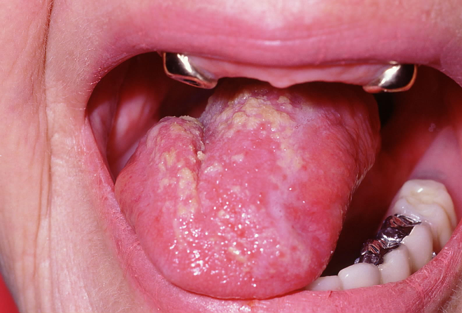
Pseudomembranous candidiasis presents as either white or red. The white presentation, commonly known as "thrush," will present with thick, white, patchy lesions throughout the mouth, most often on the tongue and buccal mucosa (see Image. Candida Albicans Fungal Infection on the Dorsal Surface of the Tongue).[19] These plaque-like deposits can be easily rubbed off with gauze or tactile pressure. Once removed, an erythematous surface is seen, which is this condition's secondary (red) presentation. These lesions may be symptomatic, with patients describing a burning sensation in the area.
Erythema Migrans
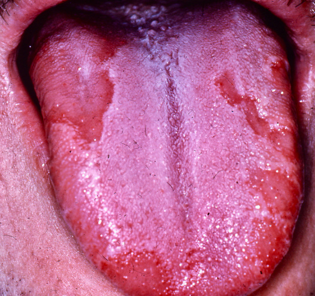
Erythema migrans presents as annular transient patches that are white and/or red on the tongue, with alternating textures, either raised or smooth. The lesions are typically located on the dorsal or lateral aspect of the tongue and will change positions and sizes over time (see Image. Geographic Tongue [Erythema Migrans] Present on the Dorsal Surface of the Tongue).[3]
Morsicatio Buccarum
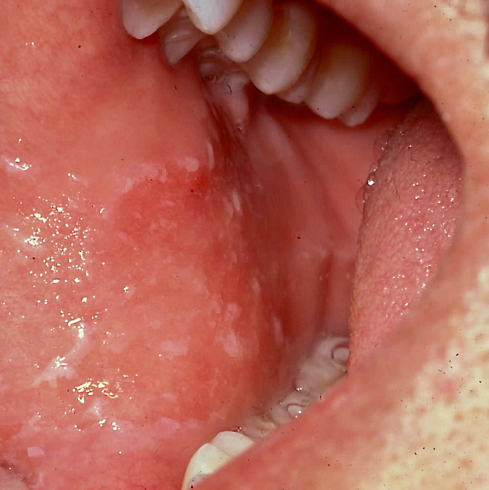
Morsicatio buccarum presents as shredded oral mucosa on the cheek or tongue (linguarum).[20] This line of disrupted tissue is typically concurrent with the plane of occlusion (see Image. Morsicatio Buccarum, aka "Cheek Biting," Present on the Buccal Mucosa).
Linea Alba
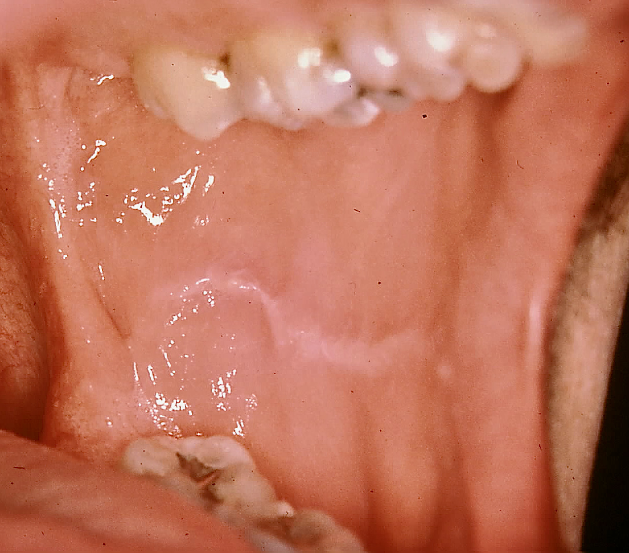
Linea alba presents unilaterally or bilaterally, depending on the patient's habits, as a white line on the buccal mucosa (see Image. Linea Alba Present on Buccal Mucosa).
Leukoedema
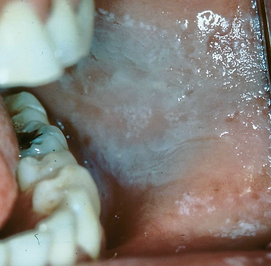
Leukoedema presents as a grayish-white lesion on the oral mucosa (see Image. Leukoedema Present on Buccal Mucosa).[15] This lesion is generally believed to be a variation of normal anatomy instead of being associated with disease.[21] Tissues associated with this lesion present in an edematous state and will resolve with stretching or manipulating the tissue.[6]
Oral Lichen Planus
Lichen planus typically presents as white striations, known as Wickham's striae, on the buccal mucosa or tongue but can also appear as white papules, plaques, or erosions.[18] Oral lichenoid reactions, which clinically resemble lichen planus, can result from a wide range of common medications, including but not limited to nonsteroidal anti-inflammatory drugs and antihypertensive agents.[22][23]
Evaluation
The evaluation of benign chronic white lesions of the oral mucosa is a meticulous process, necessitating a comprehensive approach by healthcare professionals. It begins with a thorough patient history, including inquiries into potential carcinogenic habits such as smoking. The clinical examination is a cornerstone, involving a detailed inspection of the oral cavity to identify specific characteristics and locations of white lesions. Diagnostic tools such as biopsy may be employed to confirm the nature of the lesion if necessary. Considering the diverse range of lesions, clinicians need to differentiate between benign and potentially malignant conditions. The evaluation process aims to guide clinicians in formulating an accurate differential diagnosis, allowing for timely and appropriate management strategies tailored to each patient's unique circumstances.
Pseudomembranous Candidiasis
Pseudomembranous candidiasis is easily managed after a clinical diagnosis. Since this condition can occur in patients with underlying systemic diseases, a complete medical history should be gathered to rule out any underlying cause.
Erythema Migrans
Erythema migrans is potentially an oral manifestation of psoriasis but is also seen as a normal variant.[3] Patients observed with this condition should be evaluated by their primary care provider to rule out potential systemic involvement.
Morsicatio Buccarum
Morsicatio buccarum typically results from parafunctional habits, neurological dysfunction, or patients undergoing stressful situations, such as adolescence or pregnancy.[24]
Linea Alba
Linea alba results from the negative pressure of sucking one's cheeks. Discussion with the patient regarding their potential habits is prudent for developing a differential diagnosis.
Leukoedema
Leukoedema is an idiopathic condition, possibly associated with local irritation. Leukoedema is associated with smoking and diabetes mellitus.[21] Stretching the oral tissue surrounding the lesion can aid in the diagnosis, as leukoedema will resolve upon introducing tension.
Oral Lichen Planus
Lichen planus is evaluated by histological examination and DIF to obtain a definitive diagnosis. The histological findings consist of hyperkeratosis of the epithelium, obliteration of basal epithelial cells, loss of spinous epithelial cells, saw-tooth appearance to epithelial ridges, with a band of lymphocytic infiltrate within the lamina propria.[25] A negative DIF screening differentiates oral lichen planus from other vesiculobullous lesions, such as pemphigus or pemphigoid.
Treatment / Management
Pseudomembranous Candidiasis
Pseudomembranous candidiasis is treatable using an antifungal medication, such as fluconazole or clotrimazole.[26] It is paramount to emphasize the need for regular oral hygiene, especially if the patient has a removable dental prosthesis, such as a denture, to prevent or limit recurrence.[2]
Erythema Migrans
Erythema migrans has no specific treatment recommended.[27] Since erythema migrans may be associated with underlying systemic disease, prompt referral to the primary care provider to evaluate for these conditions is indicated.
Morsicatio Buccarum
Morsicatio buccarum has no specific treatment, though a habit-changing and/or stress-reduction protocol can be beneficial to patients who present with cheek or tongue biting.
Linea Alba
Linea alba has no specific treatment; however, a habit-changing protocol or stress reduction can benefit patients with this condition.
Leukoedema
Leukoedema is considered a normal variation with no recommended treatment. Though it has been associated with smoking and diabetes mellitus, managing these underlying habits and conditions may aid in the lesion’s resolution.
Oral Lichen Planus
Lichen planus is predominately treated with topical corticosteroids.[28] If the lesion is a lichenoid drug reaction, the lesion should resolve with the termination of the offending medication. Though corticosteroids are the mainstay treatment, hydroxychloroquine, calcineurin inhibitors, and retinoids can be utilized in recalcitrant cases.[29]
Differential Diagnosis
The differential diagnosis of benign chronic white lesions of the oral mucosa includes the following:
Pseudomembranous Candidiasis
- Oral hairy leukoplakia
- Nutritional deficiencies
- Lichen planus
- Trauma
Erythema Migrans
- Evaluation for psoriasis
- Lichen planus
- Fissured tongue
- Herpes simplex infection
- Candidiasis
Morsicatio Buccarum
- Oral lichen planus
- Candidiasis
- Leukoplakia
- Chemical burn
- White sponge nevus
Linea Alba
- Leukoedema
- Morsicatio buccarum
- Lichen planus
Leukoedema
- Leukoplakia
- Hyperkeratosis
- White sponge nevus
- Morsicatio buccarum
Oral Lichen Planus
- Pemphigus
- Pemphigoid
- Leukoedema
- Leukoplakia
Prognosis
Pseudomembranous Candidiasis
Pseudomembranous candidiasis generally carries a favorable prognosis contingent on patient adherence to the prescribed antifungal regimen and maintenance of oral hygiene. It is seldom life-threatening.[30]
Erythema Migrans
Erythema migrans boasts an excellent prognosis and is amenable to palliative management.[31]
Morsicatio Buccarum
Morsicatio buccarum's prognosis is good, provided the patient engages in a stress-reduction protocol or gains awareness of their habits.
Linea Alba
Similarly, Linea alba exhibits a good prognosis when coupled with stress reduction or habit awareness.
Leukoedema
Leukoedema, a normal variation, is characterized by an excellent prognosis.
Oral Lichen Planus
While oral lichen planus generally has a good prognosis, its oral manifestations may persist for several years and may heal with scarring. Oral lichen planus is considered potentially premalignant.[32]
Complications
While benign chronic white lesions of the oral mucosa are generally non-threatening, certain complications and challenges may arise. Precise diagnosis and appropriate management are essential to address potential complications associated with these benign chronic white lesions of the oral mucosa.
Pseudomembranous candidiasis, if left untreated, can lead to persistent discomfort, difficulty in swallowing, and altered taste sensation.[30] In severe cases, the pseudomembranes formed by Candida overgrowth may lead to extensive oral tissue damage, causing bleeding and ulcerations. Immunocompromised individuals, such as those with HIV/AIDS or undergoing chemotherapy, are at an increased risk of systemic candidiasis,
Morsicatio buccarum, if the habit persists, can result in chronic irritation and potential secondary infections.[33]
In the case of lichen planus, the chronic nature of oral lesions may lead to scarring, and the condition is considered potentially premalignant, requiring vigilant monitoring. Long-standing oral lichen planus lesions, particularly erosive or atrophic variants, carry a slightly elevated risk of developing into oral squamous cell carcinoma. Aghbari et al discovered 1.1% of patients in a cohort of almost 20,000 patients developed squamous cell carcinoma from lichen planus.[34] Patients should be monitored and counseled on the risk for potential transformation to squamous cell carcinoma, even though the risk is low. Clinical documentation, histological evaluation, and DIF are paramount for definitive diagnosis and therapy.
Deterrence and Patient Education
Pseudomembranous Candidiasis
Patients presenting with pseudomembranous candidiasis should be encouraged to practice optimal oral hygiene, especially if they have an underlying systemic disease, such as diabetes, or are immunocompromised. Oral hygiene becomes even more paramount if the patient has a removable dental prosthesis.
Erythema Migrans
Erythema migrans is a harmless inflammatory process, with no treatment necessary if there is no underlying condition.[35] Documentation of this lesion is recommended.
Morsicatio Buccarum
Morsicatio buccarum should be brought to the patient’s attention, as they may be unaware of the causative habit. Patients should be encouraged not to self-mutilate and to attempt to mitigate their habit. The lesion should be documented.
Linea Alba
Linea alba should be brought to the patient’s attention, as they may be unaware they are sucking on their cheeks. Patients should be counseled on ways to reduce stress, and the lesion should be documented.
Leukoedema
Leukoedema is a variation of normal but should be brought to the patient’s attention.
Oral Lichen Planus
Lichen planus should also be brought to the patient’s attention, as there is a risk for systemic involvement or possible impact on the patient’s quality of life. It is the clinician’s responsibility to refer the patient to the appropriate specialist for evaluation.
Pearls and Other Issues
It is imperative to gather a comprehensive patient history and meticulously document findings to facilitate accurate diagnosis and therapeutic intervention for any white lesion. Any lesion that cannot be attributed to the patient's medical history or adverse habits should promptly trigger a referral for further evaluation.
Enhancing Healthcare Team Outcomes
Healthcare professionals, including dentists, physicians, advanced care practitioners, nurses, pharmacists, and others, need specialized knowledge and skills in oral pathology, clinical examination, and diagnostic proficiency to accurately identify and differentiate benign chronic white lesions of the oral mucosa.
Even for the trained dental professional, discovering a white lesion on the oral mucosa can be intimidating and a diagnostic challenge. Gathering a complete medical history is a critical first step if a clinician discovers a white lesion during routine care. The discovering physician should appropriately document all lesions noted. If the lesion can be attributed to an obvious source, subsequent follow-up is appropriate. However, if there is any doubt regarding the lesion's etiology or presentation, an appropriate referral to a dental professional is warranted. It is essential to provide the patient's history and any pertinent medical information to the consulting clinician to aid in drawing appropriate conclusions and, therefore, an appropriate diagnosis and treatment regimen.
Enhancing team performance and patient-centered care for patients with benign chronic white lesions of the oral mucosa requires healthcare professionals to engage in continuing education, training, and staying updated on the latest advancements in oral pathology.