Amegakaryocytic Thrombocytopenia

Earn CME/CE in your profession:


Continuing Education Activity

Amegakaryocytic thrombocytopenia is a severe form of thrombocytopenia with reduced or absent megakaryocytes and normal cellularity in the remaining cell lines. Infants affected by the congenital form may experience intracranial bleeding, and progression to aplastic anemia is likely. While reviewing the congenital and acquired forms, this topic will discuss the evaluation and management of amegakaryocytic thrombocytopenia. The interprofessional team's role in evaluating and treating patients with amegakaryocytic thrombocytopenia will be delineated.

Objectives:

  • Identify key clinical features and risk factors associated with amegakaryocytic thrombocytopenia to facilitate early recognition.
  • Differentiate amegakaryocytic thrombocytopenia from other causes of thrombocytopenia through understanding unique histopathologic findings and clinical presentations.
  • Implement evidence-based diagnostic protocols and treatment strategies for both congenital and acquired forms of amegakaryocytic thrombocytopenia.
  • Coordinate care across disciplines to optimize patient outcomes and provide a seamless continuum of care for those with amegakaryocytic thrombocytopenia.

Introduction

Thrombocytopenia, a platelet count of less than 150,000 cells/μL for adults and children, is a common clinical finding with multiple potential etiologies. Amegakaryocytic thrombocytopenia is a severe form marked by reduced or absent megakaryocytes and no additional abnormalities on bone marrow evaluation. First described by Greenwald and Sherman in the pediatric literature in 1929, there are both congenital and acquired forms.[1]  

Congenital amegakaryocytic thrombocytopenia (CAMT) is an autosomal recessive disorder presenting with severe thrombocytopenia at birth. An almost universal progression to bone marrow failure with impaired production of all three hematopoietic cell lines occurs.[2][3][4][5]

Acquired amegakaryocytic thrombocytopenia (AAMT) is likely immune-mediated and presents later in life.[6][7][8] Generally, the other cell lines remain intact, but progression to bone marrow failure is possible.

Etiology

CAMT

Traditionally, congenital amegakaryocytic thrombocytopenia results from a lack of thrombopoietin signaling caused by mutations in the myeloproliferative leukemia virus oncogene (MPL). This gene is responsible for encoding the thrombopoietin (THPO) receptor. Two main types of CAMT were described by Ballmaier et al in 2001.[5]

Type 1 (CAMT-1)

CAMT-1 results from a stop codon or frameshift mutation, causing a loss of the intracellular domain of the MPL receptor. This mutation completely eliminates receptor signaling and causes a complete loss of receptor function. The result is severe thrombocytopenia present at birth, increased risk of intracranial bleeding, and early progression to bone marrow failure, with the mean age being 33 months.[4][9][10][11]

Type 2 (CAMT-2)

CAMT-2 results from a splicing defect or amino acid substitution, which affects the MPL receptor's glycosylation and results in an inability to react with thrombopoietin. These mutations can also cause a loss of hydrogen bonds within the MPL receptor, causing it to be unstable. Some residual receptor function remains. Children with CAMT-2 have more mild thrombocytopenia that will transiently normalize during the first year of life. The rate of bone marrow failure is slower than that of CAMT-1 and typically occurs between ages 3 and 6 years, with the mean being 5 years.[10][11]

Not all children with a clinical picture consistent with CAMT have a c-MPL mutation. The homozygous mutation of p.R119C prevents liver cells from producing thrombopoietin.[12][13] As described by Thompson and Nguyen, abnormalities in the HOXA11 and MECOM genes (regulate megakaryocyte differentiation) cause congenital amegakaryocytosis, thrombocytopenia, and eventual bone marrow failure in most patients.[3][14] Patients with the HOXA11 and MECOM genetic mutations are differentiated from other forms of CAMT due to their association with radio-ulnar synostosis. A null mutation in the RBM8A gene causes thrombocytopenia-absent radius syndrome, which is always associated with bilateral radial aplasia. Additional proposed alternate etiologies are an X-linked variety of CAMT and an anti-HLA A2 antibody.[9][12][15] 

AAMT

The exact mechanism of acquired amegakaryocytic thrombocytopenia is unknown. Three mechanisms have been proposed:[16][17].

  1. Suppression of maturation of megakaryocytes by an exogenous agent: AAMT is seen in association with Ebstein Barr virus (EBV), parvovirus B19, hepatitis C virus, interferon therapy, cytomegalovirus, benzene exposure, alcohol use disorder, vitamin B12 deficiency, and radioiodine therapy.[18][19]
  2. Suppression of megakaryocyte maturation by endogenous stimuli due to antibody-mediated or T-cell autoimmunity: AAMT has been seen in association with thymoma with a more aggressive disease course, adult-onset Still disease, eosinophilic fasciitis, systemic lupus erythematosus, autoimmune hemolytic anemia, systemic sclerosis, Graves disease, and hyperestrogenic states.[16][20][21][22]
  3. An early manifestation of a stem cell abnormality: AAMT has been described as a precursor to acute myeloid leukemia, myelodysplastic syndrome, aplastic anemia, and non-Hodgkin lymphoma.[23][7] A case report has shown an association with large granular lymphocyte leukemia.[24] Some other reported cytogenetic abnormalities include the Philadelphia chromosome and 5q deletion.[23] These findings suggest that the defect in AAMT lies in an early progenitor cell in the megakaryocytic lineage.

Epidemiology

CAMT

With less than 100 cases reported, CAMT is rare.[9] Consanguinuity is a potential cause, and a slight female preponderance is observed.[5] The incidence is thought to be underestimated due to misdiagnosis as neonatal alloimmune thrombocytopenia. Distinguishing CAMT from primary aplastic anemia may be difficult once the patient has progressed to pancytopenia.[25] 

AAMT

Similarly, the incidence rate of AAMT is likely higher than reported, as many cases are misdiagnosed as immune thrombocytopenic purpura.[23] Affected females are typically diagnosed between 40 and 60 years of age, whereas most affected males lie at both ends of the age distribution, peaking in their sixties.[26]

Pathophysiology

Megakaryopoeisis begins with the hematopoietic stem cell (HSC) in the bone marrow. The HSC matures into a multipotent progenitor cell, a committed megakaryocyte progenitor cell, an immature megakaryocyte, and finally into a mature megakaryocyte, which gives rise to blood platelets.[27] Importantly, THPO is a cytokine needed at every step of megakaryopoiesis to increase the number, size, and ploidy of megakaryocytes and promote the expression of platelet-specific markers.[4] THPO also enhances the expression of vascular endothelial growth factor (VEGF), HoxB4, and HoxA9, promoting HSC growth and survival.[9] THPO is mainly produced in the liver but can also be produced in the kidneys' proximal tubular cells and the bone marrow stromal cells.[28] 

The THPO receptor, also called the MPL receptor, is found in the bone marrow, liver, spleen, and CD34+ cells.[4] THPO binding initiates a cascade of signaling events within the target cell via the JAK-Stat kinase family of proteins along with MAP and PI3 kinase pathways.[28] MPL signaling is vital for differentiating multipotent progenitor cells into megakaryocyte and erythrocyte progenitor cells. Problems in the signaling can lead to impaired megakaryocyte and erythrocyte production, as seen in the later stages of CAMT.[29]

CAMT

The c-Mpl gene is located in the 1p34 locus and has 12 exons.[30] CAMT can occur due to the inheritance of homozygous mutations of the c-Mpl gene, which is seen in families with consanguinity, or due to two different inherited mutations of c-Mpl resulting in a compound heterozygous state.[10] Close to 41 mutations have been defined in the literature. The most frequent location for mutations is within the first and fifth exons of the c-Mpl gene (75% of all mutations seen in CAMT), with 60% in exons 2 and 3 alone.[5][9][30] Some examples of mutations include C268T, G304C, G305C, G578A, F104S, P635L, R102P, R257C, R257L, W154R, 1,499delT, and Q186X mutations.[9][31]

AAMT

AAMT can be caused by antibody or T-cell-mediated autoimmunity.[19] Proposed pathophysiology includes anti-THPO antibodies, antibodies against antigens on megakaryocyte progenitor cells, antibodies against granulocyte monocyte colony-stimulating factor or antibodies against the megakaryocyte colony-forming unit, failure of terminal megakaryocyte differentiation, suppression of megakaryocyte colony-forming unit by T cells and adherent monocytes, monoclonal T-cell population destroying megakaryocyte lineage, or a defect in cytokine (IL 7, stem cell factor, TGF-beta 1) mediated regulation of megakaryopoiesis.[6][20][23][32]

Anti-MPL antibodies have been seen with systemic lupus erythematosus and systemic sclerosis.[19][33] Hepatitis C infection causes the generation of anti-MPL antibodies, which are first absorbed onto MPL receptors on platelets, resulting in their destruction in the spleen and creating an immune thrombocytopenic purpura-like picture. Later, these antibodies bind to the MPL receptor on megakaryocytes and block the function of THPO, resulting in AAMT.[34] Interestingly, interferon therapy used to treat patients with hepatitis C can generate anti-MPL antibodies.[34]

Histopathology

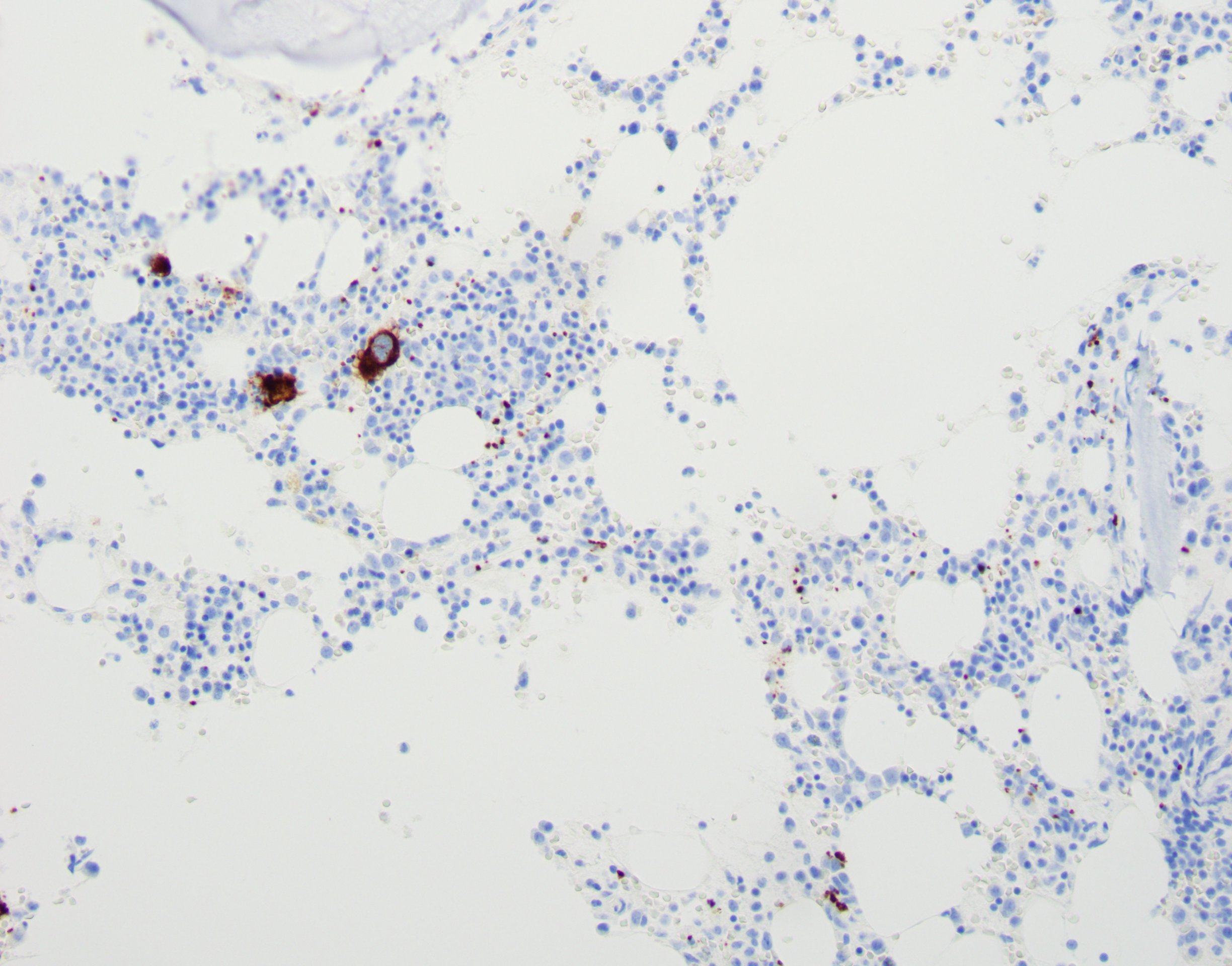
The bone marrow evaluation in patients with amegakaryocytic thrombocytopenia typically demonstrates normal overall cellularity with a reduction or absence of megakaryocytes. The present megakaryocytes may look immature or small, and no evidence of dysplasia is observed.[4][9][10] Immunohistochemical staining for CD-61 megakaryocyte antigen shows diminished megakaryocytes on core biopsy.[7] 

Patients with CAMT may have minimal findings on bone marrow evaluation early in the disease, which can be misleading. Serial bone marrow biopsies may be necessary to elucidate the diagnosis.[10] On peripheral smear, platelets appear normal in size and morphology.[3][10] Later, once pancytopenia develops, affected patients have hypocellular marrow with decreased progenitors in all lineages, making it challenging to distinguish CAMT from other causes of aplastic anemia.[9]

History and Physical

CAMT

Congenital amegakaryocytic thrombocytopenia presents with severe thrombocytopenia, typically a platelet count of <21,000 cells/μL, on the first day or within the first month of life. Occasionally, affected infants develop findings during fetal development. Common manifestations are purpura, intracranial bleeds, recurrent rectal bleeding, or pulmonary hemorrhage within hours of birth.[4][9] Notably, decreased P-selectin expression on neonatal platelets can reduce platelet activation and predispose affected infants to the high bleeding tendency pre- or perinatally in patients with CAMT.[35] 

A family history of thrombocytopenia may be present.[4] No characteristic congenital phenotypic abnormalities are associated with CAMT. Some studies have noted neurological defects like strabismus, cerebellar agenesis, hypoplasia of the corpus callosum and brainstem, facial malformations, and cortical dysplasia. The mechanism behind the neurological findings is unclear. One possibility is that the absence or deficiency of MPL in the brain, as seen in CAMT, can lead to developmental delay. The other hypothesis considers the long-term intracranial neurological sequelae.[3][5][9][10][31]

AAMT

AAMT is a diagnosis of exclusion, and patients usually present with bleeding complications after failing standard treatment for immune thrombocytopenia, including steroids or intravenous immunoglobulin therapy.[7] Patients with AAMT can present with petechiae, purpura, ecchymosis, easy bruising, epistaxis, or fatigue. Splenomegaly is absent.[8] One case report described a patient with AAMT who presented with massive hemoperitoneum due to hemorrhagic corpus luteum.[36]

Evaluation

Bone marrow biopsy is the mainstay of diagnosing CAMT or AAMT (see Histopathology section).

CAMT

Bone marrow biopsy is warranted for all children with severe thrombocytopenia (ie, platelet counts <50,000 cells/μL) at birth and platelets of normal size and morphology. When a reduced number of megakaryocytes are seen on biopsy, c-Mpl gene testing should be performed.[25] 

The c-Mpl gene analysis is performed with bidirectional sequencing of all 12 exons, including coding regions, splice sites, and intron-exon boundaries. Samples can be obtained from whole blood, bone marrow, skin fibroblasts, buccal brushings, amniotic fluid, or CD34+ cells.[4][37] Testing is conducted by GeneDx in Maryland (with 95% to 97% sensitivity) and at Prevention Genetics in Wisconsin.[4][9] Chromosome analysis and FISH studies are alternative genetic tests. 

The presence of either homozygous or compound heterozygous mutations in the c-Mpl gene confirms the diagnosis of CAMT.[5] Plasma TPO levels will be high. THPO levels rise tenfold since THPO internalization and destruction by MPL receptors do not occur. Patients should also be screened for THPO mutations because CAMT caused by these mutations can easily be reversed with an MPL agonist like romiplostim.[12] In patients with THPO mutations, the levels of THPO will be low due to a defect in the secretion of THPO.[10][12]

AAMT

The etiology of AAMT is quite varied. AAMT is considered when the patient presents with thrombocytopenia and bleeding that does not respond to corticosteroids or IVIG. AAMT should be considered when a patient presents with thrombocytopenia unresponsive to corticosteroids or IVIG, and a bone marrow biopsy reveals an isolated reduction or absence of megakaryocytes. A strong clinical suspicion for AAMT is necessary to request a bone marrow biopsy and confirm the diagnosis.

Treatment / Management

CAMT

Treatment considerations for CAMT are as follows:

1. Allogeneic hematopoietic stem cell transplant (HSCT) is the only curative option for patients with CAMT and a c-Mpl mutation (most cases).[9] HSCT should be considered as early as possible. Some studies suggest that HLA typing of patients and their siblings should be performed at the time of diagnosis. One case report demonstrated a successful transplant from the patient's father, a haploidentical donor with 5 of 10 HLA-matched alleles despite the parent carrying the MPL mutant gene [11]. The average age at which CAMT patients undergo HSCT is 38 months, ranging from 7 to 89 months.[9] Due to the risk of requiring multiple transfusions, alloimmunization, and infections, HSCT should be performed before developing pancytopenia.[9][25] 

Conditioning regimens for HSCT include the use of busulfan, cyclophosphamide, and total body irradiation.[5] The conditioning regimen for allogeneic hematopoietic stem cell transplantation (alloSCT) typically comprises either a Busulfan or Total Body Irradiation (TBI) based protocol. These regimens are fully myeloablative and carry survival rates of approximately 80% but often cause significant pulmonary, mucosal, and hepatotoxicity.[38] Infertility is a potential long-term result, and discussions regarding future fertility and fertility preservation are necessary. Recent clinical cases have shown promise for non-myeloablative protocols [38][23]. These regimens are similar to those used in acquired aplastic anemia treated with antithymocyte globulin therapy (AGT); they preserve fertility, are less toxic overall, and leave myeloid chimerism engrafted.

Unfortunately, the rarity of this disease thwarts the creation of clinical trials. HSCT has no benefit for patients with a THPO mutation because the liver produces THPO.[13] For HLA-mismatched HSCT, the 1-year incidence of graft failure is 19%; for HLA-matched HSCT, it is 7% [39]

2. Supportive treatment includes irradiated, leukocyte-reduced platelet transfusions; antifibrinolytics like tranexamic acid; avoidance of nonsteroidal anti-inflammatory drugs; and aspirin. Once pancytopenia develops, packed red blood cells and antibiotics may be needed.

3. Romiplostim, a THPO peptide mimetic, and eltrombopag, a small molecule agonist of the MPL receptor that induces conformational changes in the receptor, can be used for CAMT cases with THPO mutations.[12][28] There is no benefit to using these medications for patients with c-Mpl mutations.

4. Experimental therapies include gene therapy with lentiviral vectors, CRISPR-Cas9 gene editing, and experimental drugs, including LGD-4665, minibodies, and diabodies are experimental drugs. Gene therapy with lentiviral vectors has been used to repair the mutant c-Mpl gene. However, concerns about the leukemogenicity of this approach remain.[9] CRISPR-Cas9 gene editing to repair the mutation has been tried.[40] LGD-4665, minibodies, and diabodies are experimental drugs that stimulate some forms of mutant MPL receptors to increase megakaryocyte proliferation.[28][41]

AAMT

The goal of therapy is to treat the underlying etiology of AAMT (eg, resection of the thymus in thymoma-associated AAMT).[42] Hoffman suggested using plasmapheresis, cyclophosphamide, cyclosporine, and prednisone for antibody-mediated AAMT and cyclosporine, cytokine therapy, and ATG for T-cell–mediated AAMT.[43] If a specific thrombopoiesis inhibition site cannot be identified, empiric therapy with different immunosuppressive or immunomodulatory agents is warranted.[44] Some experts advised starting with the least toxic and most cost-effective medications with periodic treatment response monitoring.[36] 

The following medications and therapies have been used for the treatment of AMMT:

  • steroids, which suppress immune B- and T-cell–mediated autoimmunity;
  • high-dose IVIG therapy, which binds antibodies against THPO and megakaryocytes;
  • rituximab, which suppresses the production of autoantibodies by B cells;
  • cyclosporine, a calcineurin inhibitor;
  • ATG that suppresses T-cell–mediated autoimmunity and stimulates hematopoiesis directly;
  • bone marrow transplant;
  • cyclophosphamide or azathioprine, especially in patients with systemic lupus erythematosus;
  • lithium carbonate, vincristine, mycophenolate mofetil, danazol, and eltrombopag; and
  • recombinant IL-11proteins.[45]

A case report described the successful treatment of adult-onset Still disease with tocilizumab and cyclosporine.[19] Some refractory cases of AAMT have been treated with a combination therapy of ATG and cyclosporine.[23][32][46][32] Other reports have shown a positive response to avatrombopag, despite being refractory to eltrombopag.[47]

Differential Diagnosis

CAMT

The initial evaluation of thrombocytopenia in the neonate begins by determining the underlying cause. The age of onset (before or after 72 hours of life), the severity of the thrombocytopenia, and maternal history are all considered when determining the most appropriate steps. The differential diagnosis includes thrombocytopenia due to birth asphyxia or placental insufficiency, TORCH (toxoplasmosis, rubella, cytomegalovirus, and herpes simplex virus) infections, sepsis, congenital syphilis, varicella, parvovirus B19, Wiskcott-Aldrich syndrome, Fanconi anemia, and dyskeratosis congenita.[5][35] Neonatal autoimmune thrombocytopenia can be distinguished from CAMT by normal-to-increased megakaryocytes in the bone marrow.[9][10] In thrombocytopenia with absent radius, patients have skeletal or cardiac defects with a spontaneous increase in platelet counts after the first year of life.[4]

AAMT

The etiology of thrombocytopenia in adults is extensive.[45] Each cause must be painstakingly ruled out before diagnosing AAMT; diagnosis is ultimately confirmed on bone marrow biopsy. Most commonly, AAMT is misdiagnosed as immune thrombocytopenic purpura (ITP). The typical presentation is a patient with ITP who fails to improve with corticosteroids or IVIG. Other potential causes of thrombocytopenia are as follows:

  • Infections, such as Epstein–Barr virus, varicella, leptospirosis, anaplasmosis, dengue, babesiosis, and tick-borne diseases
  • Medications, including daptomycin, valproic acid, linezolid, and penicillin
  • Beverages, eg, alcohol, tonic water containing quinine, and herbal supplements
  • Vitamin deficiency, including B12 and folic acid deficiency
  • Decreased THPO production due to liver dysfunction
  • Blood cancers or solid organ cancers with bone metastasis 
  • Chemotherapy and radiation therapy for blood cancers or metastatic cancer
  • Rheumatologic conditions, such as systemic lupus erythematosus with or without the antiphospholipid syndrome
  • Peripheral destruction of platelets by shearing stress as seen in the use of intra-aortic balloon pumps and aortic aneurysms
  • Splenic sequestration of platelets
  • Consumptive coagulopathies-disseminated intravascular coagulation, heparin-induced thrombocytopenia with thrombosis, or hemolytic uremic syndrome. 

Prognosis

CAMT

Once pancytopenia develops, the prognosis for patients affected by CAMT is poor. One study revealed 30% of patients affected by CAMT died from bleeding complications, and an additional 20% died from complications related to HSCT.[4] Many patients develop bleeding issues within the first year and progress to aplasia (or secondarily myelodysplasia) by age 5.[38]

AAMT

Patients can achieve remission immediately after targeted therapy for the underlying condition or develop a long relapsing and remitting course with AAMT. Additionally, patients can progress to aplastic anemia or require HSCT to achieve remission.[7] Once AAMT progresses to aplastic anemia, patients have a poor prognosis.[8]

Complications

CAMT

Progression to bone marrow failure and aplastic anemia occurs by the mean age of 13 months for patients with CAMT-1 and 5 years for patients with CAMT-2.[9] A patient's risk of developing pancytopenia with CAMT is higher than currently reported and likely occurs in more than 90% of cases.[5] 

CAMT patients are at an increased risk of developing myelodysplastic syndrome and acute myeloid leukemia. This results from karyotype instability and the mutator effect (ie, an increased accumulation of chromosomal aberrations over time or an expansion of abnormal cell clones in the bone marrow).[2][5][9] Some studies have shown that increased amounts of myelosuppressive cytokines (ie, TNF-alpha and IFN-gamma) contribute to bone marrow failure in CAMT patients.[25]

AAMT

AAMT can progress to aplastic anemia within an average of 1 month to 2 years following diagnosis.[8] AAMT can also progress to myelodysplastic syndrome.[18] A single case report describes mononeuritis multiplex as a complication of AAMT.[18]

Deterrence and Patient Education

Amegakaryocytic thrombocytopenia is a rare disease. It can either be congenital due to genetic defects or acquired due to various underlying diseases. Early diagnosis is vital in patients with CAMT. Patients and caregivers must know the risks of hemorrhage, progression to aplastic anemia, myelodysplastic syndrome, and acute myeloid leukemia. Definitive diagnosis requires a bone marrow biopsy.

Treatments may also be invasive and involve an HSCT. Great care should be taken to void activities that will place the patient at high risk for hemorrhaging and intracranial bleeding (eg, skiing, mountain climbing, and contact sports). Treating AAMT can be challenging, and multiple immunosuppressive and immunomodulatory agents may be ineffective. Patients may ultimately require supportive platelet transfusions more than once a week.

Enhancing Healthcare Team Outcomes

Amegakaryocytic thrombocytopenia is a rare disease requiring a high degree of clinical suspicion. Serious complications like fatal hemorrhage or pancytopenia occur, and early diagnosis is essential. Collaboration among interprofessional team members to expedite care, provide individualized treatment plans, and monitor for symptoms, such as bleeding, to assess the potential for serious complications is crucial to optimize care and improve patient outcomes. 

To ensure shared decision-making, an in-depth discussion with patients and their families regarding the risks and benefits of treatment options is a priority for the interprofessional care team. Due to the rarity of the illness, enrollment of patients in national and international studies, when available, is beneficial. No large multicenter randomized control trials are possible; hence, hematologists must depend on case reports describing success with various therapies for CAMT and AAMT.[5][26]


(Click Image to Enlarge)
<p>Bone Marrow Core Biopsy

Bone Marrow Core Biopsy. The image depicts CD61 immunohistochemical staining on a bone marrow core biopsy sample from a patient with acquired amegakaryocytic thrombocytopenia, with a decreased number of megakaryocytes highlighted in brown.


Department of Pathology, Rochester General Hospital, New York

Details

Author

Ekta Tirthani

Author

Mina S. Said

Updated:

8/30/2023 6:54:46 PM

References


[1]

Balduini CL. The name counts: the case of 'congenital amegakaryocytic thrombocytopenia'. Haematologica. 2023 May 1:108(5):1216-1219. doi: 10.3324/haematol.2022.282024. Epub 2023 May 1     [PubMed PMID: 36226496]

Level 3 (low-level) evidence

[2]

Maserati E, Panarello C, Morerio C, Valli R, Pressato B, Patitucci F, Tassano E, Di Cesare-Merlone A, Cugno C, Balduini CL, Lo Curto F, Dufour C, Locatelli F, Pasquali F. Clonal chromosome anomalies and propensity to myeloid malignancies in congenital amegakaryocytic thrombocytopenia (OMIM 604498). Haematologica. 2008 Aug:93(8):1271-3. doi: 10.3324/haematol.12748. Epub 2008 Jun 2     [PubMed PMID: 18519517]


[3]

Khincha PP, Savage SA. Neonatal manifestations of inherited bone marrow failure syndromes. Seminars in fetal & neonatal medicine. 2016 Feb:21(1):57-65. doi: 10.1016/j.siny.2015.12.003. Epub 2015 Dec 24     [PubMed PMID: 26724991]


[4]

Al-Qahtani FS. Congenital amegakaryocytic thrombocytopenia: a brief review of the literature. Clinical medicine insights. Pathology. 2010 Jun 4:3():25-30     [PubMed PMID: 21151552]


[5]

Ballmaier M, Germeshausen M. Congenital amegakaryocytic thrombocytopenia: clinical presentation, diagnosis, and treatment. Seminars in thrombosis and hemostasis. 2011 Sep:37(6):673-81. doi: 10.1055/s-0031-1291377. Epub 2011 Nov 18     [PubMed PMID: 22102270]


[6]

Simkins A, Maiti A, Short NJ, Jain N, Popat U, Patel KP, Oo TH. Acquired amegakaryocytic thrombocytopenia and red cell aplasia in a patient with thymoma progressing to aplastic anemia successfully treated with allogenic stem cell transplantation. Hematology/oncology and stem cell therapy. 2019 Jun:12(2):115-118. doi: 10.1016/j.hemonc.2017.09.001. Epub 2018 Jan 31     [PubMed PMID: 29409729]


[7]

Brown GE, Babiker HM, Cantu CL, Yeager AM, Krishnadasan R. "Almost bleeding to death": the conundrum of acquired amegakaryocytic thrombocytopenia. Case reports in hematology. 2014:2014():806541. doi: 10.1155/2014/806541. Epub 2014 Feb 6     [PubMed PMID: 24649385]


[8]

Chaudhary UB, Eberwine SF, Hege KM. Acquired amegakaryocytic thrombocytopenia purpura and eosinophilic fasciitis: a long relapsing and remitting course. American journal of hematology. 2004 Mar:75(3):146-50     [PubMed PMID: 14978695]


[9]

Geddis AE. Congenital amegakaryocytic thrombocytopenia. Pediatric blood & cancer. 2011 Aug:57(2):199-203. doi: 10.1002/pbc.22927. Epub 2011 Feb 18     [PubMed PMID: 21337678]


[10]

Geddis AE. Congenital amegakaryocytic thrombocytopenia and thrombocytopenia with absent radii. Hematology/oncology clinics of North America. 2009 Apr:23(2):321-31. doi: 10.1016/j.hoc.2009.01.012. Epub     [PubMed PMID: 19327586]


[11]

Wang S, Yang X, Ai Y, Zhu Y. Haploidentical Hematopoietic Stem Cell Transplantation in a 3-Year-Old Girl with Congenital Amegakaryocytic Thrombocytopenia: A Case Report. Klinische Padiatrie. 2022 Nov:234(6):388-390. doi: 10.1055/a-1933-2583. Epub 2022 Nov 15     [PubMed PMID: 36379227]

Level 3 (low-level) evidence

[12]

Varghese LN, Defour JP, Pecquet C, Constantinescu SN. The Thrombopoietin Receptor: Structural Basis of Traffic and Activation by Ligand, Mutations, Agonists, and Mutated Calreticulin. Frontiers in endocrinology. 2017:8():59. doi: 10.3389/fendo.2017.00059. Epub 2017 Mar 31     [PubMed PMID: 28408900]


[13]

Pecci A, Ragab I, Bozzi V, De Rocco D, Barozzi S, Giangregorio T, Ali H, Melazzini F, Sallam M, Alfano C, Pastore A, Balduini CL, Savoia A. Thrombopoietin mutation in congenital amegakaryocytic thrombocytopenia treatable with romiplostim. EMBO molecular medicine. 2018 Jan:10(1):63-75. doi: 10.15252/emmm.201708168. Epub     [PubMed PMID: 29191945]


[14]

Germeshausen M, Ancliff P, Estrada J, Metzler M, Ponstingl E, Rütschle H, Schwabe D, Scott RH, Unal S, Wawer A, Zeller B, Ballmaier M. MECOM-associated syndrome: a heterogeneous inherited bone marrow failure syndrome with amegakaryocytic thrombocytopenia. Blood advances. 2018 Mar 27:2(6):586-596. doi: 10.1182/bloodadvances.2018016501. Epub     [PubMed PMID: 29540340]

Level 3 (low-level) evidence

[15]

Azuno Y, Yaga K. Successful cyclosporin A therapy for acquired amegakaryocytic thrombocytopenic purpura. American journal of hematology. 2002 Apr:69(4):298-9     [PubMed PMID: 11921030]


[16]

Mulroy E, Gleeson S, Chiruka S. Danazol: an effective option in acquired amegakaryocytic thrombocytopaenic purpura. Case reports in hematology. 2015:2015():171253. doi: 10.1155/2015/171253. Epub 2015 Apr 5     [PubMed PMID: 25945269]

Level 3 (low-level) evidence

[17]

Evans DI. Immune amegakaryocytic thrombocytopenia of the newborn: association with anti-HLA-A2. Journal of clinical pathology. 1987 Mar:40(3):258-61     [PubMed PMID: 3494047]


[18]

Khan F, Shehna A, Mukundan L. Mononeuritis multiplex in acquired amegakaryocytic thrombocytopenia. Journal of neurosciences in rural practice. 2015 Oct-Dec:6(4):588-90. doi: 10.4103/0976-3147.165351. Epub     [PubMed PMID: 26752909]


[19]

Ichikawa T, Shimojima Y, Otuki T, Ueno KI, Kishida D, Sekijima Y. Acquired Amegakaryocytic Thrombocytopenia in Adult-onset Still's Disease: Successful Combination Therapy with Tocilizumab and Cyclosporine. Internal medicine (Tokyo, Japan). 2019 Dec 1:58(23):3473-3478. doi: 10.2169/internalmedicine.2929-19. Epub 2019 Aug 6     [PubMed PMID: 31391399]


[20]

Manoharan A, Williams NT, Sparrow R. Acquired amegakaryocytic thrombocytopenia: report of a case and review of literature. The Quarterly journal of medicine. 1989 Mar:70(263):243-52     [PubMed PMID: 2690174]

Level 3 (low-level) evidence

[21]

Dahal S, Sharma E, Dahal S, Shrestha B, Bhattarai B. Acquired Amegakaryocytic Thrombocytopenia and Pure Red Cell Aplasia in Thymoma. Case reports in hematology. 2018:2018():5034741. doi: 10.1155/2018/5034741. Epub 2018 Mar 11     [PubMed PMID: 29713553]

Level 3 (low-level) evidence

[22]

Ikeda N, Hisano Y, Kamao T, Uno M, Mizushima T. Acquired Amegakaryocytic Thrombocytopenia Associated With Autoimmune Hemolytic Anemia. Cureus. 2022 Jul:14(7):e27315. doi: 10.7759/cureus.27315. Epub 2022 Jul 26     [PubMed PMID: 36042987]


[23]

Agarwal N, Spahr JE, Werner TL, Newton DL, Rodgers GM. Acquired amegakaryocytic thrombocytopenic purpura. American journal of hematology. 2006 Feb:81(2):132-5     [PubMed PMID: 16432869]


[24]

Ergas D, Tsimanis A, Shtalrid M, Duskin C, Berrebi A. T-gamma large granular lymphocyte leukemia associated with amegakaryocytic thrombocytopenic purpura, Sjögren's syndrome, and polyglandular autoimmune syndrome type II, with subsequent development of pure red cell aplasia. American journal of hematology. 2002 Feb:69(2):132-4     [PubMed PMID: 11835350]


[25]

Savoia A, Dufour C, Locatelli F, Noris P, Ambaglio C, Rosti V, Zecca M, Ferrari S, di Bari F, Corcione A, Di Stazio M, Seri M, Balduini CL. Congenital amegakaryocytic thrombocytopenia: clinical and biological consequences of five novel mutations. Haematologica. 2007 Sep:92(9):1186-93     [PubMed PMID: 17666371]


[26]

Novotný JP, Köhler B, Max R, Egerer G. Acquired Amegakaryocytic Thrombocytopenic Purpura Progressing into Aplastic Anemia. Prague medical report. 2017:118(4):147-155. doi: 10.14712/23362936.2017.16. Epub     [PubMed PMID: 29324222]


[27]

Kaushansky K. Thrombopoietin. The New England journal of medicine. 1998 Sep 10:339(11):746-54     [PubMed PMID: 9731092]


[28]

Kim AR, Sankaran VG. Thrombopoietin: tickling the HSC's fancy. EMBO molecular medicine. 2018 Jan:10(1):10-12. doi: 10.15252/emmm.201708450. Epub     [PubMed PMID: 29191946]


[29]

Hirata S, Takayama N, Jono-Ohnishi R, Endo H, Nakamura S, Dohda T, Nishi M, Hamazaki Y, Ishii E, Kaneko S, Otsu M, Nakauchi H, Kunishima S, Eto K. Congenital amegakaryocytic thrombocytopenia iPS cells exhibit defective MPL-mediated signaling. The Journal of clinical investigation. 2013 Sep:123(9):3802-14. doi: 10.1172/JCI64721. Epub 2013 Aug 1     [PubMed PMID: 23908116]


[30]

Ok Bozkaya İ, Yaralı N, Işık P, Ünsal Saç R, Tavil B, Tunç B. Severe Clinical Course in a Patient with Congenital Amegakaryocytic Thrombocytopenia Due to a Missense Mutation of the c-MPL Gene. Turkish journal of haematology : official journal of Turkish Society of Haematology. 2015 Jun:32(2):172-4. doi: 10.4274/tjh.2013.0191. Epub     [PubMed PMID: 26316487]


[31]

Ihara K, Ishii E, Eguchi M, Takada H, Suminoe A, Good RA, Hara T. Identification of mutations in the c-mpl gene in congenital amegakaryocytic thrombocytopenia. Proceedings of the National Academy of Sciences of the United States of America. 1999 Mar 16:96(6):3132-6     [PubMed PMID: 10077649]


[32]

Nishino S, Kodaka T, Sawada Y, Goka T, Gotoh Y, Tsunemine H, Takahashi T. Marked rebound thrombocytosis in response to glucocorticoids in a patient with acquired amegakaryocytic thrombocytopenia. Journal of clinical and experimental hematopathology : JCEH. 2018 Dec 13:58(4):166-170. doi: 10.3960/jslrt.18016. Epub 2018 Nov 9     [PubMed PMID: 30416171]


[33]

Katsumata Y, Suzuki T, Kuwana M, Hattori Y, Akizuki S, Sugiura H, Matsuoka Y. Anti-c-Mpl (thrombopoietin receptor) autoantibody-induced amegakaryocytic thrombocytopenia in a patient with systemic sclerosis. Arthritis and rheumatism. 2003 Jun:48(6):1647-51     [PubMed PMID: 12794833]


[34]

Ichimata S, Kobayashi M, Honda K, Shibata S, Matsumoto A, Kanno H. Acquired amegakaryocytic thrombocytopenia previously diagnosed as idiopathic thrombocytopenic purpura in a patient with hepatitis C virus infection. World journal of gastroenterology. 2017 Sep 21:23(35):6540-6545. doi: 10.3748/wjg.v23.i35.6540. Epub     [PubMed PMID: 29085203]


[35]

Germeshausen M, Ballmaier M. CAMT-MPL: congenital amegakaryocytic thrombocytopenia caused by MPL mutations - heterogeneity of a monogenic disorder - a comprehensive analysis of 56 patients. Haematologica. 2021 Sep 1:106(9):2439-2448. doi: 10.3324/haematol.2020.257972. Epub 2021 Sep 1     [PubMed PMID: 32703794]


[36]

Obi EI, Osegi N, Etu-Efeotor TP, Pughikumo OC, Toluhi H, Makinde O. Massive hemoperitoneum from hemorrhagic corpus luteum in a patient with acquired amegakaryocytic thrombocytopenic purpura. Clinical case reports. 2020 Apr:8(4):719-721. doi: 10.1002/ccr3.2767. Epub 2020 Mar 7     [PubMed PMID: 32274044]

Level 3 (low-level) evidence

[37]

Ballmaier M, Holter W, Germeshausen M. Flow cytometric detection of MPL (CD110) as a diagnostic tool for differentiation of congenital thrombocytopenias. Haematologica. 2015 Sep:100(9):e341-4. doi: 10.3324/haematol.2015.125963. Epub 2015 Apr 24     [PubMed PMID: 25911549]


[38]

Oved JH, Shah YB, Venella K, Paessler ME, Olson TS. Non-myeloablative conditioning is sufficient to achieve complete donor myeloid chimerism following matched sibling donor bone marrow transplant for myeloproliferative leukemia virus oncogene (MPL) mutation-driven congenital amegakaryocytic thrombocytopenia: Case report. Frontiers in pediatrics. 2022:10():903872. doi: 10.3389/fped.2022.903872. Epub 2022 Jul 28     [PubMed PMID: 35967582]

Level 3 (low-level) evidence

[39]

Cancio M, Hebert K, Kim S, Aljurf M, Olson T, Anderson E, Burroughs L, Vatsayan A, Myers K, Hashem H, Hanna R, Horn B, Prestidge T, Boelens JJ, Boulad F, Eapen M. Outcomes in Hematopoietic Stem Cell Transplantation for Congenital Amegakaryocytic Thrombocytopenia. Transplantation and cellular therapy. 2022 Feb:28(2):101.e1-101.e6. doi: 10.1016/j.jtct.2021.10.009. Epub 2021 Oct 17     [PubMed PMID: 34670170]


[40]

Cleyrat C, Girard R, Choi EH, Jeziorski É, Lavabre-Bertrand T, Hermouet S, Carillo S, Wilson BS. Gene editing rescue of a novel MPL mutant associated with congenital amegakaryocytic thrombocytopenia. Blood advances. 2017 Sep 26:1(21):1815-1826. doi: 10.1182/bloodadvances.2016002915. Epub 2017 Sep 22     [PubMed PMID: 29296828]

Level 3 (low-level) evidence

[41]

Fox NE, Lim J, Chen R, Geddis AE. F104S c-Mpl responds to a transmembrane domain-binding thrombopoietin receptor agonist: proof of concept that selected receptor mutations in congenital amegakaryocytic thrombocytopenia can be stimulated with alternative thrombopoietic agents. Experimental hematology. 2010 May:38(5):384-91. doi: 10.1016/j.exphem.2010.02.007. Epub 2010 Feb 24     [PubMed PMID: 20188141]

Level 2 (mid-level) evidence

[42]

Dragani M, Andreani G, Familiari U, Marci V, Rege-Cambrin G. Pure red cell aplasia and amegakaryocytic thrombocytopenia in thymoma: The uncharted territory. Clinical case reports. 2020 Apr:8(4):598-601. doi: 10.1002/ccr3.2642. Epub 2020 Mar 17     [PubMed PMID: 32274018]

Level 3 (low-level) evidence

[43]

Katai M, Aizawa T, Ohara N, Hiramatsu K, Hashizume K, Yamada T, Kitano K, Saito H, Shinoda T, Wakata S. Acquired amegakaryocytic thrombocytopenic purpura with humoral inhibitory factor for megakaryocyte colony formation. Internal medicine (Tokyo, Japan). 1994 Mar:33(3):147-9     [PubMed PMID: 8061390]


[44]

Lonial S, Bilodeau PA, Langston AA, Lewis C, Mossavi-Sai S, Holden JT, Waller EK. Acquired amegakaryocytic thrombocytopenia treated with allogeneic BMT: a case report and review of the literature. Bone marrow transplantation. 1999 Dec:24(12):1337-41     [PubMed PMID: 10627644]

Level 3 (low-level) evidence

[45]

Hussain SA, Fatima H, Faisal H, Bansal M. Acquired Amegakaryocytic Thrombocytopenia Progressing to Aplastic Anaemia. European journal of case reports in internal medicine. 2022:9(9):003479. doi: 10.12890/2022_003479. Epub 2022 Sep 5     [PubMed PMID: 36299833]

Level 3 (low-level) evidence

[46]

Gupta L, Gupta V, Pani KC, Mittal N, Agarwal V. Successful use of azathioprine in glucocorticoid refractory immune amegakaryocytic thrombocytopenia of lupus. Reumatologia clinica. 2020 May-Jun:16(3):249-250. doi: 10.1016/j.reuma.2018.03.005. Epub 2018 Apr 17     [PubMed PMID: 29678667]


[47]

Tu X, Xue A, Wu S, Jin M, Zhao P, Zhang H. Case Report: Successful Avatrombopag Treatment for Two Cases of Anti-PD-1 Antibody-Induced Acquired Amegakaryocytic Thrombocytopenia. Frontiers in pharmacology. 2021:12():795884. doi: 10.3389/fphar.2021.795884. Epub 2022 Jan 27     [PubMed PMID: 35153753]

Level 3 (low-level) evidence

[48]

Ammeti D, Marzollo A, Gabelli M, Zanchetta ME, Tretti-Parenzan C, Bottega R, Capaci V, Biffi A, Savoia A, Bresolin S, Faleschini M. A novel mutation in MECOM affects MPL regulation in vitro and results in thrombocytopenia and bone marrow failure. British journal of haematology. 2023 Dec:203(5):852-859. doi: 10.1111/bjh.19023. Epub 2023 Aug 23     [PubMed PMID: 37610030]


[49]

Capaci V, Adam E, Bar-Joseph I, Faleschini M, Pecci A, Savoia A. Defective binding of ETS1 and STAT4 due to a mutation in the promoter region of THPO as a novel mechanism of congenital amegakaryocytic thrombocytopenia. Haematologica. 2023 May 1:108(5):1385-1393. doi: 10.3324/haematol.2022.281392. Epub 2023 May 1     [PubMed PMID: 36226497]


[50]

Xue Y, Wu Q, Pu D, Xu F, Li Y. Radiation‑induced pure red cell aplasia combined with acquired amegakaryocytic thrombocytopenia in a thymoma after rapid response to radiotherapy: A case report and literature review. Experimental and therapeutic medicine. 2023 May:25(5):237. doi: 10.3892/etm.2023.11936. Epub 2023 Apr 4     [PubMed PMID: 37114181]

Level 3 (low-level) evidence