Nuclear Medicine Neuro PET Assessment, Protocols, and Interpretation

Earn CME/CE in your profession:


Continuing Education Activity

A brain PET (positron emission tomography) scan is a highly sensitive nuclear medicine imaging technique that maps the distribution of radiopharmaceuticals in physiological and pathological conditions. This activity describes the process of obtaining a brain PET scan and reviews the current clinical use of neuro PET assessments by the interprofessional team.

Objectives:

  • Summarize the PET acquisition process.
  • Review the different radiopharmaceuticals used in neurology-related PET.
  • Outline the impact of amyloid PET imaging on neurodegenerative outcomes.
  • Describe the importance of using F18 Fluorodopa (FDOPA) by the interprofessional team in the evaluation of movement disorders.

Introduction

Brain positron emission tomography (PET) scans provide functional assessments of different physiological and pathophysiological cerebral changes. PET scans allow us to clinically visualize glucose or amino acid metabolism, dopamine receptors, as well as amyloid and tau deposits in the brain. PET imaging in the context of neuroimaging is used to evaluate tumors, infection, inflammation, various neurodegenerative processes, and seizure disorders. The range of pathologies that can be evaluated for in PET neuroimaging has been gradually growing with new radiotracers showing promise in evaluating diseased brain tissue. [1]

Anatomy and Physiology

Various radiotracers act by targeting and localizing to specific biological and physiological processes in the body. [2] Every radiotracer contains a radioisotope and a biochemical compound. The biochemical compound essentially acts by localizing to a specific target. These compounds are present in radiopharmaceuticals in trace levels and hence are called radiotracers. These radiotracers have no pharmacological effect because the active compound is present at trace levels. While radiotracers act to localize to tissue, a radioisotope, on the other hand, allows us to capture an image.

For glucose metabolism evaluation, F18 Fluorodeoxyglucose (FDG) is typically used. FDG normally biodistributes to the entire cerebral and cerebellar cortical structures. It also maps subcortical structures such as the thalami and basal ganglia. FDG enters the cell through glucose transporters (GLUT), mainly GLUT1. [3] It is then phosphorylated by hexokinase and trapped in the cell. It is important to note that FDG does not go metabolic breakdown through the Krebs cycle. FDG uptake is proportional to the upregulation of GLUT transporters and hexokinase activity. On the other hand, amino acid imaging of the brain uses F18 Flourodopa (FDOPA). FDOPA crosses the blood-brain barrier and enters the cell through large neutral amino acid transporters (LAT). [4] 

FDOPA is converted to fluorodopamine by a dopa decarboxylase enzyme. [5] LAT transporters and dopa decarboxylase activity are upregulated in brain tumors resulting in increased FDOPA uptake. FDOPA also biologically accumulates and is stored in the presynaptic dopaminergic nerve terminals of the basal ganglia structures. This latter allows us to evaluate dopamine metabolism in movement disorders. Additionally, three amyloid imaging PET agents are FDA approved to evaluate amyloid buildup in the brain. Either F18 florbetapir, F18 flutemetamol, or F18 florbetaben can be used to evaluate qualitatively, and quantitatively amyloid neuritic plaque density. After injection, these radiotracers will bind to cortical beta-amyloid plaques. Tau deposition in the brain is another hallmark of certain neurodegenerative diseases. Tau neurofibrillary tangles can be evaluated qualitatively and quantitatively using the FDA-approved radiotracer. [6]

Indications

A variety of conditions can be assessed using brain PET scans:

  • Differentiating frontotemporal dementia from Alzheimer's disease (AD) [7]
  • Differentiating several neurodegenerative conditions by molecularly phenotyping patients using a combination of radiotracers
  • Differentiating tremors related to parkinsonian versus non-parkinsonian syndromes [8]
  • Evaluating the severity of disease in neurodegenerative conditions and movement disorders
  • Evaluating the seizure onset zone in medically refractory epilepsy
  • Diagnosing encephalitis [9]
  • Localizing the site of infection such as in the setting of encephalitis
  • Evaluating brain tumor prognosis
  • Defining the optimal site in preparation of biopsy (e.g., site of maximum tracer uptake)
  • Delineating tumor extent for surgery and radiotherapy planning
  • Differentiating glioma recurrence from treatment-induced changes, e.g., pseudoprogression, radionecrosis
  • Differentiating tumor response from the pseudo-response during antiangiogenic therapy
  • Intraoperative navigation for brain tumor resection [10]

Contraindications

No definite contraindications exist to the performance of a brain PET scan. Only unindicated studies would be inappropriate to perform as they may confuse the clinical picture. They would also incur to the patient unnecessary radiation exposure, albeit small. Close attention as to the need for the study should be performed with greater caution in the pediatric population.

Equipment

Today, most PET scanners are hybrid-imaging devices and are traditionally coupled to a computed tomogram (CT) scanner in a single gantry or, less commonly, to magnetic resonance imaging (MRI). PET scans acquire data from patients injected with different radiopharmaceuticals. PET scans contain crystals that will interact with photons emitted from the patient. [11] The origin of these photons in the patient’s body is deducted through advanced computer circuitry, and an image is reconstructed. This image will provide information on the biodistribution of the radiopharmaceutical in the specific patient at the time of injection. 

Personnel

Nuclear medicine technologists are intimately involved in the acquisition of PET scans. They represent qualified personnel and have been trained in radiation safety practices and elements related to safe and effective use, quality control, and handling of radiopharmaceuticals. They are also qualified in the acquisition of PET imaging and proper reconstruction. They are trained in the proper application of the PET scanner machine. Depending on the institutional policy, they are frequently also responsible for intravenous access.

Preparation

Patients need to be well hydrated before neuroimaging PET scans. When FDG is used, patients need to be fasting for 4-6 hours before the study. They also need to avoid sedatives before the uptake phase. Administration of sedatives or anxiolytics can occur after at least 30 minutes post-injection of the radiotracer. Patients usually need to be in a quiet, dimly lit room for the uptake phase. No special preparation is required before tau and amyloid PET imaging. Some reports have suggested avoiding a diet rich in proteins immediately before FDOPA scans, although this is not standard. Adequate hydration following the scans to help excrete the radiotracer is recommended.

Technique or Treatment

Following intravenous access and the injection of the appropriate radiopharmaceutical, an uptake phase occurs. During the uptake phase, the radiotracer will biodistribute to its intended target and wash out from background tissues, allowing optimal imaging. Uptake times vary based on the radiotracer and indication. The PET radiopharmaceutical is a positron emitter and, once injected, will biodistribute in tissues where it will interact with electrons. This interaction phenomenon is called an annihilation phenomenon, as the positron will annihilate with the electron and produce two 511 KeV photons that travel in two opposite directions at 180 degrees. [12] These photons will interact on their end with the PET crystal and the subsequent production of the PET image.

Complications

As PET scans use small amounts of radioactive compounds, there is a minimal amount of radiation exposure. The concern occurs with multiple repeated scans. Radiation exposure is of most concern in pregnant women and very young children. Following the radiotracer injection, patients may experience some discomfort at the injection site and bruising from the intravenous cannulation process.

Radiotracer extravasation at the site can also occasionally happen and lead to erythema and/or swelling. Other very uncommon findings experienced by patients may be stomach discomfort, nausea, or headaches, although the vast majority of patients do not feel anything at all. Anxiety and claustrophobia can be encountered in some patients and may require anxiolytics or sedatives in order to proceed with the PET scan.

One should pay great attention to the timing of the sedation in relation to the timing of the radiotracer injection in order not to affect the radiopharmaceutical biodistribution. This would then lead to inconclusive or erroneous interpretations.

Clinical Significance

A variety of clinical conditions benefit from neurological assessments with a PET scan. We will summarize them into the following categories:

Neurodegenerative Disorders

Dementias are considered a significant cause of morbidity in geriatric patients and significantly impact medical and socioeconomic outcomes. [13] Clinical diagnosis can be challenging and PET scans may play a role in providing an accurate and early diagnosis. [14][15] They are also predictive of disease progression and severity and can impact management. [16] A negative amyloid PET scan excludes the possibility of Alzheimer's type dementia. Patients with mild cognitive impairment (MCI) can be classified into their likelihood of developing dementia. [16][17] This is accomplished by using a combination of FDG, amyloid, and or tau PET scans. [18] This also allows earlier access to treatment to potentially slow disease progression and permits proper clinical decision-making. [16][17] 

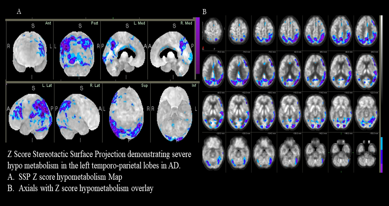
Disease-modifying therapies are currently in clinical trials. FDG PET shows early on in AD a pattern of decreased glucose metabolism in the precuneus, posterior cingulate, and temporoparietal regions translating to underlying neuronal dysfunction. [14][15][16][17] As disease progresses, the frontal lobes are also affected. One should note that an amyloid PET scan will also be positive about 10 years before the clinical onset of AD. [19] Tau PET will show increased radiotracer deposition in the temporoparietal regions and the posterior cingulate. [20][21][22][23]

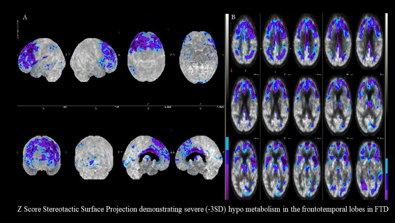
On the other hand, frontotemporal dementias will show a pattern of decreased FDG uptake in the anterior cerebral hemisphere and frontotemporal lobes. [24][20][25] The amyloid PET scan will generally be negative. [19]

In a large multicenter study, the original IDEAS (Imaging Dementia - Evidence for Amyloid Screening) study enrolled 11050 patients and showed that the use of amyloid scans changed management in 60.2% of MCI and 63.5% of dementia patients. Medication use was the most affected by the change. Amyloid scans changed the diagnosis in 35.6% of patients. [26]

Movements Disorders

Clinicians are frequently faced with patients presenting with parkinsonian-like syndromes. These patients have symptoms similar to Parkinson disease (PD) patients; however, they may not have the disease. Management of the different parkinsonian syndromes are different as well. FDOPA PET scans will be positive in parkinsonian syndromes. Decreased radiotracer uptake will be seen in the basal ganglia in patients with PD, corticobasal degeneration, multiple system atrophy, and progressive supranuclear palsy. However, patients with non-parkinsonian syndromes such as essential tremors, myoclonus, or other dystonia will have preserved FDOPA uptake. [27][28][29][30]

Neurooncological Disorders

FDG PET is no longer routinely used in assessing brain tumors and is on occasion used to evaluate brain tumor recurrence versus post-radiation necrosis changes when MRI results are equivocal. On the other hand, amino acid PET has taken a larger role in the clinical management of neurooncology patients. FDOPA can be used clinically in a variety of scenarios: [31][32]

  • Differentiation of grade III and IV tumors from non-neoplastic lesions or grade I and II gliomas
  • Prognostication of gliomas
  • Guidance of an optimal biopsy site (e.g., site of maximum tracer uptake)
  • Delineation of tumor extent for surgery and radiotherapy planning
  • Differentiation of glioma recurrence from treatment-induced changes, e.g., pseudoprogression, radionecrosis
  • Detection of malignant transformation in grade I and II gliomas
  • Response assessment during and after radiotherapy and/or chemotherapy
  • Differentiation of tumor response from pseudo-response during antiangiogenic therapy
  • Intraoperative navigation

However, FDG PET still offers high clinical accuracy in primary brain lymphoma and is of great value in the initial diagnosis, follow-up, and treatment response assessments. [33][34]

Epilepsy 

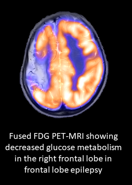
FDG PET plays a major role in the presurgical workup of medically refractory epilepsy patients. One-third of epilepsy patients are medically refractory, and their seizures are not controlled with antiepileptic drugs. FDG PET will show decreased glucose metabolism in the seizure onset zone (SOZ) and sometimes along the epilepsy network. It allows accurate localization or at least lateralization of the SOZ that needs to be resected for seizure freedom or improved seizure control. [34][35][36][37] FDG PET offers prognostic information in the presurgical workup of medically refractory epilepsy patients.

Encephalitis

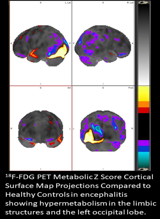
In encephalitis, FDG PET is the most sensitive imaging technique available. [38][9] A variety of patterns have been described depending on the timing of PET imaging in relation to the acute, subacute, or chronic phase of encephalitis. Acutely, increased glucose metabolism can be seen in the limbic system and the site of encephalitis in the cortex. Visual cortex and frontal lobe hypermetabolism or hypometabolism have also been described in specific pathologies. [35][39][40][41][42][43] Later on, these areas of hypermetabolism will start to exhibit decreased glucose metabolism during the chronic phase or following remission. [44][9]

Enhancing Healthcare Team Outcomes

Brain PET scans are advanced imaging techniques that require specialized expertise. Increased awareness of the clinical availability of these tools will enhance patient care. Proper utilization through integrated discussion between imaging experts and expert neurologists, epileptologists, neurosurgeons, movement disorder specialists, and neurooncologists will allow for improved diagnosis and improved patient outcomes. Multidisciplinary meetings can serve as a platform to enhance the appropriate and most effective use of these techniques.


(Click Image to Enlarge)
Alzheimer
Alzheimer
Contributed by Djekidel Mehdi MD

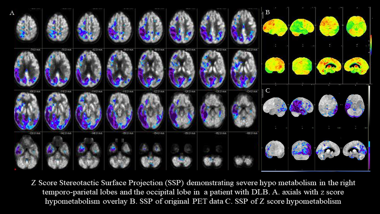
(Click Image to Enlarge)
Dementia with Lewy Bodies
Contributed by Djekidel Mehdi MD

(Click Image to Enlarge)
Frontotemporal Dementia
Contributed by Djekidel Mehdi MD

(Click Image to Enlarge)
Encephalitis FDG PET
Contributed by Djekidel Mehdi MD

(Click Image to Enlarge)
Epilepsy FDG PET
Epilepsy FDG PET
Contributed by Djekidel Mehdi MD
Details

Editor:

Joe M. Das

Updated:

4/24/2023 12:17:05 PM

References


[1]

Zimmer L,Luxen A, PET radiotracers for molecular imaging in the brain: past, present and future. NeuroImage. 2012 Jun     [PubMed PMID: 22222719]


[2]

Lameka K, Farwell MD, Ichise M. Positron Emission Tomography. Handbook of clinical neurology. 2016:135():209-227. doi: 10.1016/B978-0-444-53485-9.00011-8. Epub     [PubMed PMID: 27432667]


[3]

Zhang Y, Wang J. Targeting uptake transporters for cancer imaging and treatment. Acta pharmaceutica Sinica. B. 2020 Jan:10(1):79-90. doi: 10.1016/j.apsb.2019.12.005. Epub 2019 Dec 16     [PubMed PMID: 31993308]


[4]

Barollo S, Bertazza L, Watutantrige-Fernando S, Censi S, Cavedon E, Galuppini F, Pennelli G, Fassina A, Citton M, Rubin B, Pezzani R, Benna C, Opocher G, Iacobone M, Mian C. Overexpression of L-Type Amino Acid Transporter 1 (LAT1) and 2 (LAT2): Novel Markers of Neuroendocrine Tumors. PloS one. 2016:11(5):e0156044. doi: 10.1371/journal.pone.0156044. Epub 2016 May 25     [PubMed PMID: 27224648]


[5]

The MICAD Research Team, 4-[{sup}18{/sup}F]Fluoro-l-{i}m{/i}-tyrosine Molecular Imaging and Contrast Agent Database (MICAD). 2004     [PubMed PMID: 20641732]


[6]

Ossenkoppele R, Rabinovici GD, Smith R, Cho H, Schöll M, Strandberg O, Palmqvist S, Mattsson N, Janelidze S, Santillo A, Ohlsson T, Jögi J, Tsai R, La Joie R, Kramer J, Boxer AL, Gorno-Tempini ML, Miller BL, Choi JY, Ryu YH, Lyoo CH, Hansson O. Discriminative Accuracy of [18F]flortaucipir Positron Emission Tomography for Alzheimer Disease vs Other Neurodegenerative Disorders. JAMA. 2018 Sep 18:320(11):1151-1162. doi: 10.1001/jama.2018.12917. Epub     [PubMed PMID: 30326496]


[7]

Foster NL,Heidebrink JL,Clark CM,Jagust WJ,Arnold SE,Barbas NR,DeCarli CS,Turner RS,Koeppe RA,Higdon R,Minoshima S, FDG-PET improves accuracy in distinguishing frontotemporal dementia and Alzheimer's disease. Brain : a journal of neurology. 2007 Oct;     [PubMed PMID: 17704526]


[8]

Rinne JO. Positron emission tomography in the differential diagnosis of parkinsonism. Journal of movement disorders. 2009 Oct:2(2):53-7. doi: 10.14802/jmd.09015. Epub 2009 Oct 30     [PubMed PMID: 24868357]


[9]

Moreno-Ajona D, Prieto E, Grisanti F, Esparragosa I, Sánchez Orduz L, Gállego Pérez-Larraya J, Arbizu J, Riverol M. (18)F-FDG-PET Imaging Patterns in Autoimmune Encephalitis: Impact of Image Analysis on the Results. Diagnostics (Basel, Switzerland). 2020 May 29:10(6):. doi: 10.3390/diagnostics10060356. Epub 2020 May 29     [PubMed PMID: 32486044]


[10]

Holzgreve A,Albert NL,Galldiks N,Suchorska B, Use of PET Imaging in Neuro-Oncological Surgery. Cancers. 2021 Apr 26     [PubMed PMID: 33926002]


[11]

Shukla AK, Kumar U. Positron emission tomography: An overview. Journal of medical physics. 2006 Jan:31(1):13-21. doi: 10.4103/0971-6203.25665. Epub     [PubMed PMID: 21206635]

Level 3 (low-level) evidence

[12]

Vaquero JJ, Kinahan P. Positron Emission Tomography: Current Challenges and Opportunities for Technological Advances in Clinical and Preclinical Imaging Systems. Annual review of biomedical engineering. 2015:17():385-414. doi: 10.1146/annurev-bioeng-071114-040723. Epub     [PubMed PMID: 26643024]

Level 3 (low-level) evidence

[13]

GBD 2016 Dementia Collaborators. Global, regional, and national burden of Alzheimer's disease and other dementias, 1990-2016: a systematic analysis for the Global Burden of Disease Study 2016. The Lancet. Neurology. 2019 Jan:18(1):88-106. doi: 10.1016/S1474-4422(18)30403-4. Epub 2018 Nov 26     [PubMed PMID: 30497964]

Level 1 (high-level) evidence

[14]

Minoshima S,Mosci K,Cross D,Thientunyakit T, Brain [F-18]FDG PET for Clinical Dementia Workup: Differential Diagnosis of Alzheimer's Disease and Other Types of Dementing Disorders. Seminars in nuclear medicine. 2021 May     [PubMed PMID: 33546814]


[15]

Chételat G,Arbizu J,Barthel H,Garibotto V,Law I,Morbelli S,van de Giessen E,Agosta F,Barkhof F,Brooks DJ,Carrillo MC,Dubois B,Fjell AM,Frisoni GB,Hansson O,Herholz K,Hutton BF,Jack CR Jr,Lammertsma AA,Landau SM,Minoshima S,Nobili F,Nordberg A,Ossenkoppele R,Oyen WJG,Perani D,Rabinovici GD,Scheltens P,Villemagne VL,Zetterberg H,Drzezga A, Amyloid-PET and {sup}18{/sup}F-FDG-PET in the diagnostic investigation of Alzheimer's disease and other dementias. The Lancet. Neurology. 2020 Nov;     [PubMed PMID: 33098804]


[16]

Librizzi D, Cabanel N, Zavorotnyy M, Riehl E, Kircher T, Luster M, Hooshyar Yousefi B. Clinical Relevance of [(18)F]Florbetaben and [(18)F]FDG PET/CT Imaging on the Management of Patients with Dementia. Molecules (Basel, Switzerland). 2021 Feb 26:26(5):. doi: 10.3390/molecules26051282. Epub 2021 Feb 26     [PubMed PMID: 33652938]


[17]

Blazhenets G, Frings L, Ma Y, Sörensen A, Eidelberg D, Wiltfang J, Meyer PT, Alzheimer's Disease Neuroimaging Initiative. Validation of the Alzheimer Disease Dementia Conversion-Related Pattern as an ATN Biomarker of Neurodegeneration. Neurology. 2021 Mar 2:96(9):e1358-e1368. doi: 10.1212/WNL.0000000000011521. Epub 2021 Jan 6     [PubMed PMID: 33408150]

Level 1 (high-level) evidence

[18]

Scheltens P, De Strooper B, Kivipelto M, Holstege H, Chételat G, Teunissen CE, Cummings J, van der Flier WM. Alzheimer's disease. Lancet (London, England). 2021 Apr 24:397(10284):1577-1590. doi: 10.1016/S0140-6736(20)32205-4. Epub 2021 Mar 2     [PubMed PMID: 33667416]


[19]

Minoshima S,Drzezga AE,Barthel H,Bohnen N,Djekidel M,Lewis DH,Mathis CA,McConathy J,Nordberg A,Sabri O,Seibyl JP,Stokes MK,Van Laere K, SNMMI Procedure Standard/EANM Practice Guideline for Amyloid PET Imaging of the Brain 1.0. Journal of nuclear medicine : official publication, Society of Nuclear Medicine. 2016 Aug     [PubMed PMID: 27481605]

Level 1 (high-level) evidence

[20]

Cecchin D, Garibotto V, Law I, Goffin K. PET Imaging in Neurodegeneration and Neuro-oncology: Variants and Pitfalls. Seminars in nuclear medicine. 2021 Sep:51(5):408-418. doi: 10.1053/j.semnuclmed.2021.03.003. Epub 2021 Apr 3     [PubMed PMID: 33820651]


[21]

Yap SY, Frias B, Wren MC, Schöll M, Fox NC, Årstad E, Lashley T, Sander K. Discriminatory ability of next-generation tau PET tracers for Alzheimer's disease. Brain : a journal of neurology. 2021 Sep 4:144(8):2284-2290. doi: 10.1093/brain/awab120. Epub     [PubMed PMID: 33742656]


[22]

Ashton NJ,Leuzy A,Karikari TK,Mattsson-Carlgren N,Dodich A,Boccardi M,Corre J,Drzezga A,Nordberg A,Ossenkoppele R,Zetterberg H,Blennow K,Frisoni GB,Garibotto V,Hansson O, The validation status of blood biomarkers of amyloid and phospho-tau assessed with the 5-phase development framework for AD biomarkers. European journal of nuclear medicine and molecular imaging. 2021 Jul     [PubMed PMID: 33677733]

Level 1 (high-level) evidence

[23]

Leuzy A, Chiotis K, Lemoine L, Gillberg PG, Almkvist O, Rodriguez-Vieitez E, Nordberg A. Tau PET imaging in neurodegenerative tauopathies-still a challenge. Molecular psychiatry. 2019 Aug:24(8):1112-1134. doi: 10.1038/s41380-018-0342-8. Epub 2019 Jan 11     [PubMed PMID: 30635637]


[24]

Bastin C, Bahri MA, Bernard C, Hustinx R, Salmon E. Frontal hypometabolism in neurocognitive disorder with behavioral disturbance. Journal of nuclear medicine : official publication, Society of Nuclear Medicine. 2021 Mar 31:62(12):1783-8. doi: 10.2967/jnumed.120.260497. Epub 2021 Mar 31     [PubMed PMID: 33789936]


[25]

Dev SI, Dickerson BC, Touroutoglou A. Neuroimaging in Frontotemporal Lobar Degeneration: Research and Clinical Utility. Advances in experimental medicine and biology. 2021:1281():93-112. doi: 10.1007/978-3-030-51140-1_7. Epub     [PubMed PMID: 33433871]

Level 3 (low-level) evidence

[26]

Rabinovici GD, Gatsonis C, Apgar C, Chaudhary K, Gareen I, Hanna L, Hendrix J, Hillner BE, Olson C, Lesman-Segev OH, Romanoff J, Siegel BA, Whitmer RA, Carrillo MC. Association of Amyloid Positron Emission Tomography With Subsequent Change in Clinical Management Among Medicare Beneficiaries With Mild Cognitive Impairment or Dementia. JAMA. 2019 Apr 2:321(13):1286-1294. doi: 10.1001/jama.2019.2000. Epub     [PubMed PMID: 30938796]


[27]

Sood A, Shukla J, Shree R, Vatsa R, Modi M, Mittal BR. Comparative Performance of 99mTc-TRODAT-1 SPECT/CT and 18F-FDOPA PET/CT Imaging in Patients With Parkinson's Disease, Parkinson-Plus Syndrome, and Essential Tremor. Clinical nuclear medicine. 2021 Feb 1:46(2):95-102. doi: 10.1097/RLU.0000000000003409. Epub     [PubMed PMID: 33234920]

Level 2 (mid-level) evidence

[28]

Boertien JM, van der Zee S, Chrysou A, Gerritsen MJJ, Jansonius NM, Spikman JM, van Laar T, PPNN Study Group. Study protocol of the DUtch PARkinson Cohort (DUPARC): a prospective, observational study of de novo Parkinson's disease patients for the identification and validation of biomarkers for Parkinson's disease subtypes, progression and pathophysiology. BMC neurology. 2020 Jun 13:20(1):245. doi: 10.1186/s12883-020-01811-3. Epub 2020 Jun 13     [PubMed PMID: 32534583]

Level 2 (mid-level) evidence

[29]

Stormezand GN,Chaves LT,Vállez García D,Doorduin J,De Jong BM,Leenders KL,Kremer BPH,Dierckx RAJO, Intrastriatal gradient analyses of 18F-FDOPA PET scans for differentiation of Parkinsonian disorders. NeuroImage. Clinical. 2020     [PubMed PMID: 31981888]


[30]

Darcourt J, Schiazza A, Sapin N, Dufour M, Ouvrier MJ, Benisvy D, Fontana X, Koulibaly PM. 18F-FDOPA PET for the diagnosis of parkinsonian syndromes. The quarterly journal of nuclear medicine and molecular imaging : official publication of the Italian Association of Nuclear Medicine (AIMN) [and] the International Association of Radiopharmacology (IAR), [and] Section of the Society of.... 2014 Dec:58(4):355-65     [PubMed PMID: 25366711]


[31]

Louarn N, Kauv P, Capelle L, Mokhtari K, Itti E. Illustration of the Added Value of 18F-DOPA PET to Multimodal MRI to Distinguish Low- and High-Grade Gliomas. Clinical nuclear medicine. 2021 Jul 1:46(7):e353-e354. doi: 10.1097/RLU.0000000000003507. Epub     [PubMed PMID: 33577196]


[32]

Law I, Albert NL, Arbizu J, Boellaard R, Drzezga A, Galldiks N, la Fougère C, Langen KJ, Lopci E, Lowe V, McConathy J, Quick HH, Sattler B, Schuster DM, Tonn JC, Weller M. Joint EANM/EANO/RANO practice guidelines/SNMMI procedure standards for imaging of gliomas using PET with radiolabelled amino acids and [(18)F]FDG: version 1.0. European journal of nuclear medicine and molecular imaging. 2019 Mar:46(3):540-557. doi: 10.1007/s00259-018-4207-9. Epub 2018 Dec 5     [PubMed PMID: 30519867]

Level 1 (high-level) evidence

[33]

Marcus C, Feizi P, Hogg J, Summerfield H, Castellani R, Sriwastava S, Marano GD. Imaging in Differentiating Cerebral Toxoplasmosis and Primary CNS Lymphoma With Special Focus on FDG PET/CT. AJR. American journal of roentgenology. 2021 Jan:216(1):157-164. doi: 10.2214/AJR.19.22629. Epub 2020 Oct 28     [PubMed PMID: 33112669]


[34]

Zhang Y, Cai J, Lin Z, Yao S, Miao W. Primary Central Nervous System Lymphoma Revealed by 68Ga-FAPI and 18F-FDG PET/CT. Clinical nuclear medicine. 2021 Aug 1:46(8):e421-e423. doi: 10.1097/RLU.0000000000003517. Epub     [PubMed PMID: 33512949]


[35]

Djekidel M. (18)F-FDG PET Imaging Predicts the Epileptogenic Zone Prospectively in Recurrent Cryptogenic Meningoencephalitis with Subsequent Simple Partial Visual Seizures. Journal of nuclear medicine technology. 2021 Mar:49(1):92-94. doi: 10.2967/jnmt.120.252866. Epub 2020 Nov 20     [PubMed PMID: 33219155]


[36]

Avendaño-Estrada A, Velasco F, Velasco AL, Cuellar-Herrera M, Saucedo-Alvarado PE, Marquez-Franco R, Rivera-Bravo B, Ávila-Rodríguez MA. Quantitative Analysis of [18F]FFMZ and [18F]FDG PET Studies in the Localization of Seizure Onset Zone in Drug-Resistant Temporal Lobe Epilepsy. Stereotactic and functional neurosurgery. 2019:97(4):232-240. doi: 10.1159/000503692. Epub 2019 Nov 13     [PubMed PMID: 31722358]


[37]

Tomás J, Pittau F, Hammers A, Bouvard S, Picard F, Vargas MI, Sales F, Seeck M, Garibotto V. The predictive value of hypometabolism in focal epilepsy: a prospective study in surgical candidates. European journal of nuclear medicine and molecular imaging. 2019 Aug:46(9):1806-1816. doi: 10.1007/s00259-019-04356-x. Epub 2019 May 29     [PubMed PMID: 31144060]


[38]

Solnes LB, Jones KM, Rowe SP, Pattanayak P, Nalluri A, Venkatesan A, Probasco JC, Javadi MS. Diagnostic Value of (18)F-FDG PET/CT Versus MRI in the Setting of Antibody-Specific Autoimmune Encephalitis. Journal of nuclear medicine : official publication, Society of Nuclear Medicine. 2017 Aug:58(8):1307-1313. doi: 10.2967/jnumed.116.184333. Epub 2017 Feb 16     [PubMed PMID: 28209905]


[39]

Bordonne M, Chawki MB, Doyen M, Kas A, Guedj E, Tyvaert L, Verger A. Brain (18)F-FDG PET for the diagnosis of autoimmune encephalitis: a systematic review and a meta-analysis. European journal of nuclear medicine and molecular imaging. 2021 Nov:48(12):3847-3858. doi: 10.1007/s00259-021-05299-y. Epub 2021 Mar 7     [PubMed PMID: 33677643]

Level 1 (high-level) evidence

[40]

Ge J, Deng B, Guan Y, Bao W, Wu P, Chen X, Zuo C. Distinct cerebral (18)F-FDG PET metabolic patterns in anti-N-methyl-D-aspartate receptor encephalitis patients with different trigger factors. Therapeutic advances in neurological disorders. 2021:14():1756286421995635. doi: 10.1177/1756286421995635. Epub 2021 Feb 24     [PubMed PMID: 33717212]

Level 3 (low-level) evidence

[41]

Liu X, Shan W, Zhao X, Ren J, Ren G, Chen C, Shi W, Lv R, Li Z, Liu Y, Ai L, Wang Q. The Clinical Value of (18) F-FDG-PET in Autoimmune Encephalitis Associated With LGI1 Antibody. Frontiers in neurology. 2020:11():418. doi: 10.3389/fneur.2020.00418. Epub 2020 Jun 5     [PubMed PMID: 32581996]


[42]

Seniaray N, Verma R, Ranjan R, Belho E, Mahajan H. 18F-FDG PET/CT in Initial Diagnosis and Treatment Response Evaluation of Anti-NMDAr and Anti-GAD Dual Antibody Autoimmune Encephalitis. Clinical nuclear medicine. 2021 Jan:46(1):e63-e64. doi: 10.1097/RLU.0000000000003379. Epub     [PubMed PMID: 33181746]


[43]

Seniaray N, Verma R, Ranjan R, Belho E, Mahajan H. Metabolic Imaging Patterns on 18F-FDG PET in Acute and Subacute LGI1 Autoimmune Limbic Encephalitis. Clinical nuclear medicine. 2021 Jan:46(1):e27-e28. doi: 10.1097/RLU.0000000000003258. Epub     [PubMed PMID: 32910055]


[44]

Wei YC, Tseng JR, Wu CL, Su FC, Weng WC, Hsu CC, Chang KH, Wu CF, Hsiao IT, Lin CP. Different FDG-PET metabolic patterns of anti-AMPAR and anti-NMDAR encephalitis: Case report and literature review. Brain and behavior. 2020 Mar:10(3):e01540. doi: 10.1002/brb3.1540. Epub 2020 Jan 27     [PubMed PMID: 31985135]

Level 3 (low-level) evidence