Continuing Education Activity
Balanced ligamentous tension (BLT) is a treatment modality in osteopathic medicine that works on the principle of balancing the tension among ligaments supporting a joint to reset the normal proprioceptive feedback of that joint. While BLT can be used in any region of the body. This activity reviews the evaluation and treatment of sacroiliac and lumbosacral somatic dysfunctions and highlights the role of interprofessional teams in evaluating and treating patients with this condition.
Objectives:
Review the typical anatomy and physiology that are involved in pelvic somatic dysfunctions.
Outline the management considerations for patients with absolute contraindications to using BLT in the pelvis.
Describe the common treatments using BLT to address somatic dysfunction in the pelvis as discussed in this article.
Explain the importance of collaboration and communication amongst the interprofessional team to improve outcomes for patients affected by pelvic somatic dysfunction.
Introduction
Balanced ligamentous tension (BLT) (also referred to as ligamentous articular strain depending on geographic location), relies on the principle that the ligaments of the body provide proprioceptive feedback when tension is appropriately balanced along with the respective ligaments in a joint. According to the Sutherland model, an unequal distribution in the tension among ligaments can cause imbalances in the joint that result in a new pathological “normal”. By altering the strain on certain ligaments, the “spring” of a ligament can be returned to back to the normal physiologic range, and proper proprioceptive feedback in the joint can be re-established.
Different institutions across the country vary in their description of BLT techniques (with some distinctly separating direct and indirect methods), for example, some schools of thought describe the technique as having three primary steps which include: disengagement at the joint in question, exaggeration of the joint by carrying it into the position of its original injury, and finally balance, which involves feeling for release in the form of a “wobble point” indicating that the ligaments involved in the dysfunction have been rebalanced. When applying this concept to the pelvis, the joints of primary focus include the lumbosacral and sacroiliac.
Anatomy and Physiology
Sacrum
The pelvis, consisting of a sacrum and two innominate, provides the foundational stability necessary for proper ambulation and posture. In regards to the sacrum, the anatomy involves the fusion of sacral vertebrae S1-S5, noting that this fusion is not fully completed until age 18-30. Important landmarks include the sacroiliac (SI) joints on either wings or ala of the sacrum, which articulate with the ilium and are critical for proper sacral motion, as well as the lumbosacral joint which articulates with the L5 lumbar vertebra. Moving inferiorly, the lower part of the sacrum, termed the coccyx is the final vertebral segment. The coccyx is connected to the sacrum via a fibrocartilaginous joint known as the sacrococcygeal symphysis.[1]
The proper sacral motion requires the use of various ligaments such as the sacroiliac ligament, which connects the sacrum to the ilium and limits excessive anterior and inferior sacral motion. The sacrotuberous ligament comes together with the posterior sacroiliac ligament to function in resisting the nutation of the sacrum during normal gait.[2] The greater and lesser sciatic foramen house the sciatic nerve, superior and inferior gluteal nerve, pudendal nerve, posterior femoral cutaneous nerve, nerve to quadratus femoris, and nerve to internal obturator muscle as well as several major vessels such as the superior and inferior gluteal artery and vein along with the internal pudendal artery and vein.[3]
Important ligaments involving the ilium include the iliolumbar and ilioinguinal ligaments, both of which contribute to overall pelvic stability. Finally, the true pelvic ligaments (which form a direct bone-to-bone connection) include the sacroiliac and interosseous sacroiliac ligaments. Accessory pelvic ligaments consist of the iliolumbar ligament, inguinal ligaments, sacrotuberous ligaments, and sacrospinous ligaments.[4]
The sacral anatomy plays an important role in the proper physiological motion of the pelvis, specifically in regards to understanding the important axes of motion of the sacrum. Two diagonal oblique axes are present and go from the superior portion of the sacroiliac articulation to the contralateral inferior sacroiliac articulation and can be named either left or right, depending on their superior origin. The superior transverse axis passes through the posterior dural attachment at the second sacral segment and allows for sacral motion during the primary respiration. The middle transverse axis passes horizontally through the second sacral segment on the anterior aspect of the sacrum and is involved in postural stability that allows for appropriate sacral motion in a standing position as well as thoracic respiration. Finally, the inferior transverse axis (also at the second sacral segment) is involved in innominate motion in the way of the articulation between the ilia on the sacrum.
Innominate
The innominate is not a singular structure, but instead, the combination of three embryologically formed bones; the ilium, ischium, and pubis. Over time, these bones fuse to form the complete innominate which includes the acetabulum, or “hip socket” that houses the femoral head.[5] Points of contact between the sacrum and innominate include the ligamentous attachments at the pubic symphysis and the sacroiliac joint, both of which provide pelvic stability during ambulation. When identifying important landmarks used in the diagnosis of pelvic somatic dysfunction, the anterior and posterior superior iliac spines (ASIS and PSIS) are commonly referenced due to their relatively easy palpable nature and reliability consistent pelvic landmarks.
From a physiologic point of view, the innominate has the important feature of maintaining overall human stability when standing, walking, or running.[5] It is not an immobile structure and allows for a slight degree of mobility at its ligamentous attachments with the sacrum allowing for proper axial rotation of the pelvis with normal physical activities.
Indications
Indications include findings of pelvic somatic dysfunction with an associated complaint where other explanations of the potential source of the complaint have been ruled out.
Contraindications
Pelvic BLT is an incredibly safe treatment modality due to its passive and direct or indirect approach. Many patients with pelvic somatic dysfunction that may not be able to tolerate more active and direct treatments such as high-velocity, low-amplitude (HVLA), muscle energy, or articulatory techniques can tolerate BLT well. Some absolute contraindications include patient refusal, inability to consent to treatment, fractures at the location being treated, as well as certain bone malignancies or osteomyelitis leading to compromised bone structure.
Equipment
No special equipment is necessary to perform pelvic BLT. Providers may benefit from the use of an adjustable table and stool to aid with practitioner comfort and ensure proper ergonomics.
Personnel
The only personnel needed is the physician.
Preparation
Communication is very important, both when performing the exam and treating the patient. The physician should describe to the patient the procedure and clinical reasoning behind the treatment as well as the risks, benefits, and alternatives. Consent needs to be obtained prior to treatment. The physician then performs an osteopathic structural exam, taking note of somatic dysfunctions and areas of greatest restriction.
Technique or Treatment
SI BLT- Short Lever
The patient is lying supine on the table. Diagnose the side of restriction with the ASIS compression test. Sit facing the patient on the side of the SI restriction. Contact under the pelvis at the SI joint with the cephalad hand, so the finger pads are on the medial aspect of the SI joint and place the caudad hand on the ipsilateral ASIS. Bring the innominate through a range of motion, including superior/inferior shear, anterior/posterior rotation, and inflare/outflare. When using indirect BLT, bring the innominate into its position of ease in all the motions tested and fine-tune until a point of balanced tension between physiologic neutral and the direct articular barrier is reached. Make sure that the ligaments still feel engaged. The same is true when using direct BLT, except the innominate is brought into its restriction in all the motions tested. Anterior pressure may need to be applied to the sacrum with the hand under the SI joint, which will facilitate the release by decompressing the SI joint.
Once the point of balanced tension is found, hold this position until softening under the hand that is contacting the SI joint is felt. Warmth increased motion, or a sensation that less force is needed to hold the joint in its treatment position might also be felt. Once this happens, subtle motions can be used to find the next point of balanced tension. Repeat this process until no further change is appreciated. Reassess with the ASIS compression test. It should be noted that not all schools teach direct and indirect BLT. Those that do not may instead focus on finding a point of balanced tension without specifying whether this was achieved by first going into the direction of ease or restriction.
SI BLT- Long Lever
The patient is lying supine on the table. Diagnose the side of restriction with the ASIS compression test. Stand on the side of the SI restriction facing the patient’s head. Contact the SI joint with the hand that is away from the patient, which will be used to monitor for changes. Use the arm closest to the patient to hold the ipsilateral leg and use it as a long lever to create balanced tension at the SI joint. Internally rotate the femur in the acetabulum to engage the SI joint. Bring the leg into abduction/adduction, flexion/extension, and distraction/compression into the SI joint, noticing which motions soften and create tension at the SI joint. Stack all of the motions that create ease at the SI joint if indirect BLT is being used or all of the motions that create tension if direct BLT is being used, and fine-tune to a point of balanced tension.
Once the point of balanced tension is found to hold this position until softening under the hand that is contacting the SI joint is felt. Warmth increased motion, or a sensation that less force is needed to hold the joint in its treatment position might also be felt. Once this happens, subtle motions can be used to find the next point of balanced tension. Repeat this process until no further change is appreciated. Reassess with the ASIS compression test.
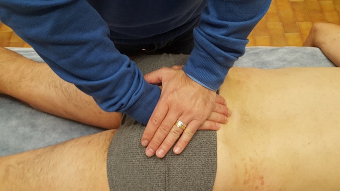
SI Decompression, direct- Supine
This technique is applicable in the treatment of SI compression and is performed in a direct manner. Diagnose the side of restriction with the ASIS compression test. With the patient lying supine and the provider seated at the side of SI restriction, the provider contacts the sacrum with the caudad hand, fingers pointing cephalad (typically by positioning the hand between the patient’s legs) in order to provide stability to the sacrum. Following this, the provider’s cephalad hand applies lateral traction to the PSIS on the affected side until a release of the joint is felt. An optional respiratory component may be included in this technique by having the patient inhale and exhale deeply while palpating for motion at the SI joint space and using this respiratory cooperation to encourage the release and balance of the affected ligaments.
A release may be felt as a slight pull of the innominate as the ligaments balance at the SI joint. Often an increase in warmth, softening of the surrounding tissues, and/or an improvement in sacral motion with either primary or secondary respiration may be noticed. An increase in motion at the SI joint is observed upon retest as an increase in mobility with the ASIS compression test.
LS Decompression- Prone
The patient is lying prone on the table. Stand on either side of the patient and face the table. Contact the sacrum with the cephalad hand, so the heel of the hand is at the sacral base. Be mindful that the fingertips are not contacting sensitive areas. Cross the other arm over and contact the lower lumbar segments with the caudad hand. Position the caudad hand perpendicular to the cephalad hand, so the hands form the letter “T,” or contact the lower lumbar segments with the palm of the caudad hand, so the fingertips point superiorly. To assess for restriction, have the patient take deep breaths and notice the motion of the sacrum and lumbar spine at the LS junction. With an inhalation, the sacrum should go into counternutation, and the lumbar lordosis should flatten. With an exhalation, the sacrum should go into nutation, and the lumbar lordosis should increase. To treat, keep the arms fairly straight with a slight bend at the elbows, and lean over the patient. The hands will naturally separate from each other and create a distraction at the LS junction. Notice the amount of restriction at the LS junction when distraction is applied at the joint, so the initial finding can be compared to the reassessment at the end. Then induce rotation of the sacrum on the lumbar spine and rotation of the lumbar spine on the sacrum until you find a point of balanced tension. Warmth, softening, increased motion, or a sensation that less force is needed to hold the joint in its treatment position might also be felt. Once this happens, subtle motions can be used to find the next point of balanced tension. Repeat this process until no further change is appreciated. Reassess by repeating your diagnostic assessment.
LS Decompression- Supine
The patient is lying supine on the table. Sit on either side of the patient-facing the patient’s head. Contact the lower lumbar region with the cephalad hand, so the finger pads are on the contralateral paraspinal muscles, and the thenar/hypothenar eminences are on the ipsilateral paraspinal muscles. Have the patient bend one or both knees and lift their pelvis off the table. Position the arm closest to the patient in-between the patient’s legs and contact the sacrum, so the fingertips are at the sacral base, and the hands are perpendicular to each other. Have the patient relax their pelvis back down on the table. They can either keep their knees bent or straighten them out. Keeping the hand and wrist relaxed will help with palpation of the tissues while it is under the weight of the patient and improve provider ergonomics. To assess the motion of the sacrum on the ilia, induce nutation/counternutation on its transverse axes by flexing and extending the wrist and right/left rotation on its vertical axes by pronating and supinating the wrist of the hand that is under the sacrum.
To assess the motion of the sacrum with respect to the lumbar spine, induce lumbar rotation by flexing and extending the wrist of the hand that is under the lumbar spine. Assess the motion at the LS junction when the patient takes deep breaths. With an inhalation, the sacrum should go into counternutation, and the lumbar lordosis should flatten. With an exhalation, the sacrum should go into nutation, and the lumbar lordosis should increase. Assess whether adding compression or distraction to the LS junction creates more freedom of motion there. To add compression bring the caudad hand superiorly and the cephalad hand inferiorly. To add distraction, bring the caudad hand inferiorly and the cephalad hand superiorly. To treat, add compression or distraction, induce motion in all the planes tested in the sacrum, and add a rotation of the lumbar spine. Depending on whether direct or indirect BLT is used, the provider will first go into the direction of ease or restriction and then fine-tune to a point of balanced tension. Warmth, softening, increased motion, or a sensation that less force is needed to hold the joint in its treatment position might also be felt. Once this happens, subtle motions can be used to find the next point of balanced tension. Repeat this process until no further change is appreciated. Reassess by repeating your diagnostic assessment.
Complications
While BLT is one of the safest techniques, patients may still experience soreness for several days following treatment and are advised to drink plenty of water after the treatment to prevent or minimize soreness.
Clinical Significance
The philosophy of osteopathic medicine is to treat the patient as a whole. The answer to the question of when OMT should be used on the pelvis can be addressed when somatic dysfunction is found on an osteopathic structural exam and when treatment would benefit the patient. While BLT is one of many osteopathic treatment modalities, the decision to use one modality over another comes down to provider preference and the responsiveness of the patient’s tissues. Here are a couple of specific examples of how OMT for pelvic dysfunction can be applied.
The pregnant patient: There are many physiologic and biomechanical changes that happen to the body during pregnancy. These changes can lead to symptoms, including back pain, pelvic pain, stiffness, and lower extremity edema that have a large impact on a person’s ability to function in daily life. Many medications normally prescribed to address these symptoms are not recommended during pregnancy, and osteopathic manipulative treatment is a nonpharmacologic option for pregnant patients looking for relief. In the Pregnancy Research on Osteopathic Manipulation Optimizing Treatment Effects (PROMOTE) study, there was not a higher conversion rate to high-risk status in third-trimester patients in the OMT group indicating that OMT is safe for this patient population. In addition to the pelvic region (pelvic diaphragm, innominate, and sacrum), attention should also be given to the occipitoatlantal joint, cervical vertebrae, clavicles, and Sibson fascia, thoracoabdominal diaphragm, ribs, hips, and cranium during the assessment and treatment with OMT in the pregnant patient.[6]
The hospitalized patient: BLT is a useful modality in treating the hospitalized patient because active participation of the patient in the treatment is not required, and it can be adapted to use in any part of the body while the patient is supine. The Multicenter Osteopathic Pneumonia Study in the Elderly (MOPSE) randomized controlled trial showed shortened length-of-stay and decreased in-hospital mortality rates in certain subgroups, which supports the role of OMT as an adjunctive treatment to standard of care in patients hospitalized with pneumonia.[7]
Another study looking at the effects of OMT on hospital length-of-stay and incidence of postoperative ileus in general surgical patients showed that OMT decreased time to flatus and decreased length-of-stay.[8] The anatomy that is targeted with OMT in the pelvis can be thought of in terms of autonomics, biomechanics, and circulation. Autonomic tone can be modulated through manipulation of the sacrum. The pelvic splanchnic nerves arise from S2-S4 nerve roots and carry afferent parasympathetic nerve fibers to the distal end of the transverse colon, descending colon, sigmoid colon, rectum, cervix, and upper vagina.[9]
Targeting the parasympathetic fibers innervating the colon could be useful in the postsurgical patient in trying to encourage the return of normal bowel function. Treating dysfunction at the lumbosacral junction and the sacroiliac joints could restore mobility and help in the process of getting the patient out of bed and walking again. Treatment of restriction in the pelvic diaphragm, which is recognized as a muscle of respiration, opens up the pathway for lymphatic and venous return and decreases any congestion in the area. A goal in treating a hospitalized patient with OMT is to help decrease the patient’s allostatic load by correcting somatic dysfunction so that they put more of their body’s energy towards healing.
Enhancing Healthcare Team Outcomes
The effectiveness of BLT primarily stems from an in-tune palpatory sense on the part of the provider. Since this is developed through hands-on practice, it is important that as a provider, one continues to hone these skills through repetition, as this will result in the highest likelihood of correctly diagnosing and treating patients with this particular modality. Practitioners are encouraged to attend workshops and meetings where they can further develop not only their palpatory skills with BLT but also many other treatment modalities within osteopathic manipulative medicine. Proper application of this technique can be very beneficial to nearly all patients as it is a gentle and very tolerable approach for even the most vulnerable patient populations. Further research into the efficacy of BLT in the treatment of ligamentous somatic dysfunctions could shed light on novel uses for this effective modality in the field of osteopathic manipulative treatment.