Learning Outcome
- Recall the causes of conjunctivitis
- Describe the presentation of conjunctivitis
- Summarize the treatment of conjunctivitis

Conjunctivitis is a common cause of eye redness and, subsequently, a common complaint in the emergency department, urgent care, and primary care clinics. It can affect people of any age, demographic or socioeconomic status. Although usually self-limiting and rarely resulting in vision loss, it is essential to rule out other sight-threatening causes of red-eye when assessing for conjunctivitis.
The conjunctiva is the transparent, lubricating mucous membrane covering the outer surface of the eye.[1] It is composed of two parts, the "bulbar conjunctiva," which covers the globe, and the "tarsal conjunctiva," which lines the eyelid's inner surface.
Conjunctivitis refers to the inflammation or infection of the conjunctiva. It can be acute or chronic and infectious or non-infectious. Acute conjunctivitis refers to symptom duration of 3 to 4 weeks from presentation (usually only lasting 1 to 2 weeks), whereas chronic is defined as lasting more than four weeks.
Conjunctivitis is the most prevalent etiology of eye redness and discharge. While there are many types of conjunctivitis, viral, allergic, and bacterial are the three most common.
Infectious conjunctivitis can result from bacteria, viruses, fungi, and parasites. However, 80% of acute cases of conjunctivitis are viral, the most common pathogen being adenovirus. Adenoviruses are responsible for 65 to 90% of cases of viral conjunctivitis.[2] Other common viral pathogens are herpes simplex, herpes zoster, and enterovirus.
Bacterial conjunctivitis is far more common in children than adults, and the pathogens responsible for bacterial conjunctivitis vary depending on the age group. Staphylococcal species, specifically Staphylococcal aureus, followed by Streptococcus pneumoniae and Haemophilus influenza, are the most common cause in adults, while in children, the disease is more often caused by H. influenza, S. pneumoniae, and Moraxella catarrhalis.[2] Other bacterial causes include Neisseria gonorrhoeae, Chlamydia trachomatis, and Corynebacterium diphtheria. N. gonorrhoeae is the most common cause of bacterial conjunctivitis in neonates.[1]
Allergens, toxins, and local irritants are responsible for non-infectious conjunctivitis.
The prevalence of conjunctivitis varies by age, sex, and time of year. There is a bimodal distribution of diagnosed cases of acute conjunctivitis NOS in the emergency department (ED). The highest diagnosis rates are among children under seven years of age, with the highest incidence occurring between the ages of 0 and 4 years. The secondary distribution peak occurs at the ages of 22 years in women and 28 years in men. Overall the rates of conjunctivitis diagnosed in the ED are slightly higher in women than in men. Seasonality is also a factor in the presentation and, thus, the diagnosis of conjunctivitis. Varying by age, there is a peak incidence in all presentations of conjunctivitis in children ages 0 to 4 years in March, followed by other age groups in May. A nationwide ED study found seasonality to be consistent for all geographic regions, regardless of changes in climate or weather patterns.[3] Allergic conjunctivitis is the most frequent cause of conjunctivitis, affecting 15 to 40% of the population, and is observed most commonly in spring and summer. Bacterial conjunctivitis rates are highest from December to April.[1][2][3]
History and physical examination are, of course, essential in the diagnosis of conjunctivitis and in determining the cause and, therefore, treatment of the condition. Important points to remember when taking the ocular history of the patient should include the timing of onset, prodromal symptoms, unilateral or bilateral eye involvement, associated symptoms, previous treatment and response, past episodes, type of discharge, the presence of pain, itching, eyelid characteristics, periorbital involvement, vision changes, photophobia, and corneal opacity.
The ocular exam should focus on visual acuity, extraocular motility, visual fields, discharge type, shape, size and response of pupil, the presence of proptosis, corneal opacity, foreign body assessment, tonometry, and eyelid swelling.
The redness of the conjunctiva in conjunctivitis is generally diffuse. It involves the entire conjunctival surface, both the bulbar and tarsal conjunctiva, which helps exclude more severe conditions such as keratitis, iritis, and angle-closure glaucoma as they involve the entire bulbar conjunctiva but spare the tarsal conjunctiva. If the redness is localized, one should consider an alternative diagnosis of foreign body, pterygium, or episcleritis.[4]
After redness, the type of discharge is an important factor in determining the cause of conjunctivitis. Bacterial conjunctivitis is typically associated with purulent discharge, which reforms immediately after removal from the eye, or mucopurulent discharge, which tends to be thicker and sticks to the eyelashes.[5][6] Compared to other causes of bacterial conjunctivitis, N gonorrhoeae is typically hyperacute in presentation, presenting with copious purulent discharge, abrupt onset, and rapid progression. Traditionally, the discharge in both viral and allergic conjunctivitis is watery. In the context of watery discharge, the additional finding of preauricular lymphadenopathy can point toward the diagnosis of viral rather than allergic conjunctivitis.[2]
Similar to redness and discharge, many other common signs and symptoms of conjunctivitis are nonspecific and can make determining the underlying cause more difficult. For example, itching has historically correlated with allergic conjunctivitis. While in the context of watery discharge and a history of atopy, this is likely true, one study found that 58% of patients with culture-positive bacterial conjunctivitis also reported itchy eyes.[7]
Comparably, papillae are a nonspecific finding in conjunctivitis. Papillae can be present in both noninfectious and infectious conjunctivitis. They are small elevations with central vessels, usually under the superior tarsal conjunctiva. Papillae are often present in bacterial conjunctivitis, allergic conjunctivitis, and contact lens intolerance. The papillae in chronic allergic conjunctivitis can lead to a cobblestone appearance of the conjunctiva.
While also nonspecific, the presence of follicles, in correlation with other findings, can help differentiate the etiology of conjunctivitis. Follicles are small, elevated yellow-white lesions found at the palpebral and bulbar conjunctiva junction, also known as the lower cul-de-sac. Follicles are a lymphocytic response often present in chlamydial and adenoviral conjunctivitis.
In a patient with a history of perioral cold sores, current skin lesions, or suspected viral conjunctivitis, a fluorescein examination should be performed as herpes simplex virus (HSV) can produce corneal dendritic lesions even in the absence of skin lesions. This exam is an important step in the physical evaluation as it may result in the only finding to differentiate HSV from other viral causes of conjunctivitis, which subsequently requires different management and follow-up. In comparison, herpes zoster ophthalmicus typically presents in patients over 60 years with a painful vesicular rash following the distribution of the fifth cranial nerve. Prodrome can include headache, fever, malaise, and photophobia. Vesicles at the tip of the nose, referred to as the Hutchinson sign, strongly predict eye involvement with herpes zoster.[8]
While presentations can often overlap, a systematic approach, thorough history, and physical exam can safely rule out any acute sight-threatening diagnoses and lead to the likely cause of conjunctivitis. The classic findings of the three most common types of conjunctivitis can be found below:
Labs and cultures are rarely indicated to confirm the diagnosis of conjunctivitis. Eyelid cultures and cytology are usually reserved for cases of recurrent conjunctivitis, those resistant to treatment, suspected gonococcal or chlamydial infection, suspected infectious neonatal conjunctivitis, and adults presenting with severe purulent discharge.[1][2][10] Rapid antigen testing is available for adenoviruses and can be used to confirm suspected viral causes of conjunctivitis to prevent unnecessary antibiotic use. One study comparing rapid antigen testing to PCR and viral culture and confirmatory immunofluorescent staining found rapid antigen testing to have a sensitivity of 89% and a specificity of up to 94%.[13]
To decrease the transmission rate, treating both viral and bacterial conjunctivitis should include patient education.
Bacterial conjunctivitis, while typically self-limiting, can be treated to help reduce the duration of symptoms. No significant difference in outcomes has been observed in trials comparing different types of ophthalmic antibiotic drops. While ointments typically last longer than drops, they tend to interfere with vision. Initial treatment for acute, non-severe bacterial conjunctivitis varies depending on the antimicrobial agent but generally is administered to the affected eye every two to 6 hours for 5 to 7 days. For mild bacterial conjunctivitis, older-generation antibiotics are generally advised. Later-generation antibiotics are reserved for more grave infections to minimize the development of resistance in the ocular surface flora.[14]
In moderate to severe cases of bacterial conjunctivitis, the latest-generation fluoroquinolones are more suitable as they provide strong gram-negative and some gram-positive coverage. Antibiotic options are available as liquid solutions and topical ointments. Liquid suspension/solutions include polymyxin B/trimethoprim, ciprofloxacin, ofloxacin, levofloxacin, moxifloxacin, gatifloxacin or azithromycin, while bacitracin, erythromycin or ciprofloxacin can be administered as an ointment. Fluoroquinolones should be prescribed for contact lens wearers to provide empiric coverage for Pseudomonas.
The recommended treatment for gonococcal conjunctivitis is ceftriaxone 1 gram intramuscular (IM), and it is recommended to treat concurrent chlamydial infection with 1 gm azithromycin PO as well. The neonatal dosing for gonococcal conjunctivitis is 25 to 50 mg/kg ceftriaxone intravenous (IV)/IM with a max dose of 125 mg, with 20 mg/kg azithromycin PO once daily for three days.
Viral conjunctivitis due to adenoviruses is self-limiting, and treatment should target symptomatic relief with cold compresses and artificial tears. Povidone-iodine 0.8% may be a potential option to decrease contagiousness in patients with adenoviral infections.[15]
Herpes simplex keratitis should receive antiviral therapy. Mild infections can have treatment with trifluridine 1% drops every 2 hours or 8 to 9 times a day for 10 to 14 days, topical ganciclovir 0.15% gel one drop five times a day until epithelium heals and then three times daily for one week, or oral acyclovir 400 mg PO 5 times a day for 7 to 10 days to limit epithelial toxicity. Patients should have a follow-up with ophthalmologists within 2 to 5 days to monitor for complications.
Treatment of herpes zoster conjunctivitis includes a combination of oral antivirals and topical steroids; however, steroids should only be part of therapy in consultation with ophthalmology. Antiviral doses differ from those used for herpes simplex and consist of oral acyclovir 800 mg five times a day, oral famciclovir 500 mg three times a day, or oral valacyclovir 1 g three times a day, each for 7 to 10 days.
A study by Wilkins et al. observed the effect of topical steroids compared with hypromellose in comforting patients with acute presumed viral conjunctivitis. It reported that a short course of topical dexamethasone in acute follicular conjunctivitis cases presumed to have viral origin was not harmful.[16]
Steroid use with antibiotics is controversial, and studies report mixed results in reducing corneal scarring.[17][18] Unfortunately, steroids may slow the rate of healing, increase the risk of corneal melting, and increase the risk of elevated IOP.
Lastly, the treatment for allergic conjunctivitis consists of allergen avoidance, artificial tears, cold compresses, and a wide range of topical agents. Topical agents include topical antihistamines alone or in combination with vasoconstrictors, topical mast cell inhibitors, and topical glucocorticoids for refractory symptoms. Oral antihistamines can also be used in moderate to severe cases of allergic conjunctivitis.
Patients with moderate to severe pain, vision loss, corneal involvement, severe purulent discharge, conjunctival scarring, recurrent episodes, lack of response to therapy, or herpes simplex keratitis should receive a prompt referral to an ophthalmologist. In addition, those requiring steroids, contact lens wearers, and patients with photophobia should also get a referral.[1][2][10]
Conjunctivitis is easily treatable and usually benign and self-limiting. Symptom duration varies depending on the type. Viral conjunctivitis typically increases in severity until day 4 or 5 and resolves within the following 1 to 2 weeks for a total duration of 2 to 3 weeks. Bacterial conjunctivitis tends to last 7 to 10 days but can be shortened by early antibiotic administration within the first six days of onset.
Viral and bacterial conjunctivitis can spread by direct contact and have high transmission rates. Patient education is crucial to prevent transmission. The importance of hand hygiene for patients, staff, family, and friends should be highlighted. One study found that when swabbing the hands of infected patients, 46% resulted in positive cultures.[2] Patients should be instructed to avoid touching their eyes, shaking hands, sharing personal items such as cosmetics or towels, and avoiding swimming pools while infected. Medical instruments should be disinfected and admitted patients with active conjunctivitis should be isolated.[1][2][3]
Viral and bacterial conjunctivitis can spread by direct contact and have high transmission rates. Patient education is crucial to prevent transmission. The importance of hand hygiene for patients, staff, family, and friends should be highlighted. One study found that when swabbing the hands of infected patients, 46% resulted in positive cultures.[2] Patients should be instructed to avoid touching their eyes, shaking hands, sharing personal items such as cosmetics or towels, and avoiding swimming pools while infected. Medical instruments should be disinfected and admitted patients with active conjunctivitis should be isolated.[1][2][3]
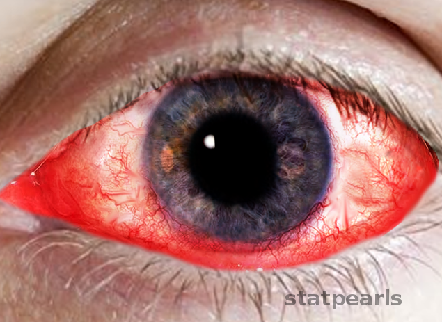
Bacterial Conjunctivitis
Contributed by O Chaigasame, MD
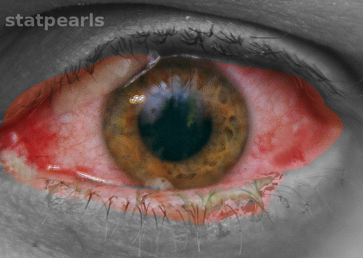
Follicular Conjunctivitis. Inflammation is noted with viral infections like herpes zoster, Epstein-Barr virus infection, infectious mononucleosis, and chlamydial infections, as well as in reaction to topical medications and molluscum contagiosum. Follicular conjunctivitis has been described in patients with COVID-19. The inferior and superior tarsal conjunctiva and the fornices show gray-white elevated swellings about 0.5 to 1 mm in diameter and have a velvety appearance.
Contributed by Prof. BCK Patel MD, FRCS

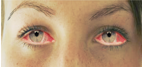
Allergic Conjunctivitis
Contributed by Katherine Humphreys
Shekhawat NS, Shtein RM, Blachley TS, Stein JD. Antibiotic Prescription Fills for Acute Conjunctivitis among Enrollees in a Large United States Managed Care Network. Ophthalmology. 2017 Aug:124(8):1099-1107. doi: 10.1016/j.ophtha.2017.04.034. Epub 2017 Jun 16 [PubMed PMID: 28624168]
Smith AF, Waycaster C. Estimate of the direct and indirect annual cost of bacterial conjunctivitis in the United States. BMC ophthalmology. 2009 Nov 25:9():13. doi: 10.1186/1471-2415-9-13. Epub 2009 Nov 25 [PubMed PMID: 19939250]
Alfonso SA, Fawley JD, Alexa Lu X. Conjunctivitis. Primary care. 2015 Sep:42(3):325-45. doi: 10.1016/j.pop.2015.05.001. Epub 2015 Jul 29 [PubMed PMID: 26319341]
de Laet C, Dionisi-Vici C, Leonard JV, McKiernan P, Mitchell G, Monti L, de Baulny HO, Pintos-Morell G, Spiekerkötter U. Recommendations for the management of tyrosinaemia type 1. Orphanet journal of rare diseases. 2013 Jan 11:8():8. doi: 10.1186/1750-1172-8-8. Epub 2013 Jan 11 [PubMed PMID: 23311542]
Sati A, Sangwan VS, Basu S. Porphyria: varied ocular manifestations and management. BMJ case reports. 2013 May 22:2013():. doi: 10.1136/bcr-2013-009496. Epub 2013 May 22 [PubMed PMID: 23704443]
Azari AA, Barney NP. Conjunctivitis: a systematic review of diagnosis and treatment. JAMA. 2013 Oct 23:310(16):1721-9. doi: 10.1001/jama.2013.280318. Epub [PubMed PMID: 24150468]
Høvding G. Acute bacterial conjunctivitis. Acta ophthalmologica. 2008 Feb:86(1):5-17 [PubMed PMID: 17970823]
Shields T, Sloane PD. A comparison of eye problems in primary care and ophthalmology practices. Family medicine. 1991 Sep-Oct:23(7):544-6 [PubMed PMID: 1936738]
Epling J. Bacterial conjunctivitis. BMJ clinical evidence. 2012 Feb 20:2012():. pii: 0704. Epub 2012 Feb 20 [PubMed PMID: 22348418]
Turaka K, Penne RB, Rapuano CJ, Ayres BD, Abazari A, Eagle RC Jr, Hammersmith KM. Giant fornix syndrome: a case series. Ophthalmic plastic and reconstructive surgery. 2012 Jan-Feb:28(1):4-6. doi: 10.1097/IOP.0b013e3182264440. Epub [PubMed PMID: 21862948]
Satpathy G, Behera HS, Ahmed NH. Chlamydial eye infections: Current perspectives. Indian journal of ophthalmology. 2017 Feb:65(2):97-102. doi: 10.4103/ijo.IJO_870_16. Epub [PubMed PMID: 28345563]
Bhosai SJ, Bailey RL, Gaynor BD, Lietman TM. Trachoma: an update on prevention, diagnosis, and treatment. Current opinion in ophthalmology. 2012 Jul:23(4):288-95. doi: 10.1097/ICU.0b013e32835438fc. Epub [PubMed PMID: 22569465]
Lansingh VC. Trachoma. BMJ clinical evidence. 2016 Feb 9:2016():. pii: 0706. Epub 2016 Feb 9 [PubMed PMID: 26860629]
Makker K, Nassar GN, Kaufman EJ. Neonatal Conjunctivitis. StatPearls. 2024 Jan:(): [PubMed PMID: 28722870]
Hoffman J. Adenovirus: ocular manifestations. Community eye health. 2020:33(108):73-75 [PubMed PMID: 32395030]
Giladi N, Herman J. Pharyngoconjunctival fever. Archives of disease in childhood. 1984 Dec:59(12):1182-3 [PubMed PMID: 6098226]
Meyer-Rüsenberg B, Loderstädt U, Richard G, Kaulfers PM, Gesser C. Epidemic keratoconjunctivitis: the current situation and recommendations for prevention and treatment. Deutsches Arzteblatt international. 2011 Jul:108(27):475-80. doi: 10.3238/arztebl.2011.0475. Epub 2011 Jul 8 [PubMed PMID: 21814523]
Wright PW, Strauss GH, Langford MP. Acute hemorrhagic conjunctivitis. American family physician. 1992 Jan:45(1):173-8 [PubMed PMID: 1309404]
Saleh D, Yarrarapu SNS, Sharma S. Herpes Simplex Type 1. StatPearls. 2024 Jan:(): [PubMed PMID: 29489260]
Yoser SL, Forster DJ, Rao NA. Systemic viral infections and their retinal and choroidal manifestations. Survey of ophthalmology. 1993 Mar-Apr:37(5):313-52 [PubMed PMID: 8387231]
Meza-Romero R, Navarrete-Dechent C, Downey C. Molluscum contagiosum: an update and review of new perspectives in etiology, diagnosis, and treatment. Clinical, cosmetic and investigational dermatology. 2019:12():373-381. doi: 10.2147/CCID.S187224. Epub 2019 May 30 [PubMed PMID: 31239742]
La Rosa M, Lionetti E, Reibaldi M, Russo A, Longo A, Leonardi S, Tomarchio S, Avitabile T, Reibaldi A. Allergic conjunctivitis: a comprehensive review of the literature. Italian journal of pediatrics. 2013 Mar 14:39():18. doi: 10.1186/1824-7288-39-18. Epub 2013 Mar 14 [PubMed PMID: 23497516]
Villegas BV, Benitez-Del-Castillo JM. Current Knowledge in Allergic Conjunctivitis. Turkish journal of ophthalmology. 2021 Feb 25:51(1):45-54. doi: 10.4274/tjo.galenos.2020.11456. Epub [PubMed PMID: 33631915]
Kaur K, Gurnani B. Vernal Keratoconjunctivitis. StatPearls. 2024 Jan:(): [PubMed PMID: 35015458]
Sobolewska B, Zierhut M. [Atopic keratoconjunctivitis]. Klinische Monatsblatter fur Augenheilkunde. 2014 May:231(5):512-7. doi: 10.1055/s-0034-1368396. Epub 2014 May 5 [PubMed PMID: 24799170]
Kari O, Saari KM, Haahtela T. [Non-allergic eosinophilic conjunctivitis]. Duodecim; laaketieteellinen aikakauskirja. 2010:126(10):1145-50 [PubMed PMID: 20597344]
Resano A, Esteve C, Fernández Benítez M. Allergic contact blepharoconjunctivitis due to phenylephrine eye drops. Journal of investigational allergology & clinical immunology. 1999 Jan-Feb:9(1):55-7 [PubMed PMID: 10212859]
Donshik PC, Ehlers WH, Ballow M. Giant papillary conjunctivitis. Immunology and allergy clinics of North America. 2008 Feb:28(1):83-103, vi. doi: 10.1016/j.iac.2007.11.001. Epub [PubMed PMID: 18282547]
Pokroy R, Marcovich A. Self-inflicted (factitious) conjunctivitis. Ophthalmology. 2003 Apr:110(4):790-5 [PubMed PMID: 12689904]
Schuster V, Seregard S. Ligneous conjunctivitis. Survey of ophthalmology. 2003 Jul-Aug:48(4):369-88 [PubMed PMID: 12850227]
Dixon MK, Dayton CL, Anstead GM. Parinaud's Oculoglandular Syndrome: A Case in an Adult with Flea-Borne Typhus and a Review. Tropical medicine and infectious disease. 2020 Jul 29:5(3):. doi: 10.3390/tropicalmed5030126. Epub 2020 Jul 29 [PubMed PMID: 32751142]
Lahoti S, Weiss M, Johnson DA, Kheirkhah A. Superior limbic keratoconjunctivitis: a comprehensive review. Survey of ophthalmology. 2022 Mar-Apr:67(2):331-341. doi: 10.1016/j.survophthal.2021.05.009. Epub 2021 May 30 [PubMed PMID: 34077767]
Sivaraman KR, Jivrajka RV, Soin K, Bouchard CS, Movahedan A, Shorter E, Jain S, Jacobs DS, Djalilian AR. Superior Limbic Keratoconjunctivitis-like Inflammation in Patients with Chronic Graft-Versus-Host Disease. The ocular surface. 2016 Jul:14(3):393-400. doi: 10.1016/j.jtos.2016.04.003. Epub 2016 May 12 [PubMed PMID: 27179980]
Xu HH, Werth VP, Parisi E, Sollecito TP. Mucous membrane pemphigoid. Dental clinics of North America. 2013 Oct:57(4):611-30. doi: 10.1016/j.cden.2013.07.003. Epub 2013 Aug 15 [PubMed PMID: 24034069]
Tolaymat L, Hall MR. Cicatricial Pemphigoid. StatPearls. 2024 Jan:(): [PubMed PMID: 30252376]
Oakley AM, Krishnamurthy K. Stevens-Johnson Syndrome. StatPearls. 2024 Jan:(): [PubMed PMID: 29083827]
Ramirez DA, Porco TC, Lietman TM, Keenan JD. Epidemiology of Conjunctivitis in US Emergency Departments. JAMA ophthalmology. 2017 Oct 1:135(10):1119-1121. doi: 10.1001/jamaophthalmol.2017.3319. Epub [PubMed PMID: 28910427]
Wong AH, Barg SS, Leung AK. Seasonal and perennial allergic conjunctivitis. Recent patents on inflammation & allergy drug discovery. 2014:8(2):139-53 [PubMed PMID: 25000933]
O'Callaghan RJ. The Pathogenesis of Staphylococcus aureus Eye Infections. Pathogens (Basel, Switzerland). 2018 Jan 10:7(1):. doi: 10.3390/pathogens7010009. Epub 2018 Jan 10 [PubMed PMID: 29320451]
Chu WK, Choi HL, Bhat AK, Jhanji V. Pterygium: new insights. Eye (London, England). 2020 Jun:34(6):1047-1050. doi: 10.1038/s41433-020-0786-3. Epub 2020 Feb 6 [PubMed PMID: 32029918]
Leung AKC, Hon KL, Wong AHC, Wong AS. Bacterial Conjunctivitis in Childhood: Etiology, Clinical Manifestations, Diagnosis, and Management. Recent patents on inflammation & allergy drug discovery. 2018:12(2):120-127. doi: 10.2174/1872213X12666180129165718. Epub [PubMed PMID: 29380707]
Azari AA, Arabi A. Conjunctivitis: A Systematic Review. Journal of ophthalmic & vision research. 2020 Jul-Sep:15(3):372-395. doi: 10.18502/jovr.v15i3.7456. Epub 2020 Aug 6 [PubMed PMID: 32864068]
Bielory L, Meltzer EO, Nichols KK, Melton R, Thomas RK, Bartlett JD. An algorithm for the management of allergic conjunctivitis. Allergy and asthma proceedings. 2013 Sep-Oct:34(5):408-20. doi: 10.2500/aap.2013.34.3695. Epub [PubMed PMID: 23998237]
Frary J, Petersen PT, Pareek M. Hutchinson's sign of ophthalmic zoster. Clinical case reports. 2020 Jan:8(1):219-220. doi: 10.1002/ccr3.2596. Epub 2019 Dec 11 [PubMed PMID: 31998523]
Leibowitz HM. The red eye. The New England journal of medicine. 2000 Aug 3:343(5):345-51 [PubMed PMID: 10922425]
Mahmood AR, Narang AT. Diagnosis and management of the acute red eye. Emergency medicine clinics of North America. 2008 Feb:26(1):35-55, vi. doi: 10.1016/j.emc.2007.10.002. Epub [PubMed PMID: 18249256]
Puri LR, Shrestha GB, Shah DN, Chaudhary M, Thakur A. Ocular manifestations in herpes zoster ophthalmicus. Nepalese journal of ophthalmology : a biannual peer-reviewed academic journal of the Nepal Ophthalmic Society : NEPJOPH. 2011 Jul-Dec:3(2):165-71. doi: 10.3126/nepjoph.v3i2.5271. Epub [PubMed PMID: 21876592]
Liesegang TJ. Herpes zoster ophthalmicus natural history, risk factors, clinical presentation, and morbidity. Ophthalmology. 2008 Feb:115(2 Suppl):S3-12. doi: 10.1016/j.ophtha.2007.10.009. Epub [PubMed PMID: 18243930]
Sambursky R, Tauber S, Schirra F, Kozich K, Davidson R, Cohen EJ. The RPS adeno detector for diagnosing adenoviral conjunctivitis. Ophthalmology. 2006 Oct:113(10):1758-64 [PubMed PMID: 17011956]
Alfonso EC, Cantu-Dibildox J, Munir WM, Miller D, O'Brien TP, Karp CL, Yoo SH, Forster RK, Culbertson WW, Donaldson K, Rodila J, Lee Y. Insurgence of Fusarium keratitis associated with contact lens wear. Archives of ophthalmology (Chicago, Ill. : 1960). 2006 Jul:124(7):941-7 [PubMed PMID: 16769827]
Mimura T, Usui T, Yamagami S, Miyai T, Amano S. Relation between total tear IgE and severity of acute seasonal allergic conjunctivitis. Current eye research. 2012 Oct:37(10):864-70. doi: 10.3109/02713683.2012.689069. Epub 2012 May 17 [PubMed PMID: 22595024]
Borkar DS, Acharya NR, Leong C, Lalitha P, Srinivasan M, Oldenburg CE, Cevallos V, Lietman TM, Evans DJ, Fleiszig SM. Cytotoxic clinical isolates of Pseudomonas aeruginosa identified during the Steroids for Corneal Ulcers Trial show elevated resistance to fluoroquinolones. BMC ophthalmology. 2014 Apr 24:14():54. doi: 10.1186/1471-2415-14-54. Epub 2014 Apr 24 [PubMed PMID: 24761794]
Monnerat N, Bossart W, Thiel MA. [Povidone-iodine for treatment of adenoviral conjunctivitis: an in vitro study]. Klinische Monatsblatter fur Augenheilkunde. 2006 May:223(5):349-52 [PubMed PMID: 16705502]
Wilkins MR, Khan S, Bunce C, Khawaja A, Siriwardena D, Larkin DF. A randomised placebo-controlled trial of topical steroid in presumed viral conjunctivitis. The British journal of ophthalmology. 2011 Sep:95(9):1299-303. doi: 10.1136/bjo.2010.188623. Epub 2011 Jan 20 [PubMed PMID: 21252084]
Srinivasan M, Mascarenhas J, Rajaraman R, Ravindran M, Lalitha P, O'Brien KS, Glidden DV, Ray KJ, Oldenburg CE, Zegans ME, Whitcher JP, McLeod SD, Porco TC, Lietman TM, Acharya NR, Steroids for Corneal Ulcers Trial Group. The steroids for corneal ulcers trial (SCUT): secondary 12-month clinical outcomes of a randomized controlled trial. American journal of ophthalmology. 2014 Feb:157(2):327-333.e3. doi: 10.1016/j.ajo.2013.09.025. Epub 2013 Oct 1 [PubMed PMID: 24315294]
Srinivasan M, Mascarenhas J, Rajaraman R, Ravindran M, Lalitha P, Ray KJ, Zegans ME, Acharya NR, Lietman TM, Keenan JD, Steroids for Corneal Ulcers Trial Group. Visual recovery in treated bacterial keratitis. Ophthalmology. 2014 Jun:121(6):1310-1. doi: 10.1016/j.ophtha.2013.12.041. Epub 2014 Mar 5 [PubMed PMID: 24612976]
Wright C, Tawfik MA, Waisbourd M, Katz LJ. Primary angle-closure glaucoma: an update. Acta ophthalmologica. 2016 May:94(3):217-25. doi: 10.1111/aos.12784. Epub 2015 Jun 27 [PubMed PMID: 26119516]
Austin A, Lietman T, Rose-Nussbaumer J. Update on the Management of Infectious Keratitis. Ophthalmology. 2017 Nov:124(11):1678-1689. doi: 10.1016/j.ophtha.2017.05.012. Epub 2017 Sep 21 [PubMed PMID: 28942073]
Sainz de la Maza M, Molina N, Gonzalez-Gonzalez LA, Doctor PP, Tauber J, Foster CS. Clinical characteristics of a large cohort of patients with scleritis and episcleritis. Ophthalmology. 2012 Jan:119(1):43-50. doi: 10.1016/j.ophtha.2011.07.013. Epub 2011 Oct 2 [PubMed PMID: 21963265]
Brandt MT, Haug RH. Traumatic hyphema: a comprehensive review. Journal of oral and maxillofacial surgery : official journal of the American Association of Oral and Maxillofacial Surgeons. 2001 Dec:59(12):1462-70 [PubMed PMID: 11732035]
Ullman S, Roussel TJ, Culbertson WW, Forster RK, Alfonso E, Mendelsohn AD, Heidemann DG, Holland SP. Neisseria gonorrhoeae keratoconjunctivitis. Ophthalmology. 1987 May:94(5):525-31 [PubMed PMID: 3601368]
Schachter J, Lum L, Gooding CA, Ostler B. Pneumonitis following inclusion blennorrhea. The Journal of pediatrics. 1975 Nov:87(5):779-80 [PubMed PMID: 1185349]
Wood M. Conjunctivitis: diagnosis and management. Community eye health. 1999:12(30):19-20 [PubMed PMID: 17491982]