Continuing Education Activity
Scalp reconstruction procedures are done for medical or cosmetic reasons. Scalp reconstruction techniques share similarities to plastic surgery techniques, including the use of free flaps for very large defects. This activity discusses the indications, contraindications, and complications of scalp reconstruction and highlights the role of the interprofessional team in the pre and postoperative management of patients undergoing this procedure.
Objectives:
Characterize the techniques used for scalp reconstruction.
Enumerate the indications for scalp reconstruction.
State the complications of scalp reconstruction.
Outline the importance of collaboration and coordination amongst the interprofessional team in optimizing the outcomes for patients undergoing scalp reconstruction.
Introduction
Scalp reconstruction procedures range from those for medical indications to those for cosmetic reasons. Koss et al. made a detailed analysis of trauma-related scalping injuries and their management.[1] The history of scalp reconstruction techniques mirrors that of plastic surgery techniques, including the use of free flaps for very large defects.[2][3][4][5]
More typical procedures include incisional and excisional biopsies, excision of benign and malignant tumors, and scalp reduction surgery. Benign lesions of the scalp that are excisable include epidermoid cysts, nevus sebaceus, and blue and melanocytic nevi. Malignant lesions that typically undergo excision include basal and squamous cell carcinoma, Bowen disease, Merkel cell carcinoma, and malignant melanoma. Wounds created from these procedures may be small and superficial and amenable to primary closure but often are large, deep (down to calvaria), and extensive, needing more complex closure and covering.
Scalp reconstruction surgery has been used in the past to treat alopecia but is rarely used nowadays due to advances in hair transplantation. Cosmetic indications are not in the remit of this article and will not be part of the discussion here.
Reconstruction of the scalp follows the reconstructive ladder of any other plastic surgical procedure: granulation (secondary intention); primary closure; advancement flap; rotational flap; use of split-thickness skin graft (STSG); or full-thickness skin graft (FTSG); and free flaps. The selection of one or a combination of methods depends on anatomical (skin laxity, wound depth, location) and patient-related factors (smoker, wound care, general health).
Traumatic scalp avulsive injuries can occur and be devastating. These can be addressed as above with other defects or require potentially significantly more extensive surgeries both in regards to the number and complexity.[6][7][8]
Anatomy and Physiology
Layers
The layers can easily be remembered by the mnemonic SCALP: S (Skin); C (sub-Cutaneous tissue); A (Aponeurotic layer); L (Loose areolar tissue); P (Pericranium). It is helpful to remember that the aponeurotic layer is also referred to as the galea and the loose areolar tissue is also known as just loose connective tissue.
The skin of the scalp is the same thickness of the rest of the body making it a potentially useful donor site for skin grafts. The vessels, lymphatics and nerves course through the subcutaneous layer.[9] The galea aponeurosis is the strength layer of the scalp and is contiguous with the frontalis muscle anteriorly, the occipitalis muscle posteriorly and the temporoparietal fascia laterally. The scalp divides into sections depending on the underlying skull anatomy. These sections are the right and left frontal, parietal, vertex and occipital scalp. The scalp represents a continuation of the face with much denser hair-bearing follicles and pilosebaceous units. Compared to the face, this can lead to faster epithelization and a more esthetic ability to hide scarring.
The majority of scalp mobility is in the layer of the loose areolar tissue layer. During dissection, this pocket can be easily dissected and is a relatively safe plane, and nerves lie superior to this plane and vasculature generally lying superiorly and in the layer of the dense connective tissue. An important area where this mobility lessens is the superior temporal septum in the lateral frontal region. This thick ligamentous area is also known as the conjoined tendon or sometimes the conjoint tendon. It represents a confluence of the frontal periosteum, the deep temporal fascia, and the temporoparietal fascia. To gain tissue from these areas, the ligamentous adhesions must be divided, for example as in the endoscopic browlift.[10]
The best replacement for scalp tissue is scalp tissue. Identical hair-bearing qualities of the scalp are difficult to reproduce. Hair transplantation can be used at a later time to reestablish hairlines or camouflage scars.
Vascular supply
Five large arteries perfuse the scalp bilaterally. Flaps rely on these arteries either via random pattern or axial blood supply. The scalp can be broadly divided into four different vascular territories[9]:
- Anterior: supratrochlear and supraorbital artery (terminal branches of the internal carotid)
- Lateral: superficial temporal artery
- Posterior: occipital artery
- Posterolateral: posterior auricular artery
The veins anastomose with each other and enter the diploic veins of the skull bones and the dural sinuses. The veins accompany the arteries and as such generally have the same names as above.
Innervation
The scalp has innervation from branches of the three divisions of the trigeminal nerve (CN V), cervical spinal nerve and branches from the cervical plexus. From anterior to posterior the nerves are:
- Supratrochlear nerve and supraorbital nerve (V1)
- Zygomaticotemporal nerve (V2)
- Auriculotemporal nerve (V3)
- Lesser occipital nerve (V3)
- Greater occipital nerve (C2, C3)
- Occipital nerve (C3)
Lymphatic drainage
The skin from the frontal part of the scalp drains into the parotid nodes, submandibular nodes, and deep cervical lymph nodes. The skin from the posterior portion drains into the posterior auricular nodes and occipital lymph nodes. This fact is important as malignancies can metastasize to these lymph nodes.
Indications
Granulation (secondary intention)
Generally speaking, the more superficial a wound is, the more appropriate granulation is a suitable method of reconstruction. Superficial wounds tend to heal within a few weeks, while wounds that extend into the aponeurosis or deeper can take 6 to 8 weeks to heal. Granulation method is usable in any location of the scalp. Good candidates include patients with poor wound healing (smokers, diabetics, previous radiotherapy to the region) and those patients who want a minimal amount of surgery. This method is commonly aided by a negative pressure wound-vac system to increase favorable results in the setting of a larger defect.[11][12][13][14]
Primary closure
The ideal wound to do primary closure is any full thickness wound where the edges can be opposed with minimal tension. Primary closure method can be used in any location of the scalp. Good candidates include any patient who has wounds that will close easily with this method with minimal surgical time.
Advancement Flap
The ideal wound for an advancement flap is any full thickness wound where the edges are not closable with primary closure. The ideal location on the scalp is where there is redundant local tissue and where hiding the incision lines within cosmetic subunits is possible. Good candidates are those patients who can undergo the additional surgical time required.
Rotational Flap
The ideal wound for a rotational flap is any full thickness wound where the edges are not closable with primary closure. The ideal location on the scalp is where there is redundant local tissue and where the incision lines are hidable within cosmetic subunits. Good candidates are those patients who can undergo the additional surgical time required.
Transpositional flap
Similar to indications with advancement and rotational flaps, but the flap moves laterally over normal intervening skin for placement into the defect. Good candidates are those patients who can undergo the additional surgical time required.
Skin Grafts
- Split-thickness skin grafts (STSG)
The ideal wound for an STSG is any full-thickness scalp wound where the wound will not easily close with primary closure or a flap. Good candidates for STSG are those patients who have large scalp defects which are devoid of a vascular bed to have a full-thickness skin graft and those who are willing to have additional surgical procedures.[15]
- Full-thickness skin grafts (FTSG)
The ideal wound for an FTSG is any full-thickness scalp wound where the wound will not easily close with primary closure or a flap. Good candidates for FTSG are those patients who have large scalp defects which have a good vascular bed, and those who are willing to have additional surgical procedures.
Some have proposed artificial dermis as an alternative to using donor skin from the patient. Its use is not widespread.[16]
Free Flaps
Free Flaps are sometimes the best reconstruction solution for very large scalp defects. This method is the most complex in the reconstruction ladder. There are rarely seen in routine dermatological practice and not concentrated on in this article.
Allograft
There are a variety of available products that can be used as part of a measure of scalp reconstruction. Allografts can be used to prepare a defect for further reconstruction from an autogenous graft or flap, or as a single stage reconstruction.[7][17]
Contraindications
There are no specific contraindications for scalp reconstruction. Previous radiotherapy to the area may prevent the graft from taking, due to poor vascularity. The patient's health may prevent a general anesthetic which can limit what reconstruction is possible. Graft failure may require debridements of non-vital tissues.
Equipment
- Clinic or operating room setting
- Local anesthetic
- Standard surgical excision equipment (fine plastics tray)
- Dermatome
- Diathermy
- Suction
- Sutures (absorbable and non-absorbable)
- Pressure bolster
- Wound dressings
Personnel
- ENT surgeon
- Plastic surgeon
- Oral and maxillofacial surgeon
- Head and neck advanced nurse specialist
- Other head and neck specialist physician
Preparation
The resulting scalp defect should merit consideration and planning in advance, especially if a dermatome and general anesthetic are required. Well, designed flaps require preservation of the natural hairline, incorporation of major vascular pedicles, and closure without tension. There should be a complete workup done by the treating team including full medical history review, clinical examination, often photographs for the record (with appropriate consent and permission), and a review and discussion of the treatment options, risks, benefits, alternatives, and procedures.
Technique or Treatment
Granulation (secondary intention)
This technique requires moist wound care. Application of moist, emollient, non-stick dressings to the wound. The dressings need regular changing. The use of antibiotic creams/ointments should be avoided due to the risk of allergic contact dermatitis, and instead, gentle cleaning and bathing of the area are advisable. The healing process normally takes anywhere from 2 to 8 weeks to occur depending on the depth of the wound. Beyond the scope of this article is management through a negative pressure wound-vac system.
Primary closure
Layered closure with appropriate wound tension. The wound should be elliptical with a length-to-width ratio of 3 to 1. Where possible the wound should be along relaxed skin tension lines and should be undermined to reduce wound closure tension and facilitate wound eversion. Appropriate strength and sized sutures should be placed both deep and superficially. Sutures should be removed at 7 to 10 days if using non-resorbable sutures. Proper wound care is necessary.
Advancement Flap
Unilateral advancement flaps are normally used in small wounds. Use the neighboring area with the most redundant skin and try and respect the cosmetic subunits. A pedicle feeds the mobilized tissue; the ratio should not exceed 3 to 1. The wound edges may develop 'dog ears' that need correction. There are many types of advancement flaps including island pedicle, V-Y,[18] single and bi-pedicle advancement flaps. The same rules for sutures and post-operative wound care applications. Flap designs and more advanced techniques are beyond the scope of this article.
Rotational Flap
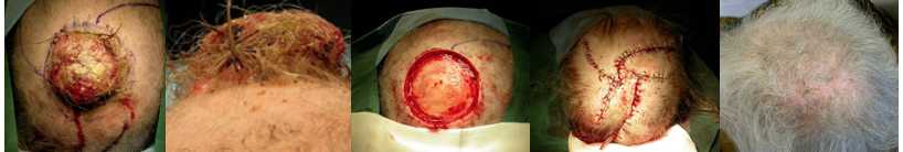
Rotational flaps are so named as the primary movement is to rotate nearby redundant skin into the skin defect. These flaps can be single, bilateral or multiple types. Because the neighboring redundant skin rotates into the defect generally the shape of this flap is an arc of a circle (see figures below a-b = pre-op, c-d = intra-op, e = 4 months post-op). A pedicle feeds the flap and not exceeding the 3 to 1 rule still applies. This technique can allow multiple rotational flaps to be raised creating a local tissue reservoir. The same rules for sutures and post-operative wound care applications. Flap designs and more advanced techniques are beyond the scope of this article.
Transpositional flap
Transpositional flaps have this name because the movement is the mobilize non-adjacent skin and transposition the skin over intervening skin into the defect. The most commonly used flaps are the rhomboid and bilobed flaps.[19] The same rules for sutures and post-operative wound care applications. Flap designs and more advanced techniques are beyond the scope of this article.
Skin Grafts
- Split-thickness skin grafts (STSG)
This variety is a graft of epidermis or partial thickness dermis. Usually, it merits consideration when the defect is large and other forms of reconstruction are not suitable. STSG harvesting is normally with a manual or powered dermatome under local or general anesthetic. Typical thickness is of the range 0.30 to 0.45mm.
STSG usually have better take success rates, and can generally cover a larger surface than FTSG, due to the STSG being thin and have reduced metabolic requirements. Meshing can be performed to the graft to allow greater wound coverage and fluid egress, helping prevent a fluid build up that can inhibit imbibition. STSG leaves a donor site that needs a dressing but no surgical closure. The main disadvantage of STSG is that they often leave a white, patch-like appearance, which can have a poor cosmetic match for the grafted scalp area. See FTSG for improving the graft take rate.
- Full-thickness skin grafts (FTSG)
FTSG are grafts of the epidermis and complete dermis with or without subcutaneous fat. Harvesting comes from an appropriate area using routine surgical equipment, e.g., neck skin (dermatome not required). Commonly a precise template is made of the wound, and the donor site will require primary closure. FTSG is thicker and has higher metabolic demand; thus they have a higher rate of failure, though that is often dependant on the surgeon and their particular skill set as well as the patient and their co-morbidities. Persistent oozing or bleeding from the wound can lead to hematoma or seroma which can cause the graft not to take. Manually fenestrating the graft can allow the blood/serum to drain. A bolster dressing is applied to the wound with silk tie-over sutures to encourage the graft to sit flush and to take. Over time plasmatic imbibition, revascularization, and contraction occur.
Complications
- Infection
- Bleeding
- Scar
- Poor cosmesis
- Wound breakdown
- Flap failure
- Graft failure (hematoma/seroma)
- Need for further surgery
Clinical Significance
From the management of total scalp avulsion injuries to advancements in microsurgery and free tissue transfer, the techniques used in scalp reconstruction have mirrored advances in plastic surgery.[20] However, most scalp defects can undergo management by local tissue rotational or advancement flaps. Well, designed flaps require preservation of the natural hairline, incorporation of major vascular pedicles, and closure without tension.
Enhancing Healthcare Team Outcomes
Pre-operative planning and proper intra-operative execution is the key to successful scalp reconstruction. The surgeon should have good knowledge of scalp anatomy, hair physiology, skin biomechanics, and available local tissue to allows for excellent aesthetic reconstruction. Nurses who look after these patients should be fully aware of the potential complications that can occur in the post-operative period. At the first sign of infection, vascular compromise or graft necrosis, the plastic surgeon should be notified. All this points to the necessity of an interprofessional team approach, to managing these cases. This interprofessional team includes surgeons, surgical nursing, and pharmacists in the event of post-procedure infection or prophylaxis. Such an approach best guarantees an optimal patient outcome.