Neuroanatomy, Pterygopalatine Ganglion


Introduction

The pterygopalatine ganglion (PPG) is one of four parasympathetic ganglia located within the head region, existing as a bilateral pair. The pterygopalatine ganglion may also be referred to as the sphenopalatine ganglion, Meckel's ganglion, or the nasal ganglion. The pterygopalatine ganglion is responsible for housing the post-ganglionic parasympathetic neuronal cell bodies, in addition to acting as a conduit for post-ganglionic sympathetic and sensory axonal fibers. The fibers that arise from the pterygopalatine ganglion regulate secretomotor functions to and provide sensation from various structures that include: the lacrimal glands, the mucous membranes of the oropharynx, nasopharynx, nasal cavity, and upper portion of the oral cavity. The fibers from the pterygopalatine ganglion are also responsible for providing innervation to the cerebral and meningeal blood vessels.[1][2][3] The pterygopalatine ganglion is a structure that is morphologically formed during the third trimester of fetal life,[2][4] with its neurons derived from Schwann cell precursors.[5] A group of headache disorders referred to as trigeminal autonomic cephalalgias (TACs), which include cluster headaches, are thought to be influenced by the pterygopalatine ganglion. There is strong evidence to support the usage of pterygopalatine ganglion blockade, radiofrequency ablation, or neurostimulation of the pterygopalatine ganglion to relieve cluster headaches.[6] The utilization of pterygopalatine ganglion blockade for the treatment of migraines, trigeminal neuralgia, and other conditions has also demonstrated varying degrees of success.[6]

Structure and Function

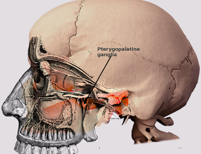
Located within the pterygopalatine fossa (PPF), the pterygopalatine ganglion comprises the largest parasympathetic ganglion and is one of four ganglia located within the head region. The pterygopalatine ganglion includes an assortment of parasympathetic, sympathetic, and somatosensory nerve fibers.[1][2][4] The pterygopalatine ganglia exist as a bilateral pair, with each pterygopalatine ganglion located posterior to the lateral insertion of the middle nasal concha, covered by a thin layer of mucosa in the pterygopalatine fossa.[1][4][7] The pterygopalatine fossa and pterygopalatine ganglion are bordered by the maxillary sinus anteriorly; the root of the pterygoid process posteriorly; the perpendicular plate of the palatine bone medially; and the pterygomaxillary fissure laterally.[8]  

The pterygopalatine ganglion houses post-ganglionic parasympathetic neurons that receive synaptic transmissions from pre-ganglionic parasympathetic fibers that arise from the superior salivatory nucleus, located within the pons of the brainstem.[1][8] These pre-ganglionic, efferent parasympathetic fibers form the nervus intermedius (intermediate nerve), a constituent of cranial nerve VII (the facial nerve), pass through the geniculate ganglion and form the greater petrosal nerve. The greater petrosal nerve merges with the deep petrosal nerve, which carries post-ganglionic sympathetic fibers, forming the nerve of the pterygoid canal, AKA the Vidian nerve. The Vidian nerve enters the posterior region of the pterygopalatine fossa to unite with the pterygopalatine ganglion. Within the pterygopalatine ganglion, the pre-ganglionic parasympathetic fibers synapse with post-ganglionic fibers. The post-ganglionic parasympathetic fibers will then traverse through the ophthalmic and maxillary divisions of the trigeminal nerve to provide secretomotor signals to the lacrimal gland, mucous membranes of the nasal cavity, nasopharynx, oropharynx, and upper oral cavity. The post-ganglionic parasympathetic fibers also provide parasympathetic signals to the meningeal and cerebral blood vessels[1][2][9][10][11]  

The source of the sympathetic neurons associated with the pterygopalatine ganglion originates from pre-ganglionic sympathetic neurons located in the intermediolateral cell column at the level of the first thoracic vertebra.[8] The pre-ganglionic fibers project axons to the superior cervical ganglia, where the post-ganglionic sympathetic neurons are situated. The post-ganglionic sympathetic neuronal axons travel through the internal carotid plexus, eventually forming the deep petrosal nerve. As stated above, the deep petrosal nerve connects with the greater petrosal nerve to form the nerve of the pterygoid canal, eventually making its way to the pterygopalatine ganglion. The post-ganglionic sympathetic fibers pass through the pterygopalatine ganglion to provide sympathetic innervation to the nasal and pharyngeal mucosa as well as the lacrimal gland.[1][12]

Somatic sensory fibers, primarily from the maxillary division of the trigeminal nerve, are another set of axons that run through the pterygopalatine ganglion without synapsing at the pterygopalatine ganglion itself. Following the maxillary nerve distal to its branches from the trigeminal nerve, it enters the posterior region of the pterygopalatine fossa via the foramen rotundum. Within the pterygopalatine fossa, the maxillary nerve gives off somatic sensory branches that pass through the pterygopalatine ganglion to predominantly form the greater and lesser palatine nerves.[1][2][8][13] The general somatic sensation from the mucosa, gingiva, and hard palate of the oral cavity is conducted through the greater palatine nerve, while the lesser palatine nerve transmits general somatic sensation signals from the tonsils, uvula, and soft palate.

Embryology

The neurons of the pterygopalatine ganglion, along with those of the otic, submandibular, and ciliary ganglia, are derived from Schwann cell precursors that migrate via associated pre-ganglionic axons.[5][14][15] These Schwann cell precursors differentiate into peripheral neurons once they reach the final location of the adult ganglionic structures that they form. The pterygopalatine ganglion completes its morphological development and establishes its neuronal connections by the third trimester of fetal life.[2][4]

Blood Supply and Lymphatics

Current evidence suggests that the pterygopalatine ganglion is responsible for providing innervation to the anterior cerebral and meningeal blood vessels through the actions of several neurotransmitters that include: acetylcholine, nitric oxide, and vasoactive intestinal peptide.[10][16][17][18][19] The current thought is that the release of these neurotransmitters has implications in the pathogenesis of various headache types.[20][21][22]

Nerves

As described in more detail above, multiple nerves converge at the pterygopalatine ganglion. This section describes which nerves arise from the pterygopalatine ganglion and the various structures innervated by these nerves. The pterygopalatine ganglion gives rise to the following nerves: the nasopalatine nerve, the lesser palatine nerve, the greater palatine nerve, the posterior superior lateral nasal nerves, the posterior inferior lateral nasal nerves, and pharyngeal nerve.

The posterior inferior lateral nasal nerves branch off from the greater palatine nerve in the pterygopalatine canal to innervate the medial and inferior nasal meatuses, along with the inferior nasal concha. The posterior superior lateral nasal nerves directly project from the pterygopalatine ganglion to go through the sphenopalatine foramen to enter the nasal cavity posteriorly to innervate the middle and superior nasal conchae, the posterior aspect of the nasal septum, and the posterior ethmoidal air cells.[2]

The maxillary nerve, with its purely sensory axonal fibers, serves as a pathway for the post-ganglionic sympathetic and parasympathetic fibers of the pterygopalatine ganglion. The autonomic fibers of the pterygopalatine ganglion traverse with branches of the maxillary nerve that include the zygomatic, posterior superior alveolar, and infra-orbital nerves. The post-ganglionic sympathetic and parasympathetic fibers that travel with the zygomatic nerve are especially important because these nerves are what ultimately provide innervation to the lacrimal gland.[2][8]

The nasopalatine nerve exits the pterygopalatine fossa from the sphenopalatine foramen and emerges within the posterior aspect of the nasal cavity. The nasopalatine nerve continues within the nasal cavity along the nasal septum in an anteroinferior direction, traversing a groove in the vomer and providing branches along its trajectory. The nasopalatine nerve ultimately passes through the incisive canal and fossa to innervate the gingiva and mucosa of the anterior hard palate, just posterior to the maxillary incisors and canine teeth.

The greater palatine nerve innervates the remaining portion of the hard palate’s gingiva and mucosa. The lesser palatine nerve is responsible for providing innervation to the soft palate, uvula, and tonsil. The soft palate communicates the sensation of taste from the lesser palatine nerve to the greater petrosal nerve.

Finally, the pharyngeal nerve, which branches off of the pterygopalatine ganglion, exits the pterygopalatine fossa via the palatovaginal canal. The pharyngeal nerve is responsible for providing innervation to the mucosa and glands of the nasopharynx.

Muscles

Neurons that originate from the pterygopalatine ganglion do not typically innervate muscular tissue.

Physiologic Variants

Rusu et al. in 2009 had analyzed the pterygopalatine ganglion of 20 adult human specimens, to which four distinct morphological forms of the pterygopalatine ganglion detailing whether the pterygopalatine ganglion was a single or partitioned unit as well as where the Vidian nerve entered the pterygopalatine ganglion, were described as follows:[4]  

  • Type A – a partitioned structure with the superior partition receiving the Vidian nerve and constituting 10% of observed specimens
  • Type B – a single structure with the superior part (base) of the pterygopalatine ganglion receiving the Vidian nerve and constituting 55% of observed specimens
  • Type C – a single structure with the inferior part (tip) of the pterygopalatine ganglion receiving the Vidian nerve and constituting 15% of observed specimens
  • Type D – a partitioned structure with the inferior partition receiving the Vidian nerve and constituting 20% of observed specimens

Various sources of the literature have detailed the shape of the pterygopalatine ganglion to be triangular,[23] conical,[4] or piriform (pear-shaped), among others.[24][25]

Clinical Significance

The pterygopalatine ganglion is theorized to be a component for a group of headache disorders classified as trigeminal autonomic cephalalgias (TACs) which present as unilateral headaches with ipsilateral autonomic features (lacrimation, rhinorrhea, nasal congestion, eyelid edema, and ptosis).[26][27] The following conditions currently classify as TACs: cluster headaches, paroxysmal hemicrania, short-lasting unilateral neuralgiform headache attacks with conjunctival injection and tearing (SUNCT), short-lasting unilateral neuralgiform headache attacks with cranial autonomic symptoms (SUNA), and hemicrania continua.[28] A proposed mechanism for the TACs occurs through a trigeminal autonomic reflex, in which afferent signals from the dura mater and cranial vessels get relayed through axonal fibers of the trigeminal ganglion to the trigeminal-cervical complex.[26][27] These signals then stimulate the superior salivatory nucleus, resulting in the parasympathetic activity that is mediated by the pterygopalatine ganglion and its constituents (lacrimation, rhinorrhea, nasal congestion, and periorbital edema) and secondary inhibition of sympathetic signals (ptosis) or a combination of both (miosis).   

 A systematic review conducted by Ho et al. found that there is substantial evidence to support the targeting of the pterygopalatine ganglion via pterygopalatine ganglion blockade, radiofrequency ablation, or neurostimulation for the treatment of cluster headaches.[6] Additionally, their analysis showed limited evidence to support the usage of pterygopalatine ganglion blockade for the treatment of trigeminal neuralgia and migraines; for the reduction of analgesics used following endoscopic sinus surgery; and for the reduction of pain associated with nasal packing removal post-nasal operations.[6] Ho et al. detail other studies that have investigated therapeutic interventions aimed at the pterygopalatine ganglion to treat other conditions, such as postherpetic neuralgia and post-dural puncture headache, but these studies lack sufficient evidence or dismissed previous claims for the usage of such interventions.[6] In conclusion, Ho et al. argue that larger, double-blinded, randomized-controlled studies pertaining to therapeutic interventions targeting the pterygopalatine ganglion need to be conducted to establish the validity of these interventions in the treatment of cluster headaches, migraines, trigeminal neuralgia, and other pathologies of the head region.[6]


(Click Image to Enlarge)
Pterygopalatine ganglia
Image courtesy O.Chaigasame
Details

Editor:

Thomas McNary

Updated:

7/24/2023 11:50:10 PM

References


[1]

Robbins MS, Robertson CE, Kaplan E, Ailani J, Charleston L 4th, Kuruvilla D, Blumenfeld A, Berliner R, Rosen NL, Duarte R, Vidwan J, Halker RB, Gill N, Ashkenazi A. The Sphenopalatine Ganglion: Anatomy, Pathophysiology, and Therapeutic Targeting in Headache. Headache. 2016 Feb:56(2):240-58. doi: 10.1111/head.12729. Epub 2015 Nov 30     [PubMed PMID: 26615983]


[2]

Piagkou M, Demesticha T, Troupis T, Vlasis K, Skandalakis P, Makri A, Mazarakis A, Lappas D, Piagkos G, Johnson EO. The pterygopalatine ganglion and its role in various pain syndromes: from anatomy to clinical practice. Pain practice : the official journal of World Institute of Pain. 2012 Jun:12(5):399-412. doi: 10.1111/j.1533-2500.2011.00507.x. Epub 2011 Sep 29     [PubMed PMID: 21956040]


[3]

Tolba R, Weiss AL, Denis DJ. Sphenopalatine Ganglion Block and Radiofrequency Ablation: Technical Notes and Efficacy. The Ochsner journal. 2019 Spring:19(1):32-37. doi: 10.31486/toj.18.0163. Epub     [PubMed PMID: 30983899]


[4]

Rusu MC, Pop F, Curcă GC, Podoleanu L, Voinea LM. The pterygopalatine ganglion in humans: a morphological study. Annals of anatomy = Anatomischer Anzeiger : official organ of the Anatomische Gesellschaft. 2009 Apr:191(2):196-202. doi: 10.1016/j.aanat.2008.09.008. Epub 2008 Nov 14     [PubMed PMID: 19124232]


[5]

Adameyko I, Fried K. The Nervous System Orchestrates and Integrates Craniofacial Development: A Review. Frontiers in physiology. 2016:7():49. doi: 10.3389/fphys.2016.00049. Epub 2016 Feb 19     [PubMed PMID: 26924989]


[6]

Ho KWD, Przkora R, Kumar S. Sphenopalatine ganglion: block, radiofrequency ablation and neurostimulation - a systematic review. The journal of headache and pain. 2017 Dec 28:18(1):118. doi: 10.1186/s10194-017-0826-y. Epub 2017 Dec 28     [PubMed PMID: 29285576]

Level 1 (high-level) evidence

[7]

Candido KD, Massey ST, Sauer R, Darabad RR, Knezevic NN. A novel revision to the classical transnasal topical sphenopalatine ganglion block for the treatment of headache and facial pain. Pain physician. 2013 Nov-Dec:16(6):E769-78     [PubMed PMID: 24284858]


[8]

Khonsary SA, Ma Q, Villablanca P, Emerson J, Malkasian D. Clinical functional anatomy of the pterygopalatine ganglion, cephalgia and related dysautonomias: A review. Surgical neurology international. 2013:4(Suppl 6):S422-8. doi: 10.4103/2152-7806.121628. Epub 2013 Nov 20     [PubMed PMID: 24349865]


[9]

Jürgens TP, May A. Role of sphenopalatine ganglion stimulation in cluster headache. Current pain and headache reports. 2014 Jul:18(7):433. doi: 10.1007/s11916-014-0433-4. Epub     [PubMed PMID: 24880803]


[10]

Elsås T, Edvinsson L, Sundler F, Uddman R. Neuronal pathways to the rat conjunctiva revealed by retrograde tracing and immunocytochemistry. Experimental eye research. 1994 Jan:58(1):117-26     [PubMed PMID: 8157097]


[11]

Tepper SJ, Caparso A. Sphenopalatine Ganglion (SPG): Stimulation Mechanism, Safety, and Efficacy. Headache. 2017 Apr:57 Suppl 1():14-28. doi: 10.1111/head.13035. Epub     [PubMed PMID: 28387016]


[12]

Rusu MC, Pop F. The anatomy of the sympathetic pathway through the pterygopalatine fossa in humans. Annals of anatomy = Anatomischer Anzeiger : official organ of the Anatomische Gesellschaft. 2010 Feb 20:192(1):17-22. doi: 10.1016/j.aanat.2009.10.003. Epub 2009 Nov 5     [PubMed PMID: 19939656]


[13]

Lovasova K, Sulla IJ, Bolekova A, Sulla I, Kluchova D. Anatomical study of the roots of cranial parasympathetic ganglia: a contribution to medical education. Annals of anatomy = Anatomischer Anzeiger : official organ of the Anatomische Gesellschaft. 2013 May:195(3):205-11. doi: 10.1016/j.aanat.2013.01.011. Epub 2013 Feb 4     [PubMed PMID: 23433588]


[14]

Espinosa-Medina I, Outin E, Picard CA, Chettouh Z, Dymecki S, Consalez GG, Coppola E, Brunet JF. Neurodevelopment. Parasympathetic ganglia derive from Schwann cell precursors. Science (New York, N.Y.). 2014 Jul 4:345(6192):87-90. doi: 10.1126/science.1253286. Epub 2014 Jun 12     [PubMed PMID: 24925912]


[15]

Dyachuk V, Furlan A, Shahidi MK, Giovenco M, Kaukua N, Konstantinidou C, Pachnis V, Memic F, Marklund U, Müller T, Birchmeier C, Fried K, Ernfors P, Adameyko I. Neurodevelopment. Parasympathetic neurons originate from nerve-associated peripheral glial progenitors. Science (New York, N.Y.). 2014 Jul 4:345(6192):82-7. doi: 10.1126/science.1253281. Epub 2014 Jun 12     [PubMed PMID: 24925909]


[16]

Hara H, Jansen I, Ekman R, Hamel E, MacKenzie ET, Uddman R, Edvinsson L. Acetylcholine and vasoactive intestinal peptide in cerebral blood vessels: effect of extirpation of the sphenopalatine ganglion. Journal of cerebral blood flow and metabolism : official journal of the International Society of Cerebral Blood Flow and Metabolism. 1989 Apr:9(2):204-11     [PubMed PMID: 2921295]


[17]

Talman WT, Nitschke Dragon D. Neuronal nitric oxide mediates cerebral vasodilatation during acute hypertension. Brain research. 2007 Mar 30:1139():126-32     [PubMed PMID: 17291465]


[18]

Toda N, Ayajiki K, Yoshida K, Kimura H, Okamura T. Impairment by damage of the pterygopalatine ganglion of nitroxidergic vasodilator nerve function in canine cerebral and retinal arteries. Circulation research. 1993 Jan:72(1):206-13     [PubMed PMID: 8417843]


[19]

Roloff EV, Tomiak-Baquero AM, Kasparov S, Paton JF. Parasympathetic innervation of vertebrobasilar arteries: is this a potential clinical target? The Journal of physiology. 2016 Nov 15:594(22):6463-6485. doi: 10.1113/JP272450. Epub 2016 Oct 5     [PubMed PMID: 27357059]


[20]

Mojica J, Mo B, Ng A. Sphenopalatine Ganglion Block in the Management of Chronic Headaches. Current pain and headache reports. 2017 Jun:21(6):27. doi: 10.1007/s11916-017-0626-8. Epub     [PubMed PMID: 28432602]


[21]

May A, Goadsby PJ. The trigeminovascular system in humans: pathophysiologic implications for primary headache syndromes of the neural influences on the cerebral circulation. Journal of cerebral blood flow and metabolism : official journal of the International Society of Cerebral Blood Flow and Metabolism. 1999 Feb:19(2):115-27     [PubMed PMID: 10027765]


[22]

Goadsby PJ, Edvinsson L. Human in vivo evidence for trigeminovascular activation in cluster headache. Neuropeptide changes and effects of acute attacks therapies. Brain : a journal of neurology. 1994 Jun:117 ( Pt 3)():427-34     [PubMed PMID: 7518321]


[23]

Alfieri A, Jho HD, Schettino R, Tschabitscher M. Endoscopic endonasal approach to the pterygopalatine fossa: anatomic study. Neurosurgery. 2003 Feb:52(2):374-78; discussion 378-80     [PubMed PMID: 12535367]


[24]

Siéssere S, Vitti M, Sousa LG, Semprini M, Iyomasa MM, Regalo SC. Anatomic variation of cranial parasympathetic ganglia. Brazilian oral research. 2008 Apr-Jun:22(2):101-5     [PubMed PMID: 18622477]


[25]

Iwanaga J, Wilson C, Simonds E, Vetter M, Schmidt C, Yilmaz E, Choi PJ, Oskouian RJ, Tubbs RS. Clinical Anatomy of Blockade of the Pterygopalatine Ganglion: Literature Review and Pictorial Tour Using Cadaveric Images. The Kurume medical journal. 2018 Dec 21:65(1):1-5. doi: 10.2739/kurumemedj.MS651001. Epub 2018 Aug 30     [PubMed PMID: 30158355]


[26]

Goadsby PJ, Lipton RB. A review of paroxysmal hemicranias, SUNCT syndrome and other short-lasting headaches with autonomic feature, including new cases. Brain : a journal of neurology. 1997 Jan:120 ( Pt 1)():193-209     [PubMed PMID: 9055807]

Level 3 (low-level) evidence

[27]

Goadsby PJ. Pathophysiology of cluster headache: a trigeminal autonomic cephalgia. The Lancet. Neurology. 2002 Aug:1(4):251-7     [PubMed PMID: 12849458]


[28]

. Headache Classification Committee of the International Headache Society (IHS) The International Classification of Headache Disorders, 3rd edition. Cephalalgia : an international journal of headache. 2018 Jan:38(1):1-211. doi: 10.1177/0333102417738202. Epub     [PubMed PMID: 29368949]