Continuing Education Activity
As per the Person with Disability Act (PDA) 1995, a person with low vision is one with impairment of visual function after treatment with regular and standard refractive correction but who is capable of using vision for planning and execution of daily tasks with appropriate assistive products. Low vision aids (LVA) are devices intended to improve visual performance in patients with low vision. These devices improve academic, social, and skilled interaction and help uplift the daily experience. The various functional effects of low vision include loss of central vision, loss of peripheral vision, and cloudy media. The main goals of low vision management include education, increasing functionality, and providing a link to the community and social services. The major strategies for improving low vision include using appropriate technology, cost-effective interventions, orientation for daily living activities, focusing on the target groups, and utilizing proper educational and vocational adaptation. LVA can be optical devices like magnifiers or telescopes and non-optical devices like lamps, stands, and large prints. This activity deals with the classification, principle, issues of concern, and clinical significance of LVA.
Objectives:
- Describe the functions of low vision aids.
- Summarize the clinical significance of low vision aids.
- Identify the concerns of low vision aids.
- Explain the classification of low vision aids.
Introduction
Low vision aids (LVA) or low vision assistive products (LVAPs) are devices that aid people with low vision and allow them to use their residual vision for better living.[1] LVAP work by making the objects appear bigger, brighter, and blacker or more closely, with improved contrast.[2] These can be broadly divided into optical or non-optical.
According to the World Health Organization, there are at least 2.2 billion people globally with visual impairment.[3] Out of these, at least 1 billion could have been prevented from vision impairment with timely intervention. Among these 1 billion, uncorrected refractive errors are responsible for most cases, accounting for 88.4 million.[4] Cataracts that can be reversed with a minor surgery comprised another 94 million, glaucoma 7.7 million, corneal opacity 4.2 million, diabetic retinopathy 3.9 million, trachoma 2 million, and uncorrected presbyopia 826 million.[5]
World health organization (WHO) international classification of diseases (ICD-10) defines a person with low vision as one with visual acuity between 6/18-3/60 in the good eye and field of vision between 20 and 30 degrees.[6] The WHO working definition of LVA (Bangkok, 1992) defines a person with low vision as one who has a visual impairment or visual functional impairment even after treatment of the ocular pathology and or refractive error correction with a vision of less than 20/60 to a perception of light or a visual field of fewer than 10 degrees from the point of fixation.[7]
Vision Impairment as Per the International Classification of Diseases-11 (ICD-11) {2018}
|
Distance vision impairment
|
|
Mild
|
VA <6/12 – 6/18
|
|
Moderate
|
VA >6/18 – 6/60
|
|
Severe
|
VA <6/60 – 3/60
|
|
Blindness
|
VA <3/60
|
|
Near vision impairment
|
|
Near VA at 40 cm
|
VA <N6 or M.08
|
WHO Classification of Low Vision
|
S. No
|
Grading
|
Corrected Visual Acuity in Better Eye
|
WHO Definition
|
Working Vision
|
Definition as Per Indian Standards
|
|
1
|
0
|
6/6-6/18
|
Normal
|
Normal
|
Normal
|
|
2
|
1
|
<6/18-6/60
|
Visual impairment
|
Low vision
|
Low vision
|
|
3
|
2
|
<6/60-3/60
|
Severe visual impairment
|
Low vision
|
Blind
|
|
4
|
3
|
<3/60-1/60
|
Blind
|
Low vision
|
Blind
|
|
5
|
4
|
<1/60-PL
|
Blind
|
Low vision
|
Blind
|
|
6
|
5
|
No PL
|
Blind
|
Total blindness
|
Total blindness
|
|
S. No
|
Grading
|
Good Eye
|
Worse Eye
|
Percent Blindness
|
|
1
|
1
|
6/9-6/18
|
6/24-6/36
|
20%
|
|
2
|
2
|
6/18-6/36
|
6/60-Nil
|
40%
|
|
3
|
3
|
6/60-4/60
|
3/60-Nil
|
75%
|
|
4
|
4
|
3/60-1/60
|
CF 1 feet-Nil
|
100%
|
|
5
|
5
|
CF at 1 foot- Nil
|
CF 1 feet- Nil
|
100%
|
|
6
|
6
|
6/6
|
Nil
|
30%
|
Function
Low vision aids utilize the angular magnification effect by increasing the relative size and relative distance. Angular magnification is apparent object size compared to the actual object size when seen without the device, e.g., a telescope. The relative size makes the object larger and bigger (no accommodation is needed), e.g., CCTV. Relative distance is achieved by bringing the object closer (accommodation is required), e.g., magnifiers.[8]
Classification
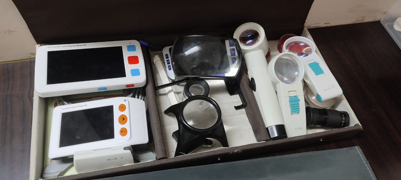
LVAPs can be broadly classified as:
Optical Devices for Distance
- Handheld telescope
- Mounted telescope [9]
Optical Devices for Near
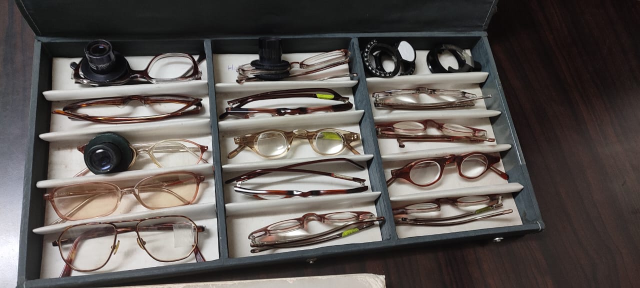
Spectacles
- Prismatic half eyes
- Bifocals
Magnifiers –Handheld
- Stand
- Illuminated
- Non-illuminated
Electronic Devices
- Video magnification systems like closed circuit television, portable video magnification
- Computer hardware and software that provides screen magnification, synthesized speech, and tactile display[10]
Non-Optical Devices
- Reading lamp
- Reading stand
- Writing guide
- Reading guide
- Signature guide
- Bold line notebooks
- Black ink bold tip pens
- Soft lead pencil – 2B,4B,6B, etc
- Needle threader
- Notex
Others
- Talking scales
- Talking glucometers
- Color identifiers
- Talking compasses
- Walking sticks
Principle
Low vision aids help in utilizing the vision to the maximum. These are useful in clinical conditions like retinitis pigmentosa, glaucoma, macular degeneration, albinism, aniridia, retinal detachment, diabetic retinopathy, optic atrophy, and chorioretinitis.[11] LVAP's function is based on the principle of angular magnification. This is achieved by increasing the relative size and decreasing the relative distance. Angular magnification is defined as an apparent increase in the object's size compared to its original size, as, for example, with a telescope.[12] Relative size increase makes the object visibility better even with a relatively lower vision. This does not require any accommodation, such as with closed circuit television (CCTV). Relative distance decrease assists by bringing the image of the object relatively closer. This requires good accommodation, as, for example, with magnifiers.[13]
For non-optical devices, the concept of illumination is extremely important. The light source is kept near to the eye, and moving the light source closer will provide higher illumination. A higher level of illumination is required in patients with retinal pathologies like loss of cone function (age-related macular degeneration), glaucoma, diabetic retinopathy, retinitis pigmentosa, and chorioretinitis. Reduced illumination is needed in albinism and aniridia.[14]
Low Vision Optical Devices for Near
Magnifying Spectacles
Optical Principle
A convex lens produces a magnified image once the object is within the focal length. This results in an erect, virtual, and magnified image. The magnification produced is 1/4 the power of the lens. This is suitable for near and intermediate distances.[2]
Advantages
- Allows a larger field of vision
- Handsfree
- Can be used monocular or binocular
- Improves near and intermediate distance
- Cosmetically better as compared to other LVAPs
Disadvantages
- Suffers from spherical aberration
- The more the power, the shorter the focal length, thus closer the reading distance.
- Close reading leads to fatigue and unacceptable posture
- Not suitable for those with eccentric fixation[15]
Hand Magnifiers
Optical Principle
A convex lens from +4.0D to +40.0D produces an erect, virtual, and magnified image. This is useful for short-time tasks in patients with the field of vision reduced to 10 degrees or more. Hand magnifiers are available in three designs – aspheric, aplanatic, and biaspheric.
Advantages
- No accommodation required
- Working distance is more
- Useful for eccentric viewing
- The additional light source may enhance vision further.
Disadvantages
- Occupies both hands
- Limited field of vision
- Manual dexterity is a prerequisite[16]
Stand Magnifiers
Optical Principle
A convex lens forms a virtual, magnified image at a short distance from the lens. The magnifier needs to be held over the reading material and moved across it.
Advantages
- Technically simple to use
- Very useful for patients with tremors, arthritis, or constricted visual fields
Disadvantages
- A small field of vision
- A flat surface is always needed for comfortable reading
- Uncomfortable posture because of close reading posture[17]
Closed Circuit Television Systems (CCTVS)
CCTV comprises a television, a camera, and a platform to put the readable text. We can control the brightness, contrast as well as polarity, and it can magnify the text from 3x-60x.[18]
Low Vision Optical Devices for Distance
Telescope
These devices employ the principle of angular magnification. The magnification can range from 2 to 10x. They can be prescribed for distant, intermediate, and near viewing, reducing the field of view with magnification.
Types
- Hand held monocular
- Clip-on
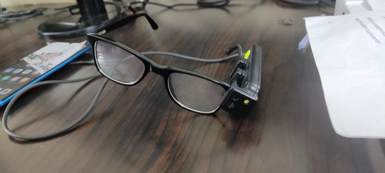
- Bioptics- Mounted on spectacles[19]
Principal
A telescope has two lenses (two optical systems) mounted in such a manner that the focal point of the objective coincides with that of the focal point of the ocular. The objective lens act as a converging lens. The magnification of the telescope is denoted by the formula M=f/f. The telescopes focus on the near object by altering the distance between the objective and the eyepiece lens or by increasing the power of the lens.
Galilean Telescope
In the Galilean telescope, the eyepiece is a negative lens, and the objective lens is a positive lens. The image formed is virtual and erect. The loss of light causes a reduction in brightness, and the field quality is poor.[15]
Keplerian Telescope
In the Keplerian telescope, the eyepiece and objective are both positive lenses. The image formed is real and inverted. Prisms are mounted to erect the image. There is more loss of light, and field quality is good. The major advantage of the telescope is that it is the only device that increases distance vision. The disadvantages are a restricted field of view, higher cost, reduced depth perception, and apprehension.[20]
Non-Optical Devices
Writing Guide
This is made of black cards having horizontally oriented rectangular cutouts along the cards. A patient with low vision can feel the empty cutout space and write.[8]
Signature Guide
This is made up of a black card with a white rectangular sheet. The white area act as a signature guide.[21]
Typoscope or Reading Guide
This is a masking device with a line cut out from an opaque area, a non-reflecting black plastic paper, or a paper of thicker consistency.[14]
Notex
This is a rectangular piece of cardboard having steps in the right upper corner, which help in identifying the currency of the note. The first cut indicates Rs 500; the second indicates Rs 100. The third indicates Rs 50, and so on.[22]
Relative Size Devices
These work on the principle that a larger object will subtend a larger visual angle and is easier to visualize. Examples include
- Large printing material
- Larger playing cards
- Computer keyboard
- A large clock, telephone, calendar, etc.[8]
Computer Softwares
Various interactive software allows the read-out of documents. Examples of these include
- SuperNova screen reader
- MAGic LVS
- Zoom Text magnifier/reader
Glare Alleviating Devices
These devices reduce unwanted light. These are useful in ocular pathologies like cataracts, corneal opacity, retinitis pigmentosa, and albinism. Various glare-reducing devices include
- Sunglasses
- Polaroid glasses
- Corning photochromatic filters (CPF glasses)
- Cap
- Umbrella
- NoIR filters[11]
Corning Photochromatic Filters (CPF Glasses)
They block approximately 99% UV-A wavelength and reduce 100% UV-B wavelength. They attenuate 98% of the blue wave light except CPF 450 and 96% of high-energy ambient blue light. The number in the CPF glass indicates the wavelength in nanometres above which the light is transmitted. Examples include
- CPF 550 (red) – useful in retinitis pigmentosa and albinism
- CPF 527 (orange) – useful in retinitis pigmentosa and diabetic retinopathy
- CPF 450 (yellow) - useful in optic atrophy, pseudophakia, and albinism
- CPF 511 (yellow-orange) - useful in optic atrophy, pseudophakia, aphakia, glaucoma, developmental cataract, and macular degeneration.[23]
NoIR Filters
These filters absorb short wavelengths in the visible spectrum, which scatter within the ocular media. These filters absorb infrared rays and ultraviolent light up to 4000nm. These filters help in filtering out excessive light and allow the optimal light to reach the eyes.
Examples of NoIR filters lenses include
- Dark amber (2%) - Useful during bright days and protect 100% from ultraviolet and infrared rays
- Standard grey (13%) - Helpful in patients with glaucoma, diabetic retinopathy, cataract, and corneal transplant patients
- Medium plum (20%) - Good in dim light and also helpful indoors
- Light grey (58%) - Reduces indoor glare light under fluorescent light
- Yellow (65%) - Helpful in patients with retinitis pigmentosa and macular degeneration[24]
Color and Contrast Sensitivity Improvement
These devices improve contrast by utilizing a light color against black or dark color. The colors with high contrast should be chosen in the room.[25]
Pinhole Spectacles
These spectacles have multiple pinholes of approximately 1 mm in size, and the holes should be at least 3-3.5 mm distance, which is the pupil's approximate size. The pinhole spectacles are helpful in patients with corneal opacities or patients having irregular astigmatism. These are less helpful in patients with reduced visual acuity.[26]
Mobility Training Devices
The mobility assisting devices for low vision patients help navigate the distance. Examples include long canes and strong portable lights.[27]
Field Expansion Devices
With the increase in magnification, the field of view decreases. The various methods by which field of view increases is
- Compression of an existing image to include more area available
- Addition of an image that relocates the image from a non-seeing area to a visible area
- Reflection of the image with the help of a mirror from a non-seeing area
Reverse telescopes and Fresnel prisms are other examples of field expansion devices, but reverse telescopes are less accepted due to the minification effect, and prisms are used in the direction of field loss with a power range of 10 to 15 D.[28]
Future of Low Vision Aids
Bionic Eye
This special LVA device is specially designed for patients who are blind due to retinitis pigmentosa and AMD. A bionic eye can also serve the purpose in patients with severe vision loss. The prime functioning depends on a healthy Optic nerve and well-developed visual cortex and association areas. This device is not helpful in patients who have congenital blindness. The device consists of a digital camera inbuilt into a pair of glasses, a video processing microchip unit, a radio transmitter, a receiver implanted in the ear, and a retina implant with electrodes on a chip behind the retina.[29]
Mechanism of Action
The camera in the eyeglasses captures the image and then sends the signals to the microchip, which converts the image into electric impulses of light and dark pixels. The image is also sent to a radio transmitter which transmits pulses wirelessly to the receiver. This, in turn, sends impulses to the retinal implant by a hair-thin walled implanted wire. The electrode generates electrical signals, which are, in turn, transmitted to the visual cortex. Bionic eye application needs training by patients. The patients should learn to interpret the objects' dark and white dots array. This device is still under clinical trial and evaluation.[30]
Issues of Concern
There are various issues of concern with LVA. These include the following:
Non-availability of Low Vision Aids Services
The presence of LVA services in a region does not necessarily mean good coverage of services. A survey in 195 countries revealed that only 115 had coverage of LVA services; among 115, 39% had <10% coverage, 22 had coverage between 11 to 50%, and 8 had coverage of more than 50%. Only 23 countries had coverage in the Asia Pacific region, and five countries had >10% coverage.
The countries or areas with >10% coverage had a higher proportion of older people and a more urbanized population. The survey also revealed that LVA services were primarily monodisciplinary and were available at secondary and tertiary eye care levels.[31]
Poor Socioeconomic Strata
Patients with poor income, daily wage earners, rural areas, small children, women, patients with disabilities, ethnic origin, refugees, and the elderly are also the vulnerable age group who miss LVA services.
Other Barriers
- Long traveling distance to access the services
- Access to cost and affordability
- Lack of awareness
- Lack of referral services
- The communication gap between patients and doctors
- Lack of motivation for visual rehabilitation
- Patient misconceptions and wrong perceptions[32]