Principles and Technique of Fogging During Subjective Refraction

Earn CME/CE in your profession:


Continuing Education Activity

This activity reviews the fogging procedure as part of the subjective refraction testing method. Fogging is used to control accommodation and elicit any hidden or latent hyperopia. Fogging involves a plus power build-up until the patients' visual acuity is reduced by one to two lines. This is followed by defogging, where the plus lenses are reduced until there is no further improvement in vision. This activity also outlines indications and contraindications for the fogging procedure. Common errors encountered while fogging are discussed in addition to preferred solutions in special cases. The activity also highlights the role of the interprofessional team in caring for patients with refractive errors, especially hyperopes.

Objectives:

  • Summarize the fogging process.
  • Identify the influence of accommodation on the refractive states of the eye.
  • Outline the importance of fogging as a tool for adjusting refraction findings.
  • Review the relationship between asthenopia and refractive errors.

Introduction

Fogging refers to using plus powers to bring the optical point of focus in front of the retina (into the vitreous) to ensure that accommodation is adequately relaxed. Accommodation is the ability of the eye to change its total dioptric power to bring objects at different distances into focus.[1][2]

This has been demonstrated in vivo and in vitro.[3][4] The stimulus to accommodation is a retinal blur.[5] The accommodative process is one of the triads of processes that occur when an eye focuses on near in addition to miosis and convergence.[6][7][8]

The principle of fogging involves using spherical powers to create artificial myopia, thereby moving the entire area of focus in the eye in front of the retina to create a situation where an attempt at accommodating will blur the vision, which further causes the patient to relax accommodation. Fogging is effective irrespective of the inherent refractive state of the eye.

Proper accommodation control is probably the most important factor in the refraction process. Without proper control of accommodation, the refraction endpoint will fluctuate, leading to wrong spectacle prescriptions.[9] 

This chapter details the principles of the fogging process and a step-by-step explanation of how to execute it properly.

Anatomy and Physiology

The crystalline lens is a spherical-shaped body in the anterior part of the eye. Together with the cornea, these ocular structures account for almost all the refractive powers of the eye.[10] The refractive index of the human crystalline lens ranges from 1.406 to 1.426.[11] The average power of the human crystalline lens is about 24.96 +/- 2.18 diopters (DS).[12]

An Asian study reported this index to range from 20.34 +/- 2.24 DS in a Chinese population.[13] The crystalline lens can adjust its power by a process known as accommodation. This is achieved by the action of the zonular fibers attached to the equator of the crystalline lens, which, when relaxed, causes the lens to assume a more spherical dimension, thereby increasing dioptric power. The opposite of this process is called dis-accommodation.

Indications

Indications for fogging include the following:

  • To control accommodation during retinoscopy.[14]
  • Refraction of pediatric patients who have excessive accommodation.[15]
  • Refraction of patients with asthenopic symptoms, which tend to be more significantly prevalent in hyperopes.[16][15]
  • When the clinician has a reasonable suspicion of a patient with an over-minus prescribed lens correction.
  • Refraction of a patient with latent hyperopia (such patients will report the same visual acuity over a range of powers).[17]
  • in certain cases of ocular deviation and amblyopia.[18][19][18]

Contraindications

Contraindications for Fogging include:

Poor Visual Acuity; Patients with very poor visual acuity may be difficult to fog as they may be unable to differentiate between the pre and post-fogging vision states, i.e., low vision patients.  

Aphakia; aphakia refers to an absence of the crystalline lens in the eye. This can be due to surgical interventions (pseudophakia) or congenital or acquired conditions. In the absence of the crystalline lens, there is no fogging effect during the Subjective refraction.

Cycloplegia; refers to the use of chemical agents to paralyze the ciliary body by blocking the acetylcholine receptors. This, in turn, renders the eye unable to accommodate. Cycloplegia is a common procedure used in the examination process for the pediatric population. Since this state of rest has been achieved chemically by cycloplegia, it is improbable that fogging will affect the ocular accommodative system.

Varying monocular endpoints; one requirement for the binocular balancing step of the refraction process is that both eyes must have similar visual acuity endpoints after monocular subjective refraction. A situation where the visual acuity is significantly different (by one or more full lines) is a contraindication for the fogging procedure and the entire binocular balancing process.

Equipment

  1. Trial frame with trial lens set or a phoropter
  2. Visual acuity charts (preferably Bailey-Lovey or ETDRS to eliminate the crowding phenomenon).[20]
  3. An occluder (for trial frames)

Personnel

  • Refraction nurses
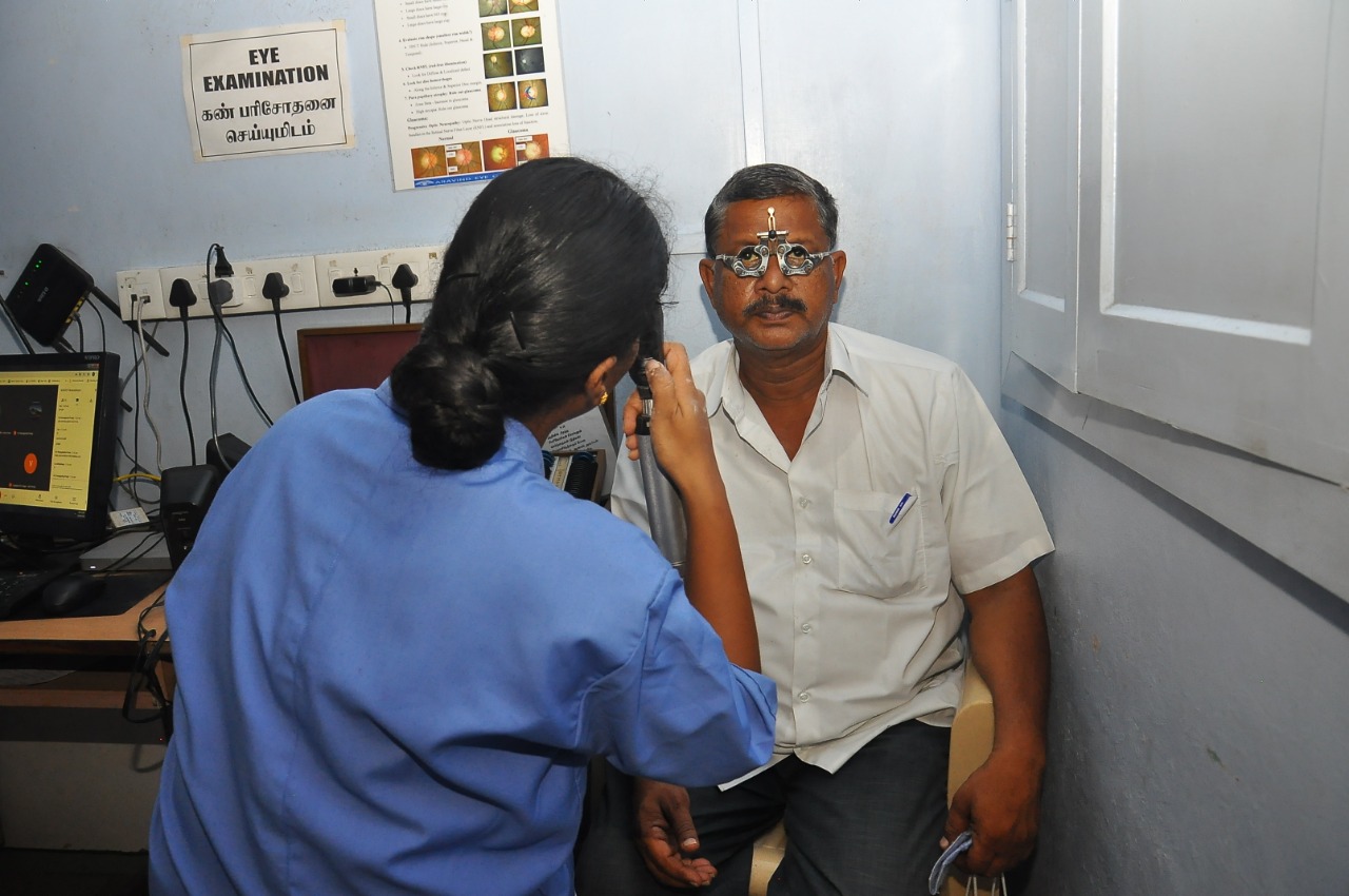
  • Optometrists (See image of optometrist assessing the subjective refraction in a patient).
  • Orthoptists
  • Ophthalmologists

Preparation

The clinician carries out fogging at three points during the refraction process, which includes the following:

  1. Before retinoscopy.[14]
  2. At the beginning of the subjective refraction process during the initial MPMVA (Maximum plus to maximum visual acuity step).
  3. At the point of the binocular MPMVA step.[17]

Note that the above steps ii and iii fall under subjective refraction.

The equipment used should be adequately cleaned and disinfected before this procedure is started. This takes special relevance in the current post-covid environment. The risk of cross infection has caused some clinicians to develop alternative techniques to the fogging process.[21] A smudged lens will make a patient report blurred vision and prolong the refraction process.

The examiner should explain to the patient what to expect during the procedure. For example, the examiner may say, " I am going to put in some lenses that will make your vision blur, then I will attempt to clear it." This ensures the patient does not get worried that the examiner is off track whenever a fogging lens is introduced.

Technique

Fogging During the Initial MPMVA

This procedure is carried out monocularly with the contralateral eye being occluded. By convention, the right eye is done first and the left eye second.  The fogging process is preceded by inserting the objective refraction findings. This could be based on:

  1. The retinoscopy results
  2. The autorefractometry results
  3. Former spectacle or contact prescription; the patient's visual acuity is noted immediately after this step.  

The examiner then puts sufficient plus power over the objective findings (See fogging images). It has been advocated in the past that +0.75 DS is adequate for this. However, practice shows this may be insufficient to fog certain patients, especially those with significant latent hyperopia. It is expected that the patient's visual acuity should be 1 or 2 lines worse than the earlier visual acuity taken with the objective findings in place.

The patient should be asked to read the optotypes on the examination chart stepwise from the largest to the smallest possible. If the patient is found to be sufficiently fogged, the examiner moves to the defogging process. If the patient is found to be insufficiently fogged by correctly reading the optotypes to a level deemed too close to the pre-fogging levels, the examiner should increase the fogging and repeat this process until the patient is satisfactorily fogged.  

The examiner then begins the defogging process by directing the patient's attention to the largest optotype and encouraging him to read downwards as the optotypes transition from larger to smaller ones. As expected, the patient should not get to the pre-fogging visual acuity. The examiner then proceeds to reduce the plus spherical power (or increase the minus powers) in the trial frame or phoropter.

The examiner should expect the patient to report an improved visual acuity with every +0.25 DS reduction (or addition of -0.25 DS) to the trial frame phoropter. The defogging process ends when the examiner notices that reducing the plus power does not improve visual acuity. For some acuity charts, one can stop the defogging process once the patient has correctly read all the letters on the smallest line, as long as that chart reaches a minimum of the 20/20 line.     

Fogging During the Binocular Balancing

Binocular balancing tests help to balance the accommodation in the two eyes under simultaneous viewing with both eyes. Therefore, binocular balancing is only indicated for patients with binocular vision and similar visual acuities. The fogging process is again employed in binocular balancing using the modified Humpriss and the Humphriss immediate contrast techniques. 

The distance monocular subjective findings are put into place before performing the modified Humpriss test. The left eye is traditionally fogged with enough plus power to blur the vision by about three lines worse than the eye being examined (right eye). The examiner then defogs the left eye to its MPMVA. This process is repeated for the left eye with the right eye fogged.

The final prescription obtained is taken to be a subjective refraction endpoint.

Complications

Common Mistakes

Under-fogging the patient: In this situation, the examiner puts in plus spherical powers (or reduced minus spherical powers) at a level that is not sufficient to reduce the visual acuity or keep accommodation in check by bringing the conoid of Sturm forward into the hypothetical vitreous space in front of the retina. The patient, therefore, continues to accommodate to bring the conoid of Sturm closer to the retina behind it.  

Over-fogging the patient: in this case, the examiner puts in too much plus spherical powers (or reduces too much minus spherical powers), thereby pushing the conoid of Sturm too far in front of the retina into the hypothetical vitreous space with a resulting consequence of unnecessarily prolonging the defogging process.  

Allowing too much time between removal and insertion of lenses. This commonly happens when using loose lenses with the trial frame. Removing the fogging lens to replace it with another one should be done as quickly as possible without causing the patient any discomfort. This is achievable by having the replacement lens in hand while removing the fogging lens.

Clinical Significance

The refraction process aims to correct any ametropia present in the eye. The amount of ametropia in the eye is significantly determined by the dioptric power of the cornea and the crystalline. While the power of the cornea is fixed, the power of the crystalline lens varies with accommodation, resulting in fluctuating amounts of ametropia. This can prolong the refraction process and lead to asthenopic symptoms if the ametropia is not adequately corrected.

It is also important to note that at several points during the refraction process, the eye is exposed to different stimuli for accommodation, including the retinoscope light, the occluder, and the testing prisms. This necessitates the fogging, carried out at the latter stages of the refraction process during binocular balancing.

Headaches can be caused by uncorrected hypermetropic patients, especially if not diagnosed, and the patient without corrective lenses does not complain of decreased vision.[22] Clinicians should request a complete eye examination before neurologically assessing these patients. Fogging can help determine hypermetropic conditions and proper lens prescription, which can attenuate symptoms in some patients.

Enhancing Healthcare Team Outcomes

The success of the subjective refraction process depends on the collaboration of different health workers attending to the patient. A patient who has been found to have high blood sugar is not a good candidate for refraction. It is therefore indicative that the nurses are available to conduct proper vital examinations like blood pressure and blood sugar before refraction. Attending clinicians should also take note of the patient's medical and social histories before refraction, as several drugs and food items affect the accommodative system.[23][24] [Level 2]

An interprofessional team can help achieve the best possible visual outcomes. Collaboration, shared decision-making with patients and family members for minors, and communication are critical elements for good visual outcomes. The interprofessional care provided to the patient must use an integrated care pathway combined with an evidence-based approach. The earlier the signs and symptoms of uncorrected hypermetropia and amblyopia, the better the prognosis and outcome. [Level 3]


(Click Image to Enlarge)
Digital image depicting the optometrist assessing the subjective refraction in a patient
Contributed by Dr. Kirandeep Kaur, MBBS, DNB, FPOS, FICO, MRCS Ed, MNAMS

(Click Image to Enlarge)
The fogging process involves examining the patient with plus power correction until visual acuity reaches 20/20.
Contributed by Marco Zeppieri, MD, PhD.
Article Details

Article Author

Mutali J. Musa

Article Editor:

Marco Zeppieri

Updated:

1/30/2023 7:36:50 AM

References

[1]

Martinez-Enriquez E,Pérez-Merino P,Velasco-Ocana M,Marcos S, OCT-based full crystalline lens shape change during accommodation in vivo. Biomedical optics express. 2017 Feb 1;     [PubMed PMID: 28270993]

[2]

Pérez-Merino P,Velasco-Ocana M,Martinez-Enriquez E,Marcos S, OCT-based crystalline lens topography in accommodating eyes. Biomedical optics express. 2015 Dec 1;     [PubMed PMID: 26713216]

[3]

Marussich L,Manns F,Nankivil D,Maceo Heilman B,Yao Y,Arrieta-Quintero E,Ho A,Augusteyn R,Parel JM, Measurement of Crystalline Lens Volume During Accommodation in a Lens Stretcher. Investigative ophthalmology     [PubMed PMID: 26161985]

[4]

Momeni-Moghaddam H,Ng JS,Cesana BM,Yekta AA,Sedaghat MR, Accommodative amplitude using the minus lens at different near distances. Indian journal of ophthalmology. 2017 Mar;     [PubMed PMID: 28440251]

[5]

Cholewiak SA,Love GD,Banks MS, Creating correct blur and its effect on accommodation. Journal of vision. 2018 Sep 4;     [PubMed PMID: 30193343]

[6]

Ghoushchi VP,Mompeán J,Prieto PM,Artal P, Binocular dynamics of accommodation, convergence, and pupil size in myopes. Biomedical optics express. 2021 Jun 1     [PubMed PMID: 34221660]

[7]

Goering M,Drennan KB,Moshirfar M, Convergence Insufficiency StatPearls. 2022 Jan;     [PubMed PMID: 32119277]

[8]

Motlagh M,Geetha R, Physiology, Accommodation StatPearls. 2022 Jan;     [PubMed PMID: 31194346]

[9]

Richdale K,Bullimore MA,Sinnott LT,Zadnik K, The Effect of Age, Accommodation, and Refractive Error on the Adult Human Eye. Optometry and vision science : official publication of the American Academy of Optometry. 2016 Jan;     [PubMed PMID: 26703933]

[10]

Lu T,Song J,Wu Q,Jiang W,Tian Q,Zhang X,Xu J,Wu J,Hu Y,Sun W,Bi H, Refractive lens power and lens thickness in children (6-16 years old). Scientific reports. 2021 Sep 29     [PubMed PMID: 34588558]

[11]

Chang YC,Mesquita GM,Williams S,Gregori G,Cabot F,Ho A,Ruggeri M,Yoo SH,Parel JM,Manns F, {i}In vivo{/i} measurement of the human crystalline lens equivalent refractive index using extended-depth OCT. Biomedical optics express. 2019 Feb 1;     [PubMed PMID: 30800489]

[12]

Jongenelen S,Rozema JJ,Tassignon MJ,EVICR.net and Project Gullstrand Study Group., Distribution of the Crystalline Lens Power In Vivo as a Function of Age. Investigative ophthalmology     [PubMed PMID: 26523387]

[13]

He J,Lu L,He X,Xu X,Du X,Zhang B,Zhao H,Sha J,Zhu J,Zou H,Xu X, The Relationship between Crystalline Lens Power and Refractive Error in Older Chinese Adults: The Shanghai Eye Study. PloS one. 2017;     [PubMed PMID: 28114313]

[14]

Chiu NN,Rosenfield M,Wong LC, Effect of contralateral fog during refractive error assessment. Journal of the American Optometric Association. 1997 May;     [PubMed PMID: 9170797]

[15]

Esteves Leandro J,Meira J,Ferreira CS,Santos-Silva R,Freitas-Costa P,Magalhães A,Breda J,Falcão-Reis F, Adequacy of the Fogging Test in the Detection of Clinically Significant Hyperopia in School-Aged Children. Journal of ophthalmology. 2019;     [PubMed PMID: 31467692]

[16]

Hashemi H,Saatchi M,Yekta A,Ali B,Ostadimoghaddam H,Nabovati P,Aghamirsalim M,Khabazkhoob M, High Prevalence of Asthenopia among a Population of University Students. Journal of ophthalmic     [PubMed PMID: 31875103]

[17]

Kaur K,Gurnani B, Subjective Refraction Techniques StatPearls. 2022 Jan;     [PubMed PMID: 35593807]

[18]

Economides JR,Adams DL,Horton JC, Bilateral Occlusion Reduces the Ocular Deviation in Intermittent Exotropia. Investigative ophthalmology     [PubMed PMID: 33393972]

[19]

Li T,Qureshi R,Taylor K, Conventional occlusion versus pharmacologic penalization for amblyopia. The Cochrane database of systematic reviews. 2019 Aug 28;     [PubMed PMID: 31461545]

[20]

Lovie-Kitchin JE, Is it time to confine Snellen charts to the annals of history? Ophthalmic     [PubMed PMID: 26497296]

[21]

Pérez González D,Loewenstein A,Gaton DD, Avoiding Diagnostic Lens Fogging During the COVID-19 Era. Clinical ophthalmology (Auckland, N.Z.). 2020;     [PubMed PMID: 33384557]

[22]

Majumdar S,Tripathy K, Hyperopia StatPearls. 2022 Jan;     [PubMed PMID: 32809551]

[23]

Kosehira M,Machida N,Kitaichi N, A 12-Week-Long Intake of Bilberry Extract ({i}Vaccinium myrtillus{/i} L.) Improved Objective Findings of Ciliary Muscle Contraction of the Eye: A Randomized, Double-Blind, Placebo-Controlled, Parallel-Group Comparison Trial. Nutrients. 2020 Feb 25;     [PubMed PMID: 32106548]

[24]

Kim J,Kang H,Choi H,Jo A,Oh DR,Kim Y,Im S,Lee SG,Jeong KI,Ryu GC,Choi C, Aqueous Extract of {i}Perilla frutescens{/i} var. acuta Relaxes the Ciliary Smooth Muscle by Increasing NO/cGMP Content In Vitro and In Vivo. Molecules (Basel, Switzerland). 2018 Jul 19;     [PubMed PMID: 30029520]