Retinal Macroaneurysm

Earn CME/CE in your profession:


Continuing Education Activity

Retinal macroaneurysm is an uncommon entity affecting the posterior segment of the eye, and it has variable presentations. Most macroaneurysms undergo spontaneous resolution. Visual acuity is affected because of edema, hemorrhage, exudates, or other secondary complications. These changes can be identified at an early stage using different investigative modalities, which include FFA (fundus fluorescein angiography), ICG (indocyanine green angiography), OCT (optical coherence tomography), OCT-A (OCT- angiography), and USG B scan (brightness scan). Proper management can avoid vision-threatening complications. This activity reviews the etiology, pathophysiology, evaluation, and management as well as highlights the role of the interprofessional team in managing the condition.

Objectives:

  • Outline the etiology and pathophysiology of retinal artery macroaneurysm.
  • Describe the clinical findings and investigations required for the evaluation of retinal artery macroaneurysm.
  • Review the available management options as well as outline the various differentials and complications of retinal artery macroaneurysm.
  • Summarize the importance of patient education and interprofessional team strategies to improve the outcomes of retinal artery macroaneurysm.

Introduction

The term Retinal macroaneurysm was coined by Robertson.[1]  It is a retinal condition defined as an acquired, focal dilation of a retinal artery (ranging between 100 to 250 μm in diameter) occurring in the posterior fundus within the first three orders of bifurcations of the central retinal arterioles or at the level of arteriovenous crossings.[2] The superotemporal arteriole is most commonly affected.[3] RAM (retinal artery macroaneurysm) may be associated with systemic hypertension and cardiovascular arteriosclerotic disease.[4] 

The patient is usually asymptomatic, and a decline in visual acuity occurs due to macular edema, end-arteriole occlusion due to thrombosis, or hemorrhage caused by rupture of the aneurysm.[5] Some lesions can resolve spontaneously by natural thrombosis; however, some require treatment.[6] 

Identifying RAM is pivotal for proper management and requires a precise approach to control the underlying systemic condition. The diagnostic modalities for identification and management include FFA (fundus fluorescein angiography), ICG-A (indocyanine green angiography), OCT (optical coherence tomography), OCT-A (optical coherence tomography angiography), and USG (ultrasonogram) B scan (brightness scan). Procedures like laser photocoagulation, intravitreal Anti VEGF (anti-vascular endothelial growth factor) agents, YAG (yttrium-aluminum-garnet) hyaloidotomy, pneumatic displacement, and pars plana vitrectomy can be used to treat complicated RAMs.

Etiology

The most important risk factors associated with RAM are chronic systemic hypertension (75% of patients), arteriosclerosis, abnormal lipid levels, and inherent structural defects in blood vessels. These lead to focal ischemia of the blood vessel wall bringing about increased vessel permeability and intimal collagen remodeling. This predisposes to saccular (sac-like or blowout, i.e., localized outpouching of arterial wall) or fusiform (spindle-shaped or cuffed, i.e., uniform widening of retinal arteriole) dilatation of vessels.[1] Other important risk factors include Coat disease, von Hippel-Lindau disease, diabetic retinopathy, and radiation retinopathy.[2][7]

Retinal arteriolar aneurysms may also be seen in association with ocular sarcoidosis, peripheral multifocal chorioretinitis, and IRVAN (idiopathic retinal vasculitis, aneurysms, and neuroretinitis).

Retinal capillary macroaneurysms or microaneurysms are a result of ischemia due to venous obstruction. These are commonly associated with diabetes or venous occlusive diseases.[8] 

Retinal venous macroaneurysm may be seen in branch retinal venous occlusion in patients with chronic hypertension. However, isolated non-occlusive venous macroaneurysms have also been reported.[9]

Epidemiology

RAM is more common in the elderly (66 to 74 years) and females (70% of cases).[10] The incidence of retinal arterial macroaneurysms as reported in the adult Chinese population is 1 per 9000 eyes as per the Beijing Eye Study.[11] Most of the cases are unilateral solitary. However, multiple macroaneurysms may occur in 15 to 20 % of cases, with bilateral occurrence in about 10 %.[12] As per Nangia et al., there is 1 case of retinal artery macroaneurysm in Central India per 1500 subjects of more than 30 years of age.[13]

Pathophysiology

In RAM, there is a weakness in the muscularis layer of the arteriolar wall causes thinning, fibrosis, and decreased elasticity of the artery.[7] This makes the artery susceptible to aneurysmal arterial dilatation. The aneurysmal dilatation is fragile and can rupture to cause hemorrhage with or without exudation of proteinaceous and lipid-rich exudates in any layer of the retina leading to visual loss.[7]

Histopathology

It is important to differentiate a macroaneurysm from a microaneurysm. Microaneurysms typically arise from capillaries and are small, red spots with sizes ranging from 15 to 100 μm in diameter.[14][15] Microaneurysm formation is associated with basement membrane thickening, pericyte loss, and endothelial cell death.[16][17][18] Leakage of fluid from microaneurysm leads to retinal edema, and their rupture can cause retinal hemorrhages.[19][20] 

The formation of a microaneurysm is usually associated with diabetic retinopathy. However, other systemic conditions include ischemic, infectious, inflammatory, hematologic, and radiation-related disorders.[21][22] Small microaneurysms have Type-III collagen, and their combined width of the focal bulge and the associated capillary is less than twice the width of the adjacent nonaffected capillary region. Large microaneurysms have a size more significant than the capillary width. Additionally, there is increased expression of matrix-metalloproteinase (MMP)-9 in large microaneurysms, which cause the breakdown of the basement membrane proteins.[8]

Retinal artery macroaneurysms arise from the muscular arterioles. Size of a macroaneurysm ranges between 100 to 250 μm in diameter.[3] There occurs distension of the affected retinal arteriole surrounded by fibrin, foamy macrophages, or even hemosiderin. There is also vessel wall thickening and variable degrees of hyalinization of the arterial wall and retinal exudate or hemorrhage as described by Fichte et al.[23] 

Histopathological examination reveals the presence of both thrombus and cholesterol within the macroaneurysm.[7] Other abnormalities include an increase in the width of the periarterial capillary free zone around the area of an aneurysm, dilatation, nonperfusion of capillaries, presence of microaneurysms, and formation of artery-to-artery collaterals.[23]

History and Physical

Retinal artery macroaneurysm can be isolated/simple (only vascular ectasia) or complex (associated with hemorrhage).[24][25]

The temporal part of the retina within the first three orders of bifurcations of the central retinal arterioles or at the level of arteriovenous crossings is usually affected.[2] It most commonly involves the superotemporal vessel (50%) followed by the inferotemporal arcade (45%).[5]

The patient is usually asymptomatic unless there is macular involvement. Symptoms of metamorphosis, including decreased visual acuity, or floaters occur due to hemorrhage or macular edema. Sometimes acute and severe visual loss may also occur. The clinical symptoms and severity of visual loss depend upon the type of RAM, which can be quiescent, hemorrhagic, or exudative, as suggested by Lavin and colleagues.[2]

Quiescent form - In this form, the visual symptoms are relatively infrequent. There is either no exudation/hemorrhage, or there is sparing of the macula. Hemorrhage/exudate, if present, measures less than one disc diameter.

Hemorrhagic form - It accounts for 50 % of the total retinal artery macroaneurysms.[7] There occurs an acute and rapid vision loss caused by associated with hemorrhage of an area more than one disc diameter.

These are usually present close to the disc because the arteries closer to the disc have a larger diameter and an increased flow rate that may rupture due to increased transmural stress. It is associated with multilayered hemorrhage involving preretinal, intraretinal, subretinal,  or vitreous spaces. Both preretinal and subretinal hemorrhage may be simultaneously present (hourglass hemorrhages) in some instances. The occurrence of hemorrhages at multiple levels is a result of sizeable aneurysmal dehiscence.[26] Intra or subretinal hemorrhage is usually a result of posterior or posterolateral aneurysmal rupture.[5] Subretinal hemorrhage is usually extensive, dense, overlying an artery, not centered at the macula, and has vitreous extensions.

Antiplatelet and anticoagulant drugs like aspirin, warfarin, and clopidogrel can also lead to subretinal bleed.[27]

Exudative form - There occurs a gradual deterioration in visual acuity in exudative retinal macroaneurysm.

Here, exudation measuring more than one disc diameter is seen. Lipid deposition in a circinate pattern around the aneurysm along with intraretinal edema and subretinal fluid accumulation can be seen caused by leakage from the damaged vessels surrounding the aneurysm. This leads to a collection of long-standing exudates and chronic macular edema.[2][28] Sometimes, only perianeurysmal exudates may also be seen, which usually do not affect the vision and have a good prognosis.[5]

Spontaneous pulsations contiguous with the arterial wall (better demonstrated using indocyanine green angiography) may be seen in the case of an isolated macroaneurysm.[29] The presence of spontaneous pulsations suggests pending rupture. However, this statement is still debatable[30] as there are no available indicators to predict an impending rupture.

The hemorrhagic form is usually associated with hypertension or cardiovascular disorder, whereas the exudative form is associated with retinal vein occlusion.[31] 

There may also be mixed variants having either a predominant hemorrhagic or an exudative component.[5]

Retinal capillary macroaneurysms are usually associated with edema of the surrounding retinal tissue.[8]

Retinal venous macroaneurysm may be associated with a premacular hemorrhage.[32]

Evaluation

This includes a complete clinical examination (including fundus examination) aided by routine examinations such as blood pressure measurement and lipid profile to assess systemic associations and imaging studies.

Fundus Fluorescein Angiography (FFA) - This is the investigation of choice to diagnose and outline the lesion. FFA is beneficial when the aneurysm is obscured (due to hemorrhage or exudation) or when the aneurysm gets involuted. Aneurysms may appear as pulsating (seen in 10% of cases with the help of dynamic FFA) round or ovoid pooling of dye which may be seen filling rapidly in the early arterial phase in case of fusiform RAMs or minimal early filling with complete filling in the middle to late phases in case of saccular RAMs. Filling defects in the early arterial phase may be due to the formation of clots or scar formation caused by blockage of the lumen as a result of thrombosis or endothelial cell formation.[28] The rate of filling depends upon the neck size of the aneurysm.[33][5] 

Areas of microangiopathies can leak, and lead can contribute to cystoid macular edema, which manifests as areas of fluorescein leakage from the dilated capillaries around the aneurysm.[7] Blocked or marked hypofluorescence occurs due to overlying or underlying dense hemorrhage. Lipid present in the macular area does not block fluorescein until a large amount of lipid is present.[5] 

FFA additionally gives information regarding capillary microaneurysms, nonperfusion areas, intraretinal microvascular abnormalities, and telangiectasias.[34] Resolved microaneurysm shows a "Z-shaped" kink appearance. This occurs due to a balance between fibrous changes in media of the artery's wall and tensile strength of the adventitial portion of the internal limiting membrane, which counters the traction applied by fibrotic changes.[26]

Indocyanine Green Angiography (ICG-A) - It is particularly significant when a hemorrhage blocks visualization of the aneurysm because of greater penetration of its near-infrared light spectrum. ICG angiography shows areas of hyperfluorescence and can suggest the accurate location of the macroaneurysm, further helping in management. It can be helpful in the presence of concomitant exudates and retinal pigment epithelial changes.[34] It can also be used to rule out peripapillary idiopathic polypoidal choroidal vasculopathy, which can cause similar subretinal hemorrhage which is not centered at the fovea.[35]

Optical Coherence Tomography (OCT) - Spectral Domain OCT (SD-OCT) is a helpful tool in diagnosing retinal artery macroaneurysm. RAMs appear as round or oval hyper-reflective lesions in the inner retinal layers and cause shadowing of the deeper tissues by elevating the internal limiting membrane and ganglionic cell layer.[36] It is also helpful in determining the accumulation and extent of intraretinal or subretinal fluid, which appears hypo-reflective. Intraretinal lipid deposits/retinal exudates appear hyper-reflective, which can be quantified using this modality. Additionally, OCT can be used to identify epiretinal membrane formation and also monitor the treatment.[37] Hence, it can quantify the amount of macular edema, differentiate various levels of blood accumulation, and can show features of subretinal changes such as pigment epithelial detachment (PED) seen in idiopathic polypoidal choroidal vasculopathy.

OCT Angiography (OCT-A) - This investigative modality can be used to demonstrate focal vascular outpouchings and delineate changes in the fluid and vasculature with time, further helping in management.[38] It shows circular regions of an increased signal where fluid is present in the case of exudative retinal artery macroaneurysms (suspended scattering particles in motion).[39]

USG B Scan - This is helpful when the fundus is not visible or in case of dense vitreous hemorrhage. It also helps differentiate RAM from retinal tears/detachments or polypoidal choroidal vasculopathy, causing breakthrough hemorrhage.

The above-mentioned investigative modalities can also be used to identify retinal capillary and retinal venous macroaneurysms. The most important differentiating feature of RAM is direct communication with an arteriole and the presence of hemorrhage in multiple retinal layers.[40]

Treatment / Management

Most of the retinal artery macroaneurysms essentially undergo spontaneous resolution and involution, and the patients can be observed. A thorough, structured workup for the systemic association should be advised with a proper follow-up for at least six months.[41] It is important to note that proper systemic control of associated risk factors (such as hypertension) does not cause resolution of the aneurysm. Still, it may decrease the risk of rupture. Hemorrhagic RAMs not involving macula can be subjected to treatment to prevent the risk of recurrent hemorrhage even though they usually undergo spontaneous resolution.[2] 

RAMs that undergo spontaneous resolution do not usually have any sequelae. In case of ruptured RAM leading to hemorrhage and exudation but not involving the macula, a close follow-up at first one month followed by every 1-3 monthly is to be done until complete involution.[4][34][41] Patients with vision-threatening retinal artery macroaneurysm associated with intraretinal, preretinal, or vitreous hemorrhages may also be closely observed for three months due to spontaneous resolutions and limited consequences.[7][34][26]

Treatment is recommended for complicated or non-resolving macroaneurysms.

1 ) Laser Photocoagulation

It includes direct or indirect laser application or a combination of both in case of a hemorrhage or exudation involving the macula.

A ) Direct laser photocoagulation- It helps in sealing the aneurysm using minimal power (100 to 300 mW) with a large spot size (200-500 microns) given for a longer duration (200-500 milliseconds) to create a light burn.[7] Direct photocoagulation means the application of laser directly to the macroaneurysm to speed up involution and decrease leakage.[41] Argon green laser can be applied directly to the lesion. In contrast, the Nd YAG (Neodymium-doped Yttrium Aluminum Garnet) laser can be used to treat a preretinal hemorrhage secondary to the retinal artery macroaneurysm.[42] Nd YAG laser photodisruption (power 1 mJ-50 mJ) causes dispersion of blood into the vitreous cavity by creating an opening in the internal limiting membrane or the posterior hyaloid membrane.[43]

B ) Indirect laser photocoagulation - Reduces the consumption of oxygen and blood flow to the aneurysm along with decreasing exudation from the surrounding capillaries. Direct thermal energy delivery to the lesion is avoided, and rather confluent burns for a shorter duration (100-200 milliseconds) with a power of 400mW at 200 μm spot size with medium darkness are given around the aneurysm and more specifically to the retinal capillaries that are damaged surrounding the lesion.[7][44]

Complications include arteriolar occlusions, retinal break, artery to vein shunting, a breakthrough of hemorrhage, capillary dropouts, choroidal neovascular membrane formation, an extension of exudation, traction at the retina, scarring of the subretinal region, or even symptomatic scotomas may occur post laser application.[45] Even a notable decline in vision may occur. Hence, evaluation before the laser is critical, especially when a portion of the arteriole supplying the macula is to be treated by laser.[46]

C ) Subthreshold laser - It is used in the management of fovea involving exudative RAMs. It does not form a visible burn mark is not formed. It causes increased expression of the heat shock protein resulting in selective retinal pigment epithelial cell damage with a better balance between angiogenic factors and cytokine release. Hence, it causes retinal hyperthermia below the cell death threshold.[47][48] 

Battaglia Parodi and colleagues conducted a pilot study to compare subthreshold (exposure time of 0.3 seconds, power of 1400 mW, spot size of 125 nm, and a duty cycle of 15%) with threshold laser treatment reported similar anatomical and visual outcomes. Still, the incidence of epiretinal membrane formation was decreased when the subthreshold laser was used.[49][50] Here, laser energy is applied indirectly to cause a gentle retinal whitening around the aneurysm but not more than that. Hence, a combined indirect and direct technique was used.

Yellow dye laser and indocyanine green dye-enhanced photocoagulation can also be used to treat retinal artery macroaneurysm.[51][52]

2 ) Intravitreal Injection of Anti VEGF (Anti-Vascular Endothelial Growth Factor) Agents

It was first used in the management of retinal artery macroaneurysm in 2009.[53] Anti-VEGFs reduce nitric oxide levels followed by vasoconstriction and decreased vascular permeability, thereby reducing leakage and edema.[54] These may also lead to a more compact alignment of the endothelial cells.[55] 

Chanana et al. reported that intravitreal bevacizumab (1.25 mg) expedited a decrease in the retinal thickness and is also well tolerated.[53] 

Jonas et al. reported the use of bevacizumab for the management of retinal artery macroaneurysm induced macular edema.[56] Javey et al. showed the successful use of intravitreal bevacizumab for the management of ruptured retinal artery macroaneurysm.[57] 

Wenkstern et al. reported the use of intravitreal ranibizumab with focal laser photocoagulation to close the retinal artery macroaneurysm with subsequent reduction in the macular edema.[58] Golan S et al. reported that bevacizumab was beneficial for reducing macular edema and improving the visual outcome, the effect of which lasted up to 13 months. The authors suggested that bevacizumab should be considered only in case of extensive macular edema or when laser photocoagulation cannot be done.[59] 

Tsakpinis et al. used intravitreal bevacizumab to treat multilevel hemorrhage in the retina and observed a faster visual rehabilitation.[60] Zweifel et al. used both intravitreal bevacizumab (1.25 mg) and ranibizumab (0.5 mg) to treat macular exudation secondary to retinal artery macroaneurysm. They concluded that intravitreal anti-VEGF agents could be used as an alternative to laser photocoagulation.[55] 

Cho et al. suggested the use of bevacizumab for both macular edema and hemorrhage secondary to retinal artery macroaneurysm.[61] Pichi et al., in a case series evaluating 38 eyes, suggested that bevacizumab causes a reduction in macular edema and hard exudates in patients with RAM.[62] 

Erol et al., in a case series of 7 patients, concluded that intravitreal ranibizumab is effective in the treatment of symptomatic retinal artery macroaneurysm and also proposed for more prospective randomized trials to study the exact role of ranibizumab in the treatment of symptomatic RAM.[63] 

Chatziralli et al. also demonstrated the use of ranibizumab for the resolution of macular edema.[64] Leung et al. in 2015 and Bormann et al. in 2017 advocated the use of intravitreal bevacizumab and laser photocoagulation to treat macular edema in patients of retinal artery macroaneurysm. Bormann et al., in 2017, suggested that ranibizumab and aflibercept can be considered a potential treatment modality to treat macular edema in complex RAM.[65] 

Lin Z et al. in 2019 suggested the use of ranibizumab and conbercept for the treatment of patients with symptomatic retinal artery macroaneurysm.[66]

3 ) YAG Laser Hyaloidotomy

This method is mainly used for pre-retinal hemorrhages. It acts by causing focal disruption of the inner limiting membrane and allowing better absorption by causing hemorrhage to dissolve into the vitreous. However, complications like epiretinal membrane formation, macular hole formation, retinal detachment, and secondary non-clearing vitreous hemorrhage may occur.[42] Yttrium aluminum garnet (YAG) laser also effectively releases the trapped sub-ILM (sub-Internal Limiting Membrane) hemorrhage into the vitreous for improving its resolution. Still, recurrent hemorrhage has been reported in such cases.[43][67]

4 ) Pneumatic Displacement with or without  intravitreal tissue plasminogen activator (tPA) injection

This can be used for submacular hemorrhages. This involves the use of perfluorocarbon gas with or without intravitreal tissue plasminogen activator (tPA) with subsequent prone position to displace hemorrhage out of the macular area by using the effect of gravity.[68] Here, pure intravitreal C3F8 gas (0.3 ml) is injected via a 30 gauge needle 4 mm posterior to the limbus in phakic eyes and 3.5 mm behind the limbus in pseudophakic eyes. Postoperatively, a prone position is advised, and the intraocular pressure is controlled with glaucoma medications. This method usually provides good visual recovery, but the presence of subepithelial hemorrhage, proliferation, or exudate can limit the recovery.[69] 

Intravitreal tPA, 0.1 to 0.2 ml (12.5 μg/0.1 ml or 25 μg/0.1 ml), can cause thrombolysis and clearance of subretinal hemorrhage.[68][70] A dose of more than 25 μg/0.1 ml is not recommended due to possible retinal toxicity. Johnson used intravitreal 0.1 to 0.2 ml of tPA at a concentration of 25 μg/0.1 ml (total dose of 25-50 μg) followed by anterior chamber paracentesis to reduce the intraocular pressure. After this, 0.3 ml of C3F8 or SF6 was injected intravitreally, and the patient was asked to start prone positioning 6 hours after the procedure and continue prone positioning for 24 to 48 hours. After this period, if the thick submacular blood persisted on examination, the prone position was continued for two more days.[69] If the intraocular pressure is higher than 25 mmHg, it is managed with oral acetazolamide or intravenous mannitol.[68] 

 5) Pars Plana Vitrectomy (PPV)

PPV may be needed in patients with non-clearing or breakthrough vitreous hemorrhage. PPV may also be used for clearing submacular hemorrhage, for faster visual recovery, or in case of poor visibility preventing recognition of other pathologies of the macula. Complications of PPV include active bleeding, cataract progression, formation of retinal breaks/detachment, macular hole, or even endophthalmitis.[71]

Hence, tPA, pneumatic displacement, and vitrectomy should be considered in cases with more extensive or non-resolving hemorrhages.

Kumar A et al. described intraoperative OCT (optical coherence tomography) guided subretinal cocktail injection for treating a ruptured retinal artery macroaneurysm with multilevel bleed, which caused the complete displacement of the subretinal bleed along with partial recovery of the outer retinal architecture as early as the first day postoperatively. The technique was first described by Martel and Mahmoud to treat massive submacular bleed secondary to the choroidal neovascular membrane. The method involves initial core vitrectomy and posterior vitreous detachment followed by injection of 0.1 ml recombinant tissue plasminogen activator (12.5 μg/0.1 ml) with 0.1 ml bevacizumab (1.25 mg/0.05 ml) and 0.3 ml air in subretinal space using an extendible 41 gauge needle attached to a 1 ml tuberculin syringe followed by filling 20% SF6 (Sulfur hexafluoride) gas in the vitreous cavity and propped up the positioning of the patient.[72][73]

Hillenkamp and colleagues compared PPV with intravitreal tPA 0.2 ml (40µg) and gas (20% SF6) versus PPV with subretinal tPA [10–20µg (0.05–0.1 ml) of rtPA using 41G subretinal cannula] and gas (20% SF6) in cases with submacular hemorrhage. They found that the subretinal tPA arm was associated with a higher rate of complete displacement of submacular hemorrhage. Final visual acuity was similar in both arms, and complications were more common in the subretinal tPA arm.[74]

Retinal capillary and retinal venous macroaneurysms associated with edema can be treated with laser photocoagulation or anti-VEGF injections.[9][8]

Retinal arterial macroaneurysms associated with uveitis need investigation and management of uveitis, especially to rule out ocular sarcoidosis and peripheral multifocal chorioretinitis.

Differential Diagnosis

In the case of retinal macroaneurysm, it is important to rule out diabetic retinopathy, retinal telangiectasia, von Hippel-Lindau disease, radiation retinopathy, retinal capillary angioma, cavernous hemangioma, malignant melanoma, polypoidal choroidal vasculopathy, Valsalva retinopathy, and hemorrhagic pigment epithelial detachment of age-related macular degeneration.[75][76][77] 

Vitreous hemorrhage in RAM can be confused with retinal vein occlusion, acute posterior vitreous detachment, or proliferative diabetic retinopathy. In contrast, Coats disease, Leber miliary aneurysms, and angiomatosis retinae should be excluded in case of predominant exudation. Similarly, causes of submacular hemorrhage include choroidal melanoma, age-related macular degeneration, polypoidal choroidal vasculopathy, presumed ocular histoplasmosis, high myopia, trauma, and Valsalva retinopathy.[78][7][77][79][80][79] 

Conditions associated with subretinal hemorrhage are angioid streaks, coagulopathies, tumors, diabetic retinopathy, CRVO (central retinal vein occlusion), CNV (choroidal neovascularization), or drugs like aspirin, warfarin, and clopidogrel.[27][81][82][83][82]

Besides retinal artery macroaneurysm, hemorrhage at multiple levels in the retina can be seen in cases of trauma (shaken baby syndrome), anemia, leukemia, and high altitude retinopathy.[84]

Retinal capillary macroaneurysms are associated with diabetic retinopathy and ischemia secondary to veno-occlusive disorders. Other systemic associations include infectious, inflammatory, hematologic, and radiation-related disorders.[21][22] Perifoveal Exudative Vascular Anomalous Complex (PEVAC) is an important differential seen in otherwise healthy individuals where a unilateral, isolated perifoveal aneurysm is seen.[85]

Retinal venous macroaneurysm is usually coexistent with branch retinal venous occlusion, although isolated venous macroaneurysm with no ocular or systemic history may also be seen.[32]

Retinal arterial aneurysms are also seen in IRVAN (idiopathic retinal vasculitis, aneurysms, and neuroretinitis) or 'idiopathic retinal arteriolar aneurysms (IRAA)' with a macular star.[86] Usually, multiple retinal arterial aneurysms are seen, especially at the posterior pole (first several orders of the retinal artery) and at the point of bifurcation of the artery. Such aneurysms may also be found over the optic disc. Typically, the retinal arterial macroaneurysms in IRVAN are not seen anterior to the retinal equator. The retinal artery usually shows a variation in the caliber in the segment between the aneurysms. The various shapes of the aneurysms include spheroid, Y-shaped, and fusiform. Usually, the same artery has aneurysms at multiple places giving the involved artery an appearance of 'multiple knots of a cord.' A macular star is seen due to the deposition of hard exudates. Severe capillary nonperfusion may be present and may involve the macula resulting in a poor visual prognosis.

Multiple retinal arterial macroaneurysms in an inflamed eye may occur in ocular sarcoidosis.[87][88] Yokoi, Oshita, and Goto reported such aneurysms in 9 eyes (7 patients, 7.2%) from a database of 97 patients with sarcoidosis with intraocular inflammation. The mean age was 61 years, and the retinal arterial aneurysms were not seen in the acute phase. Macroaneurysms were noted in the chronic phase of ocular sarcoidosis and were multiple in 5 of 9 eyes. In this series, a maximum of five macroaneurysms was present in a single eye. The macroaneurysms involved both eyes in 2 of 7 patients.[87] 

Yamanaka and colleagues noted that 14 of the total 1007 patients with uveitis had macroaneurysms. Peripheral multifocal chorioretinitis (PMC) was seen in 12 of these 14 patients. Among 12 patients with PMC, sarcoidosis was noted in 7 patients. In 2 patients without PMC, sarcoidosis was noted in 1 patient. Most (94.4%) of the macroaneurysms in this series were exudative, and only one macroaneurysm was hemorrhagic. Two patients had systemic hypertension.[89] Retinal macroaneurysm may be seen in ocular sarcoidosis associated with optic disc granuloma, arteritis, and uveitis.[88]

Prognosis

The visual prognosis depends upon:

  • Type of RAM- Quiescent retinal macroaneurysm has the best visual prognosis. Hemorrhagic macroaneurysm with intact macular integrity has a better visual prognosis than exudative forms, as they usually resolve spontaneously and do not require treatment.
  • Presence of exudates/edema - Permanent visual loss occurs due to the collection of long-standing hard exudates and chronic macular edema resulting from vascular leakage directly from the aneurysm or the damaged vessels surrounding the aneurysm.[28]
  • Severity - A hemorrhagic macroaneurysm more extensive than any other exudate is responsible for visual loss. Cases with hemorrhagic macroaneurysm combined with an exudative macroaneurysm or even a single extensive exudative macroaneurysm have a poorer visual prognosis.
  • Duration - Long-standing edema/ exudates have the least chances to improve.
  • Position - Pre retinal and vitreous hemorrhage have good visual outcomes upon clearing of hemorrhage. Subretinal hemorrhage predisposes to poorer prognosis due to damage to the outer photoreceptor layer caused by prevention of metabolic diffusion between outer retina and choriocapillaris, iron toxicity, and damage to the retina by fibrin. Submacular hemorrhage has the poorest visual outcome.[90] Submacular hemorrhages can even predispose to macular hole formation and subretinal neovascularization, further amounting to a poorer prognosis.[91] Permanent and irreversible damage can occur as early as two days to up to 2 weeks. Subfoveal hemorrhage may also be associated with neurosensory detachment. Furthermore, subsequent fibrosis and atrophy can cause permanent visual loss.

Complications

Besides causing multilayer (preretinal, intraretinal, subretinal, or vitreous) hemorrhage and chronic macular edema, retinal artery macroaneurysm can also cause secondary angle-closure glaucoma, retinal vein occlusion, serous or hemorrhagic retinal detachment, subretinal neovascularization, epiretinal membrane, or even macular hole formation.[92][91]

Persistent retinal capillary macroaneurysms can lead to Type 3 macular neovascularization.

Deterrence and Patient Education

Patients with RAM need evaluation for hypertension, dyslipidemia, and arteriosclerosis control with their primary treating physician to prevent this condition.[4] They also need to be educated regarding the condition, the various complications that may occur, and the available treatment options. The patient should be made to realize that the condition is preventable but warrants the need for compliance to the advice given by both the primary treating physician and the ophthalmologist.

Enhancing Healthcare Team Outcomes

Approach to a patient with retinal artery macroaneurysm has to be both extensive and synergetic. For example, patients suffering from hypertension should be advised of blood pressure control by referring them to a primary care physician, and those with macular involvement require prompt treatment by the ophthalmologist depending upon the characteristics, location, and duration of the lesion. Other associated systemic conditions, including diabetes mellitus, dyslipidemia, kidney disease, ischemic heart disease, may need interprofessional coordination with various specialists, including endocrinologists, nephrologists, and cardiologists.

The condition hence requires an interprofessional team approach which includes initiation of treatment and motivation by the physician for the taking advice of the ophthalmologist for early detection and prevention of vision-threatening complications, validation of proper dosage of the medication by the pharmacist, monitoring the vital signs as well as assisting in educating the patient for compliance to the treatment by the nurses, complete evaluation of visual acuity by the optometrist, and finally proper counseling and laying down the various treatment options available for this condition by the ophthalmologist.

This interprofessional teamwork is pivotal in successfully treating and managing retinal artery macroaneurysm and securing favorable patient outcomes.


(Click Image to Enlarge)
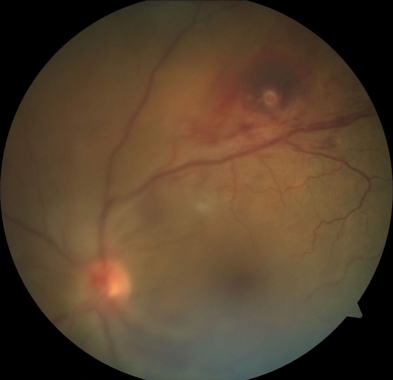
Color fundus image of the left eye shows media haze due to vitreous hemorrhage
Color fundus image of the left eye shows media haze due to vitreous hemorrhage. Retinal artery macroaneurysm is seen superotemporally.
Contributed by Koushik Tripathy, MD
Details

Updated:

8/25/2023 3:04:40 AM

References


[1]

Robertson DM, Macroaneurysms of the retinal arteries. Transactions - American Academy of Ophthalmology and Otolaryngology. American Academy of Ophthalmology and Otolaryngology. 1973 Jan-Feb;     [PubMed PMID: 4730083]


[2]

Lavin MJ,Marsh RJ,Peart S,Rehman A, Retinal arterial macroaneurysms: a retrospective study of 40 patients. The British journal of ophthalmology. 1987 Nov;     [PubMed PMID: 3689733]

Level 2 (mid-level) evidence

[3]

Lee EK,Woo SJ,Ahn J,Park KH, Morphologic characteristics of retinal arterial macroaneurysm and its regression pattern on spectral-domain optical coherence tomography. Retina (Philadelphia, Pa.). 2011 Nov;     [PubMed PMID: 21716167]


[4]

Adamczyk DT,Olivares GE,Petito GT, Retinal arterial macroaneurysm: a longitudinal case study. Journal of the American Optometric Association. 1989 Nov;     [PubMed PMID: 2607078]

Level 3 (low-level) evidence

[5]

Moosavi RA,Fong KC,Chopdar A, Retinal artery macroaneurysms: clinical and fluorescein angiographic features in 34 patients. Eye (London, England). 2006 Sep;     [PubMed PMID: 16138114]


[6]

Asdourian GK,Goldberg MF,Jampol L,Rabb M, Retinal macroaneurysms. Archives of ophthalmology (Chicago, Ill. : 1960). 1977 Apr;     [PubMed PMID: 557969]


[7]

Rabb MF,Gagliano DA,Teske MP, Retinal arterial macroaneurysms. Survey of ophthalmology. 1988 Sep-Oct;     [PubMed PMID: 3055391]

Level 3 (low-level) evidence

[8]

Spaide RF,Barquet LA, RETINAL CAPILLARY MACROANEURYSMS. Retina (Philadelphia, Pa.). 2019 Oct     [PubMed PMID: 30489449]


[9]

Kaiser HM,Pagani JM, Isolated retinal venous macroaneurysm. Optometry (St. Louis, Mo.). 2005 Sep     [PubMed PMID: 16198992]


[10]

Panton RW,Goldberg MF,Farber MD, Retinal arterial macroaneurysms: risk factors and natural history. The British journal of ophthalmology. 1990 Oct;     [PubMed PMID: 2285682]


[11]

Xu L,Wang Y,Jonas JB, Frequency of retinal macroaneurysms in adult Chinese: the Beijing Eye Study. The British journal of ophthalmology. 2007 Jun;     [PubMed PMID: 17510482]


[12]

Lewis RA,Norton EW,Gass JD, Acquired arterial macroaneurysms of the retina. The British journal of ophthalmology. 1976 Jan;     [PubMed PMID: 1268157]


[13]

Nangia V,Jonas JB,Khare A,Sinha A,Lambat S, Prevalence of retinal macroaneurysms. The Central India Eye and Medical Study. Acta ophthalmologica. 2013 Mar;     [PubMed PMID: 22690702]


[14]

Wang H,Chhablani J,Freeman WR,Chan CK,Kozak I,Bartsch DU,Cheng L, Characterization of diabetic microaneurysms by simultaneous fluorescein angiography and spectral-domain optical coherence tomography. American journal of ophthalmology. 2012 May;     [PubMed PMID: 22300473]


[15]

Talbot L,Berger SA, Fluid-mechanical aspects of the human circulation. American scientist. 1974 Nov-Dec;     [PubMed PMID: 4440940]


[16]

Roy S,Ha J,Trudeau K,Beglova E, Vascular basement membrane thickening in diabetic retinopathy. Current eye research. 2010 Dec;     [PubMed PMID: 20929292]


[17]

Beltramo E,Porta M, Pericyte loss in diabetic retinopathy: mechanisms and consequences. Current medicinal chemistry. 2013;     [PubMed PMID: 23745544]


[18]

Stitt AW,Gardiner TA,Archer DB, Histological and ultrastructural investigation of retinal microaneurysm development in diabetic patients. The British journal of ophthalmology. 1995 Apr;     [PubMed PMID: 7742285]


[19]

Ito H,Horii T,Nishijima K,Sakamoto A,Ota M,Murakami T,Yoshimura N, Association between fluorescein leakage and optical coherence tomographic characteristics of microaneurysms in diabetic retinopathy. Retina (Philadelphia, Pa.). 2013 Apr;     [PubMed PMID: 23190917]


[20]

Lee SN,Chhablani J,Chan CK,Wang H,Barteselli G,El-Emam S,Gomez ML,Kozak I,Cheng L,Freeman WR, Characterization of microaneurysm closure after focal laser photocoagulation in diabetic macular edema. American journal of ophthalmology. 2013 May;     [PubMed PMID: 23394906]


[21]

ASHTON N, Retinal micro-aneurysms in the non-diabetic subject. The British journal of ophthalmology. 1951 Apr;     [PubMed PMID: 14830727]


[22]

Bloodworth JM Jr, A re-evaluation of diabetic glomerulosclerosis 50 years after the discovery of insulin. Human pathology. 1978 Jul;     [PubMed PMID: 711223]


[23]

Fichte C,Streeten BW,Friedman AH, A histopathologic study of retinal arterial aneurysms. American journal of ophthalmology. 1978 Apr;     [PubMed PMID: 655232]


[24]

Evan Goldhagen B,Goldhardt R, Retinal Arterial Macroaneurysms: Updating your Memory on RAM Management. Current ophthalmology reports. 2019 Jun;     [PubMed PMID: 31827984]


[25]

Bopp S,Joussen AM, [Retinal Arterial Macroaneurysms (RAM)--pathology, differenzial diagnoses, and therapy]. Klinische Monatsblatter fur Augenheilkunde. 2014 Sep;     [PubMed PMID: 25181506]


[26]

Abdel-Khalek MN,Richardson J, Retinal macroaneurysm: natural history and guidelines for treatment. The British journal of ophthalmology. 1986 Jan;     [PubMed PMID: 3947596]


[27]

Ying GS,Maguire MG,Daniel E,Grunwald JE,Ahmed O,Martin DF,Comparison of Age-Related Macular Degeneration Treatments Trials Research Group., Association between Antiplatelet or Anticoagulant Drugs and Retinal or Subretinal Hemorrhage in the Comparison of Age-Related Macular Degeneration Treatments Trials. Ophthalmology. 2016 Feb;     [PubMed PMID: 26545320]


[28]

Speilburg AM,Klemencic SA, Ruptured retinal arterial macroaneurysm: diagnosis and management. Journal of optometry. 2014 Jul-Sep;     [PubMed PMID: 25000868]


[29]

Schneider U,Wagner AL,Kreissig I, Indocyanine green videoangiography of hemorrhagic retinal arterial macroaneurysms. Ophthalmologica. Journal international d'ophtalmologie. International journal of ophthalmology. Zeitschrift fur Augenheilkunde. 1997;     [PubMed PMID: 9097320]


[30]

Palestine AG,Robertson DM,Goldstein BG, Macroaneurysms of the retinal arteries. American journal of ophthalmology. 1982 Feb;     [PubMed PMID: 7199821]


[31]

Cousins SW,Flynn HW Jr,Clarkson JG, Macroaneurysms associated with retinal branch vein occlusion. American journal of ophthalmology. 1990 May 15;     [PubMed PMID: 2333919]


[32]

Khairallah M,Ladjimi A,Messaoud R,Ben Yahia S,Hmidi K,Jenzeri S, Retinal venous macroaneurysm associated with premacular hemorrhage. Ophthalmic surgery and lasers. 1999 Mar     [PubMed PMID: 10100260]


[33]

Cahuzac A,Scemama C,Mauget-Faÿsse M,Sahel JA,Wolff B, Retinal arterial macroaneurysms: clinical, angiographic, and tomographic description and therapeutic management of a series of 14 cases. European journal of ophthalmology. 2016 Jan-Feb;     [PubMed PMID: 26165327]

Level 3 (low-level) evidence

[34]

Kester E,Walker E, Retinal arterial macroaneurysm causing multilevel retinal hemorrhage. Optometry (St. Louis, Mo.). 2009 Aug;     [PubMed PMID: 19635433]


[35]

Moorthy RS,Lyon AT,Rabb MF,Spaide RF,Yannuzzi LA,Jampol LM, Idiopathic polypoidal choroidal vasculopathy of the macula. Ophthalmology. 1998 Aug;     [PubMed PMID: 9709746]


[36]

Goldenberg D,Soiberman U,Loewenstein A,Goldstein M, Heidelberg spectral-domain optical coherence tomographic findings in retinal artery macroaneurysm. Retina (Philadelphia, Pa.). 2012 May;     [PubMed PMID: 22127222]


[37]

Tsujikawa A,Sakamoto A,Ota M,Oh H,Miyamoto K,Kita M,Yoshimura N, Retinal structural changes associated with retinal arterial macroaneurysm examined with optical coherence tomography. Retina (Philadelphia, Pa.). 2009 Jun;     [PubMed PMID: 19516118]


[38]

Chang VS,Schwartz SG,Flynn HW Jr, Optical Coherence Tomography Angiography of Retinal Arterial Macroaneurysm before and after Treatment. Case reports in ophthalmological medicine. 2018;     [PubMed PMID: 29692939]

Level 3 (low-level) evidence

[39]

Kashani AH,Green KM,Kwon J,Chu Z,Zhang Q,Wang RK,Garrity S,Sarraf D,Rebhun CB,Waheed NK,Schaal KB,Munk MR,Gattoussi S,Freund KB,Zheng F,Liu G,Rosenfeld PJ, Suspended Scattering Particles in Motion: A Novel Feature of OCT Angiography in Exudative Maculopathies. Ophthalmology. Retina. 2018 Jul;     [PubMed PMID: 30221214]


[40]

Rehmani AS,Banaee T,Makkouk F, Subretinal leakage of a retinal capillary macroaneurysm - a case report. BMC ophthalmology. 2021 May 17;     [PubMed PMID: 34001046]

Level 3 (low-level) evidence

[41]

Gurwood AS,Nicholson CR, Retinal arterial macroaneurysm: a case report. Journal of the American Optometric Association. 1998 Jan     [PubMed PMID: 9479935]

Level 3 (low-level) evidence

[42]

Ulbig MW,Mangouritsas G,Rothbacher HH,Hamilton AM,McHugh JD, Long-term results after drainage of premacular subhyaloid hemorrhage into the vitreous with a pulsed Nd:YAG laser. Archives of ophthalmology (Chicago, Ill. : 1960). 1998 Nov     [PubMed PMID: 9823347]


[43]

Iijima H,Satoh S,Tsukahara S, Nd:YAG laser photodisruption for preretinal hemorrhage due to retinal macroaneurysm. Retina (Philadelphia, Pa.). 1998;     [PubMed PMID: 9801038]


[44]

Leung EH,Reddy AK,Vedula AS,Flynn HW Jr, Serial bevacizumab injections and laser photocoagulation for macular edema associated with a retinal artery macroaneurysm. Clinical ophthalmology (Auckland, N.Z.). 2015;     [PubMed PMID: 25897199]


[45]

Brown DM,Sobol WM,Folk JC,Weingeist TA, Retinal arteriolar macroaneurysms: long-term visual outcome. The British journal of ophthalmology. 1994 Jul     [PubMed PMID: 7918263]


[46]

Ghassemi F,Majidi AR, Development of a second retinal artery macroaneurysm within a few months. Graefe's archive for clinical and experimental ophthalmology = Albrecht von Graefes Archiv fur klinische und experimentelle Ophthalmologie. 2011 Sep     [PubMed PMID: 20967456]


[47]

Sramek C,Mackanos M,Spitler R,Leung LS,Nomoto H,Contag CH,Palanker D, Non-damaging retinal phototherapy: dynamic range of heat shock protein expression. Investigative ophthalmology     [PubMed PMID: 21087969]


[48]

Xiao M,Sastry SM,Li ZY,Possin DE,Chang JH,Klock IB,Milam AH, Effects of retinal laser photocoagulation on photoreceptor basic fibroblast growth factor and survival. Investigative ophthalmology     [PubMed PMID: 9501874]


[49]

Parodi MB,Iacono P,Ravalico G,Bandello F, Subthreshold laser treatment for retinal arterial macroaneurysm. The British journal of ophthalmology. 2011 Apr;     [PubMed PMID: 20956278]


[50]

Battaglia Parodi M,Iacono P,Pierro L,Papayannis A,Kontadakis S,Bandello FM, Subthreshold laser treatment versus threshold laser treatment for symptomatic retinal arterial macroaneurysm. Investigative ophthalmology & visual science. 2012 Apr 2     [PubMed PMID: 22395893]


[51]

Russell SR,Folk JC, Branch retinal artery occlusion after dye yellow photocoagulation of an arterial macroaneurysm. American journal of ophthalmology. 1987 Aug 15;     [PubMed PMID: 3618718]


[52]

Steigerwalt RD Jr,Pascarella A,Arrico L,Librando A,Plateroti R,Plateroti AM,Plateroti P,Nebbioso M, Idiopathic juxtafoveal retinal telangiectasis and retinal macroaneurysm treated with indocyanine green dye-enhanced photocoagulation. Panminerva medica. 2012 Dec;     [PubMed PMID: 23241941]


[53]

Chanana B,Azad RV, Intravitreal bevacizumab for macular edema secondary to retinal macroaneurysm. Eye (London, England). 2009 Feb     [PubMed PMID: 18388958]


[54]

Maderna E,Corsini E,Franzini A,Giombini S,Pollo B,Broggi G,Solero CL,Ferroli P,Messina G,Marras C, Expression of vascular endothelial growth factor receptor-1/-2 and nitric oxide in unruptured intracranial aneurysms. Neurological sciences : official journal of the Italian Neurological Society and of the Italian Society of Clinical Neurophysiology. 2010 Oct     [PubMed PMID: 20635108]


[55]

Zweifel SA,Tönz MS,Pfenninger L,Becker M,Michels S, Intravitreal anti-VEGF therapy for retinal macroaneurysm. Klinische Monatsblatter fur Augenheilkunde. 2013 Apr;     [PubMed PMID: 23629789]


[56]

Jonas JB,Schmidbauer M, Intravitreal bevacizumab for retinal macroaneurysm. Acta ophthalmologica. 2010 Nov;     [PubMed PMID: 20977692]


[57]

Javey G,Moshfeghi AN,Moshfeghi AA, Management of ruptured retinal arterial macroaneurysm with intravitreal bevacizumab. Ophthalmic surgery, lasers     [PubMed PMID: 21728252]


[58]

Wenkstern AR,Petersen H, Intravitreal ranibizumab in retinal macroaneurysm. Graefe's archive for clinical and experimental ophthalmology = Albrecht von Graefes Archiv fur klinische und experimentelle Ophthalmologie. 2010 Nov;     [PubMed PMID: 20508944]


[59]

Golan S,Goldenberg D,Goldstein M, Long-term follow-up of intravitreal bevacizumab in retinal arterial macroaneurysm: a case report. Case reports in ophthalmology. 2011 Sep;     [PubMed PMID: 22220164]

Level 3 (low-level) evidence

[60]

Tsakpinis D,Nasr MB,Tranos P,Krassas N,Giannopoulos T,Symeonidis C,Dimitrakos SA,Konstas AG, The use of bevacizumab in a multilevel retinal hemorrhage secondary to retinal macroaneurysm: a 39-month follow-up case report. Clinical ophthalmology (Auckland, N.Z.). 2011;     [PubMed PMID: 22069349]

Level 3 (low-level) evidence

[61]

Cho HJ,Rhee TK,Kim HS,Han JI,Lee DW,Cho SW,Kim JW, Intravitreal bevacizumab for symptomatic retinal arterial macroaneurysm. American journal of ophthalmology. 2013 May;     [PubMed PMID: 23385203]


[62]

Pichi F,Morara M,Torrazza C,Manzi G,Alkabes M,Balducci N,Vitale L,Lembo A,Ciardella AP,Nucci P, Intravitreal bevacizumab for macular complications from retinal arterial macroaneurysms. American journal of ophthalmology. 2013 Feb;     [PubMed PMID: 23111179]


[63]

Erol MK,Dogan B,Coban DT,Toslak D,Cengiz A,Ozel D, Intravitreal ranibizumab therapy for retinal arterial macroaneurysm. International journal of clinical and experimental medicine. 2015;     [PubMed PMID: 26379984]


[64]

Chatziralli I,Maniatea A,Koubouni K,Parikakis E,Mitropoulos P, Intravitreal ranibizumab for retinal arterial macroaneurysm: long-term results of a prospective study. European journal of ophthalmology. 2017 Mar 10     [PubMed PMID: 27646333]


[65]

Bormann C,Heichel J,Hammer U,Habermann A,Hammer T, Intravitreal Anti-Vascular Endothelial Growth Factor for Macular Edema due to Complex Retinal Arterial Macroaneurysms. Case reports in ophthalmology. 2017 Jan-Apr;     [PubMed PMID: 28638337]

Level 3 (low-level) evidence

[66]

Lin Z,Hu Q,Wu Y,Xu J,Zhang Q, Intravitreal ranibizumab or conbercept for retinal arterial macroaneurysm: a case series. BMC ophthalmology. 2019 Jan 15;     [PubMed PMID: 30646868]

Level 2 (mid-level) evidence

[67]

Chen YY,Lin LY,Chang PY,Chen FT,Mai ELC,Wang JK, Laser and Anti-Vascular Endothelial Growth Factor Agent Treatments for Retinal Arterial Macroaneurysm. Asia-Pacific journal of ophthalmology (Philadelphia, Pa.). 2017 Sep-Oct;     [PubMed PMID: 28828763]


[68]

Wu TT,Sheu SJ, Intravitreal tissue plasminogen activator and pneumatic displacement of submacular hemorrhage secondary to retinal artery macroaneurysm. Journal of ocular pharmacology and therapeutics : the official journal of the Association for Ocular Pharmacology and Therapeutics. 2005 Feb     [PubMed PMID: 15718829]


[69]

Johnson MW, Pneumatic displacement of submacular hemorrhage. Current opinion in ophthalmology. 2000 Jun;     [PubMed PMID: 10977228]

Level 3 (low-level) evidence

[70]

Tan CS,Au Eong KG, Surgical drainage of submacular haemorrhage from ruptured retinal arterial macroaneurysm. Acta ophthalmologica Scandinavica. 2005 Apr;     [PubMed PMID: 15799740]


[71]

Zhao P,Hayashi H,Oshima K,Nakagawa N,Ohsato M, Vitrectomy for macular hemorrhage associated with retinal arterial macroaneurysm. Ophthalmology. 2000 Mar;     [PubMed PMID: 10711904]


[72]

Kumar A,Sundar MD,Chawla R,Agarwal D,Hasan N, Intraoperative optical coherence tomography-guided subretinal cocktail injection in a case of ruptured retinal artery macro-aneurysm with multilevel bleed. Indian journal of ophthalmology. 2020 Jul;     [PubMed PMID: 32587201]

Level 3 (low-level) evidence

[73]

Martel JN,Mahmoud TH, Subretinal pneumatic displacement of subretinal hemorrhage. JAMA ophthalmology. 2013 Dec;     [PubMed PMID: 24337559]


[74]

Hillenkamp J,Surguch V,Framme C,Gabel VP,Sachs HG, Management of submacular hemorrhage with intravitreal versus subretinal injection of recombinant tissue plasminogen activator. Graefe's archive for clinical and experimental ophthalmology = Albrecht von Graefes Archiv fur klinische und experimentelle Ophthalmologie. 2010 Jan;     [PubMed PMID: 19669780]


[75]

Shukla UV, Tripathy K. Diabetic Retinopathy. StatPearls. 2024 Jan:():     [PubMed PMID: 32809640]


[76]

Tripathy K, Sharma YR, R K, Chawla R, Gogia V, Singh SK, Venkatesh P, Vohra R. Recent advances in management of diabetic macular edema. Current diabetes reviews. 2015:11(2):79-97     [PubMed PMID: 25801496]

Level 3 (low-level) evidence

[77]

Hochman MA,Seery CM,Zarbin MA, Pathophysiology and management of subretinal hemorrhage. Survey of ophthalmology. 1997 Nov-Dec;     [PubMed PMID: 9406367]

Level 3 (low-level) evidence

[78]

Fritsche PL,Flipsen E,Polak BC, Subretinal hemorrhage from retinal arterial macroaneurysm simulating malignancy. Archives of ophthalmology (Chicago, Ill. : 1960). 2000 Dec;     [PubMed PMID: 11115272]


[79]

Spalter HF, Retinal macroaneurysms: a new masquerade syndrome. Transactions of the American Ophthalmological Society. 1982;     [PubMed PMID: 7182954]


[80]

Ahmed F, Tripathy K. Posterior Vitreous Detachment. StatPearls. 2024 Jan:():     [PubMed PMID: 33085420]


[81]

Shields CL,Shields JA, Subretinal hemorrhage from a retinal arterial macroaneurysm simulating a choroidal melanoma. Ophthalmic surgery and lasers. 2001 Jan-Feb;     [PubMed PMID: 11195753]


[82]

Jena S, Tripathy K. Vitreous Hemorrhage. StatPearls. 2023 Jan:():     [PubMed PMID: 32644557]


[83]

Gupta A, Paulbuddhe VS, Shukla UV, Tripathy K. Exudative Retinitis (Coats Disease). StatPearls. 2022 Jan:():     [PubMed PMID: 32809517]


[84]

Kanukollu VM,Ahmad SS, Retinal Hemorrhage StatPearls. 2021 Jan;     [PubMed PMID: 32809612]


[85]

Sacconi R,Freund KB,Yannuzzi LA,Dolz-Marco R,Souied E,Capuano V,Semoun O,Phasukkijwatana N,Sarraf D,Carnevali A,Querques L,Bandello F,Querques G, The Expanded Spectrum of Perifoveal Exudative Vascular Anomalous Complex. American journal of ophthalmology. 2017 Dec;     [PubMed PMID: 29079450]


[86]

Tripathy K, Pathogenesis of idiopathic retinal vasculitis, aneurysms, and neuroretinitis (IRVAN) or 'idiopathic retinal arteriolar aneurysms (IRAA)' with macular star. Medical hypotheses. 2018 Mar     [PubMed PMID: 29447942]


[87]

Yokoi K,Oshita M,Goto H, Retinal macroaneurysm associated with ocular sarcoidosis. Japanese journal of ophthalmology. 2010 Sep     [PubMed PMID: 21052899]


[88]

Mahmoudzadeh R,Gopal A,Soares R,Dunn JP, Unilateral Retinal Arteritis and Macroaneurysm in Sarcoidosis. Ocular immunology and inflammation. 2021 Aug 31     [PubMed PMID: 34464228]


[89]

Yamanaka E,Ohguro N,Kubota A,Yamamoto S,Nakagawa Y,Tano Y, Features of retinal arterial macroaneurysms in patients with uveitis. The British journal of ophthalmology. 2004 Jul;     [PubMed PMID: 15205230]


[90]

Tonotsuka T,Imai M,Saito K,Iijima H, Visual prognosis for symptomatic retinal arterial macroaneurysm. Japanese journal of ophthalmology. 2003 Sep-Oct;     [PubMed PMID: 12967867]


[91]

Murhty K,Puri P,Talbot JF, Retinal macroaneurysm with macular hole and subretinal neovascular membrane. Eye (London, England). 2005 Apr;     [PubMed PMID: 15319787]


[92]

Wu TT,Chuang CT,Sheu SJ,Chiou YH, Non-vitrectomizing vitreous surgery for premacular haemorrhage. Acta ophthalmologica. 2011 Mar;     [PubMed PMID: 19758402]