Primary Congenital Glaucoma

Earn CME/CE in your profession:


Continuing Education Activity

Primary congenital glaucoma is a rare genetic congenital ocular disorder that affects children at birth. It is characterized by abnormally high intraocular pressures. This activity describes the etiology, risk factors, varied presentations, investigations, management guidelines, differential diagnosis, and prognosis for primary congenital glaucoma. This activity highlights the role of the interprofessional team in early diagnosis, and referrals have also been described in detail.

Objectives:

  • Describe the clinical presentations of patients with primary congenital glaucoma.
  • Summarize the examination findings in cases of primary congenital glaucoma.
  • Outline the medical and surgical management guidelines for treating patients with primary congenital glaucoma.
  • Explain the post-operative considerations in patients with primary congenital glaucoma.

Introduction

Hippocrates first described the term primary congenital glaucoma when he noted abnormally enlarged eyes in infants. Later on, the pathogenesis of increased intraocular pressure and angle abnormalities were correlated with this entity. Primary congenital glaucoma is also referred to as developmental glaucoma, as it might not always present immediately after birth. Depending on the age of onset, the primary congenital glaucomas have been divided into the following types.[1]

  1. True congenital glaucoma – Also known as newborn glaucoma. In this type, the child is either born with ocular enlargement or enlargement of eyes is noticed within one month of life. It is believed that the IOP is raised in the intrauterine life itself. These cases account for approximately 25 percent.
  2. Infantile glaucoma – This type includes patients who manifest between 1 and 36 months of life. It accounts for approximately 65 percent of patients with primary congenital glaucoma.
  3. Juvenile glaucoma – This group includes patients with signs of raised intraocular pressure manifest after three years of age but before adulthood. This accounts for about 10 percent of cases.

Another classification system has been defined based on the area of dysgenesis known as Hoskin classification.[2]

  1. Trabeculodysgenesis – Defect lies in the development of trabecular meshwork
  2. Iridotrabeculodysgenesis – Includes hypoplasia or hyperplasia of the stroma, anomalous iris vessels, or structural defects in the form of coloboma or aniridia.
  3. Corneotrabeculodysgenesis – This includes complex cases of congenital glaucoma like Axenfield, Rieger, or Peters anomaly.

Hoskin's classification type 1 is labeled as primary congenital glaucoma, while types 2 and 3 are labeled as secondary congenital glaucoma.

Etiology

Most cases of primary congenital glaucoma are sporadic in origin, with no family history. The significant risk factors are consanguineous marriage, genetic predisposition, and first-degree relatives, including siblings. Approximately 90 percent of cases belong to this category. Another 10 percent cases are familial, which show an autosomal recessive pattern of inheritance with incomplete penetrance ranging from 40-100 percent. Five gene loci have been linked to primary congenital glaucoma. These include GLC3A, GLC3B, GLC3C, GLC3D, and GLC3E. Locus GLC3A has been linked to the CYP1B1 gene.[3]

Mutations in this gene are most commonly responsible for autosomal recessively inherited cases.[4] Another gene latent transforming growth factor-beta – LTBP2 gene adjacent to GLC3C has also been closely related to primary congenital glaucoma.[5]

Epidemiology

The incidence of PCG varies widely across different ethnic groups. Studies from western countries have reported incidence ranging from 1/10,000 to 30,000 live births.[6] The incidence is as high as 1/2500 has been reported from countries like Saudi Arabia. Another study found that glaucoma accounted for 7% of children registered in blind schools.[7]

The higher incidence in particular countries and ethnic groups is related to the higher prevalence of consanguineous marriages. Approximately 65 to 80% of cases are bilateral.[8] A male to female ratio of 3:2 has been reported in studies from the United States and Europe.[9] A very high incidence has been reported from Slovakia of about 1:1250. A Japanese study quoted the male-to-female ratio of 6:5 in patients with CYP1BI mutation and 19:2 without the same gene mutation.[10]

Pathophysiology

The main pathophysiology is the defect in the trabecular meshwork development and the anterior chamber angle. This hampers the aqueous outflow through the anterior chamber, thus leading to increased intraocular pressures. Another theory given by Barkan, which was later disproved, was the presence of an imperforate membrane at the angle of the anterior chamber, which was proposed to impede the aqueous outflow.[11] 

Presently, the most accepted theory is that proposed by Anderson, which states that excessive collagen meshwork within the trabeculum prevents normal insertion of the ciliary body and iris.[12] This results in anteriorly inserted iris root resulting in obstruction of the trabecular meshwork, thus resulting in elevated intraocular pressures.

Histopathology

Recently histopathological and electron microscopic studies have demonstrated obstruction through the outflow pathway. Frequently, the ciliary muscle is inserted high on the trabecular meshwork. Moreover, a detailed framework analysis has shown an excessive amount of collagen in the trabecular meshwork. Other studies have demonstrated fibrillary collagen fibers, elastin fibers, and ground substances in the intervening trabecular meshwork and the canal of Schlemm.[12][13]

History and Physical

The family history of glaucoma and consanguineous marriage between parents is mandatory and should never be missed.[14] The presentation can vary from unilateral/bilateral involvement. The classic triad described for primary congenital glaucoma includes watering, photophobia, and blepharospasm. Occasionally, parents can present with complaints of bluish discoloration of eyes or abnormally enlarged eyeballs, or sudden whitening of the cornea. The child can also significantly reduce visual acuity, myopia, astigmatism, anisometropia, and amblyopia. 

The clinical examination must include the following:

  • Fixation of light – The patient’s ability to fixate and follow light should be tested with each eye separately. There may be exotropia due to poor fixation and nystagmus in long-standing cases.
  • Sclera – might give bluish discoloration because of high myopia, scleral thinning, and exposure of underlying uveal tissue.
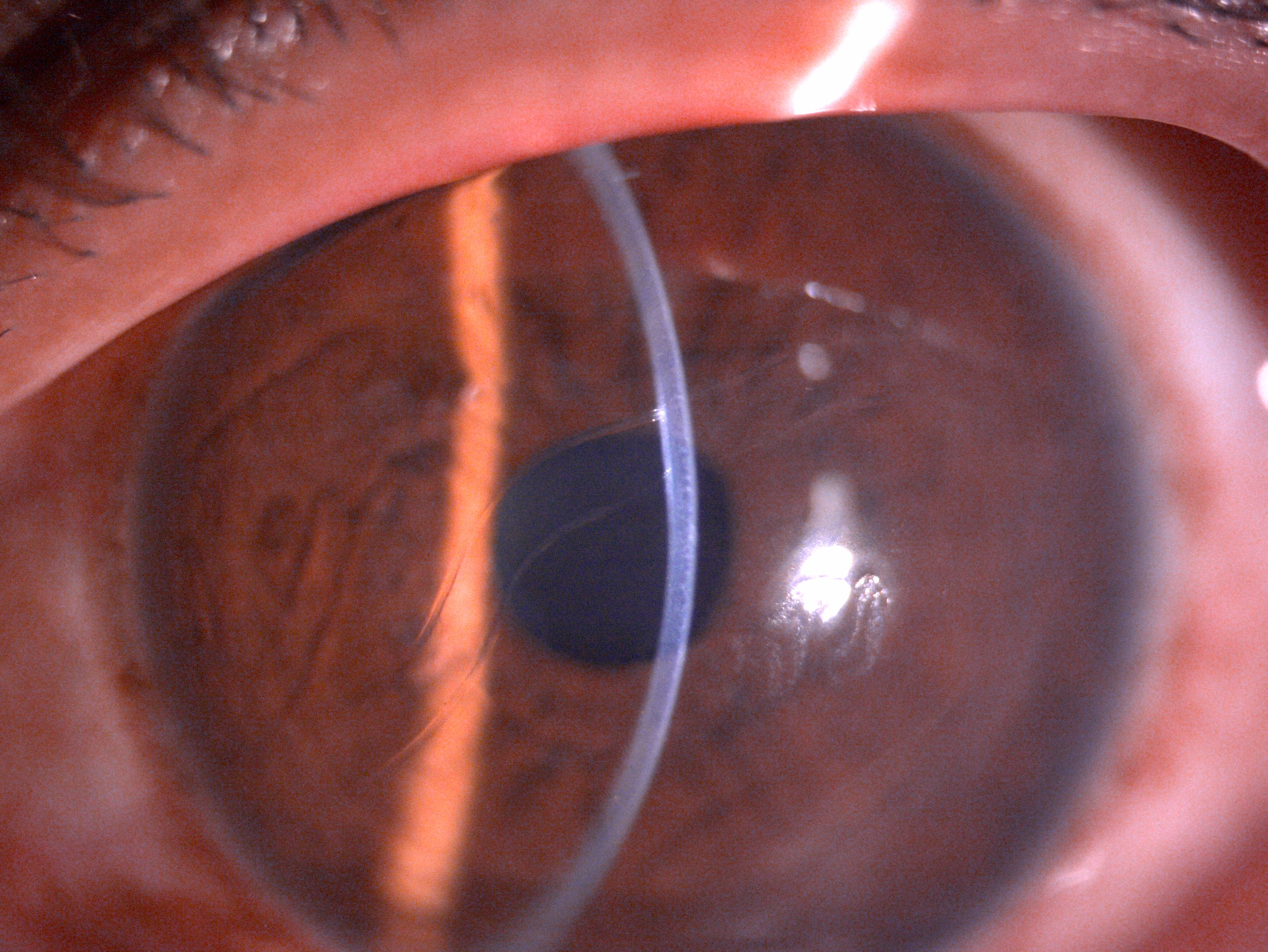
  • Cornea – Corneal examination might reveal signs of corneal enlargement, also called buphthalmos. Normal corneal size from birth to up to 6 months should be between 9.5 to 11.5 mm. A size of more than 12 mm should raise the suspicion of glaucoma. A corneal diameter of more than 13 mm in any child thereafter is also an indicator of corneal enlargement. The slit-lamp examination might reveal tears and breaks in the Descemet’s membrane called Haab’s striae (horizontal or oblique tears in the Descemet membrane). Another important finding is corneal edema. This usually starts as epithelial edema and then gradually involves the deeper layers of the cornea, occasionally causing permanent opacities impairing vision profoundly.
  • Anterior chamber- The anterior chamber is usually deep.
  • Iris – should be looked for iridodonesis, ectropion uvea, hypoplasia, or any atrophic patches.
  • Pupil - May be oval, dilated, and ischemic.
  • Lens – the clinician should look for lenticular opacities or any subluxation of the lens due to excessive stretching of zonules.
  • Optic disc- this typically shows reversible cupping in the early stages. Later stages may present with an enlarged cup disc ratio or even atrophy.
  • Intraocular pressure – IOP is usually raised at presentation and can be measured using a pneumotonmeter in the outpatient department.

Evaluation

Any suspicion of congenital glaucoma warrants an evaluation under anesthesia for detailed examination and planning of further management.[15] An examination under anesthesia should include the following

  • Cornea – Detailed corneal evaluation as explained above should be done along with measuring corneal diameter with eth help of calipers. In addition, a hand help slit lamp can be used to evaluate the cornea and anterior segment details.
  • Ophthalmoscopy – Detailed dilated fundus evaluation should be done. The size of the optic disc, cupping should be represented diagrammatically for future reference. Any abnormal vessels or other significant findings should be well documented.
  • Intraocular pressure (IOP) – IOP can be measured using a Schiotz or handheld Perkin’s applanation tonometer, or a pneumotonometer/tonopen can also be used. The effect of the anesthesia used on IOP should be kept in mind. These children usually have IOP within the range of 30 to 40 mmHg, resulting in corneal epithelial edema.
  • Gonioscopy – Gonioscopic examination can be done with the help of Koeppe’s lens; this may reveal trabeculodysgenesis with a flat or concave iris insertion.
  • Pachymetry – Corneal thickness should be measured in patients as this can affect the intraocular pressure readings. The corneal thickness will be increased in patients with central corneal edema.
  • Axial length – This can be measured using an ultrasound A. At birth, the axial length is around 18 mm, increasing to 22 mm around two years of age. In patients with primary congenital glaucoma, an increased axial length can be secondary to stretch due to increased intraocular pressure.
  • Cycloplegic retinoscopy – An increased axial length might lead to axial myopia. Thus it is important to evaluate for any underlying refractive errors in these patients.

In children around the age of 7 to 8 years, with reasonably good visual acuity, good fixation, and no nystagmus, perimetry can be tried to assess for peripheral visual field loss. SITA-FAST algorithm is preferred in these children.

When the child becomes cooperative and eligible for slit lamp examination, optical coherence tomography (OCT) can be attempted in these children to evaluate the retinal nerve and ganglion cell layer. Handheld and spectral-domain OCT are upcoming modalities for retinal nerve fiber and ganglion cell layer assessment while performing an evaluation under anesthesia.

Genetic screening, analysis, and pedigree chart evaluation can be performed in these cases.

Treatment / Management

Management of patients diagnosed with primary congenital glaucoma depends on age and severity of disease at the time of diagnosis. Though the mainstay of treatment is a surgical intervention, an initial medical course may be needed in few patients depending on their presentation.

The gold standard treatment for primary congenital glaucoma is angle surgery in the form of either goniotomy or trabeculotomy to alleviate IOP by enhancing aqueous outflow. When the angle surgery fails, trabeculectomy with mitomycin C or a glaucoma drainage device like Ahmed or Baerveldt should be attempted. In recalcitrant cases, cycloablation can be attempted by using diode cyclophotocaogulation, Nd YAG, or cryoablation. Topical or oral antiglaucoma drugs are used for decreasing corneal edema, control of IOP, and improve visualization for performing goniotomy 

Medical Treatment 

This approach is often an initial treatment measure to lower the IOP to decrease corneal edema, improve angel visualization, or until any surgical procedure is planned, or during the waiting period until fitness is obtained from a pediatrician or anesthetist for the procedure. Medical treatment is in the form of topical drugs, which act by either decreasing the aqueous production or increasing the aqueous outflow. Medications that decrease aqueous humor production include alpha-adrenergic agonists like brimonidine, apraclonidine, or beta-blockers like timolol, betaxolol, and levobetaxolol, or carbonic anhydrase inhibitors like brinzolamide.

Drugs that act by increasing aqueous outflow include parasympathomimetic drugs or prostaglandin analogs like travoprost. Alfa agonists, particularly brimonidine, should be avoided in children younger than two years of age and used with caution in children younger than six years of age, as these can cross the blood-brain barrier and may cause respiratory depression, apnea, and drowsiness.[16]

Surgical Treatment

Surgery is the mainstay of treatment of patients with primary congenital glaucoma. The type of surgical procedure depends on the disease severity, cornea clarity, and surgeon’s choice and experience.

Surgical procedures for primary congenital glaucoma can be broadly divided into 

Angle Procedures

  • These procedures work by increasing the aqueous outflow by decreasing the resistance in the aqueous pathway due to angle anomaly. The angle procedures are further of two types based on internal or external approach.
  • Internal approach using goniotomy - This was initially described by Barkan.[17] In this technique, a Barkan’s knife is passed through the limbus into the angle approximately between the root of the iris and Schwalbe’s ring, under a gonioscopic view, which is then extended through 75 degrees. The pre-requisite for this procedure is a relatively clear cornea which enables clear visualization of angle structures. Success rates depend on cornea clarity, the severity of disease, and the number of goniotomies performed. Studies have reported variable success rates from 70 to 90%.[18]
  • External approach – Trabeculotomy. This technique was first described by Smith.[19] This is useful in cases with hazy cornea due to corneal clouding or where Goniotomy procedures have failed. In this technique, a conjunctival flap is made, followed by a partial thickness scleral flap. A scleral incision is made, and the Schlemm canal is exposed. The lower prong of Harm’s trabeculotome is passed along the Schlemm’s canal on the inner side, and the upper prong visible outside serves as a guide. The trabeculotome is then rotated towards the anterior chamber to break the inner wall of the canal. A similar procedure is repeated on the other side through the 12 o‘clock opening, thus opening 120 to 180 degrees of the trabeculum.
  • A new modification using a prolene suture has been described as a modification procedure. This allows 360-degree trabeculotomy and allows to open the entire angle in a single sitting.[20] The studies have reported a reduction in intraocular pressure by 75 to 90% following trabeculotomy.[21]

Filtration Procedures

  • Trabeculectomy – This was first described by Cairns.[22] Trabeculectomy procedures alone are not much performed in PCG due to its limited success rates due to exaggerated healing responses experienced in children.
  • The use of antifibrotic agents like Mitomycin- C, 5-fluorouracil has shown promising results in improving the success rates. Success rates from 52 to 82% have been reported following anti-metabolite-assisted trabeculectomy procedures in PCG patients.[23]
  • Deep sclerectomy – This procedure involves elevation of partial-thickness scleral flap and removal of the external portion of Schlemm’s canal and outer part of the trabecular meshwork, including juxtacanalicular tissue without full penetration inside the eye.
  • Glaucoma Drainage Devices (GDD) – The role of GDD in primary congenital glaucoma as a primary procedure is limited. GDD implantation in primary congenital glaucoma is considered in cases where primary angle procedures have failed or in advanced refractory cases. Studies have shown a reduction of intraocular pressure by 28 to 49% following one year post-surgery.[24]

Combined Trabeculotomy and Trabeculectomy (CTT)

  • This procedure is attempted when the previous trabeculectomy has failed, or there is difficulty in cannulating the Schlemm canal. In this, the trabeculectomy is added to the trabeculotomy. A block of tissue is removed from the sclera aided by surgical peripheral iridectomy. Care is necessary when using mitomycin C.

Cyclodestructive Procedures

  • This procedure is reserved for eyes with poor or nil visual prognosis. The success rate in these cases is close to 30% only.[25] A newer modification in the form of a micropulse transscleral diode laser is a better and safer alternative.[26]

Differential Diagnosis

 The differential diagnosis of primary congenital glaucoma can be remembered by the following pneumonic STUMPED. 

  • S - Sclerocornea
  • T - trauma, tears in Descemet membrane
  • U - ulcer- viral, fungal, bacterial, neurotrophic, pythium[27][28][29]
  • M - Metabolic disorders - mucolipidoses, mucopolysaccharidosis, tyrisinosis
  • P - Peters anomaly
  • E - Endothelial dystrophy, congenital hereditary endothelial dystrophy, Posterior polymorphous dystrophy, Fuch dystrophy
  • D - Dermoid

Other significant differentials which should be kept in mind include:

  • Interstitial keratitis
  • High myopia 
  • Megalocornea
  • Corneal abrasion
  • Messman dystrophy
  • Reis Buckler dystrophy
  • Retinoblastoma
  • Retinopathy of prematurity 
  • Persistent primary hyperplastic vitreous
  • Traumatic glaucoma[30]
  • Congenital rubella syndrome
  • Sturge Weber syndrome 
  • Aniridia
  • Optic disc pit
  • Optic atrophy[31] 
  • Coloboma

Prognosis

The overall prognosis in primary congenital glaucoma depends on time and severity of presentation, age of presentation, and corneal clarity. Early diagnosis and management are the keys to good visual potential in these cases. Corneal edema and optic nerve head changes are reversible if timely intervention is done. Children need to be screened for any associated refractive errors from axial elongation of the eyeball and appropriately managed for the same. Intraocular pressures and optic nerve head need evaluation at regular intervals for lifelong. Late presentations can lead to sight-threatening complications.

A study from the United States showed that there was a lack of progression following adequate treatment in 90.3% at 1 year, 83.1% at 5 years, 70.8% at 10 years, and 58.3% at 34 years. Thus, highlighting the importance of appropriate management and follow-ups of these patients.[32] Another study showed that angle procedures were 90% successful among patients presenting between 2 months and 1 year of age, compared to 50% among those presenting either in infantile or late-onset or late-recognized cases.[33]

Complications

Complications in primary congenital glaucoma can be related to disease or surgery or anesthesia-related. Patients having delayed presentations may develop significant visual impairment or glaucomatous optic atrophy, or visual field defects. Development of corneal and Haab’s striae may later lead to permanent opacification of the cornea, thus hampering vision significantly. Stretching of the eyeball due to elevated pressures may lead to axial myopia or even astigmatism. Refractive correction needs to be given and followed up lifelong, as untreated cases can develop refractive amblyopia.[34] Further, these patients are at risk of lens dislocation or retinal complications.

Surgical Complications 

  • Hyphema 
  • Shallow anterior chamber[35]
  • Peripheral anterior synechiae
  • Iridodialysis
  • Cyclodialysis
  • Cataract[36][37][38]
  • Epithelial ingrowth
  • Choroidal detachment
  • Retinal detachment
  • Phthisis bulbi

Filtering Procedure-related Complications

  • Over or under filtration
  • Blebitis
  • Vitreous loss
  • Scleral collapse
  • Scleral flap leak
  • Tube lens touch
  • Endothelial decompensation from tube cornea touch
  • Tube erosion
  • Implant migration
  • Diplopia from implant-related restrictions
  • Endophthalmitis

Cyclodestructive Procedure-related Complications

  • Hypotony
  • Retinal detachment[39]
  • Phthisis

Anesthesia-related Complications 

  • Oculocardiac reflex
  • Anaphylaxis
  • Malignant hyperthermia
  • Cardiovascular collapse
  • Hepatic porphyria
  • Hypoxic brain injury

Postoperative and Rehabilitation Care

The children undergoing surgery should be started on topical steroids, either prednisolone 1% or dexamethasone 0.1% 8/7/6/5/4/3/2/1 for one week each in tapering doses. In addition, a topical antibiotic in the form of tobramycin 0.3% or 0.3% moxifloxacin or gatifloxacin four times per day for 20 days should be supplemented to prevent secondary infection. These patients will need close follow-ups post-surgery to look for any signs of hypotony, inflammation, or infection. Moreover, the intraocular pressures need to be recorded every 3 to 4 months for at least two years post-surgery.

Cycloplegic refraction will be needed every six months for these patients. Moreover, lifelong regular follow-up is needed every six months for intraocular pressure monitoring and early detection of any surgery-related complications. Cases failing angle or filtration surgery should be counseled for the need of GDD and risk of subsequent failure, amblyopia, blindness, and phthisis bulbi.

Consultations

Any child with clinical suspicion of primary congenital glaucoma presenting to the outpatient department must be evaluated meticulously by an ophthalmologist. The general ophthalmologist should start the child on antiglaucoma medications and must refer the child to a glaucoma specialist for confirmation of diagnosis and targeted treatment. The glaucoma specialist should decide for evaluation under anesthesia and surgical intervention of the child in the form of goniotomy or trabeculectomy.

Deterrence and Patient Education

Primary congenital glaucoma is a treatable disease, provided the condition is diagnosed early, and timely intervention is done. Thus, it is very important to educate the parents, general clinicians, and optometrists to ensure early suspicion and timely referral to the ophthalmologists. Any signs of enlarged eyes, watering, redness, shying away from light should be reported to the health care team at the earliest. Proper parental education about early signs and symptoms can prevent treatable blindness from primary congenital glaucoma.

Management options should be explained to the parents, and a well-informed decision should be made. It is also essential to create awareness about the need for regular follow-ups and compliance with the treatment advised. The need for follow-up with optometrists for regular refraction should also be emphasized. In patients with low vision, appropriate mobilization for low vision clinics should be ensured at an early age to help the child and parents understand the need for lifestyle and educational modifications.

Pearls and Other Issues

To sum up, primary congenital glaucoma is a vision-threatening ocular pathology in the pediatric age group. Children are highly vulnerable due to lack of communication, difficulty performing an examination, compliance to treatment, and regular follow-up. Any pediatric patient presenting to the outpatient department with lacrimation, photophobia, blepharospasm, buphthalmos, and corneal clouding should undergo a detailed clinical evaluation to diagnose the condition.

While managing these children, all the other differentials must be carefully ruled out for targeted treatment and solving the eye with good visual outcomes. The parents should be counseled regarding the importance of genetic and family screening in these cases. Angle and filtration surgery are the treatment of choice for good anatomical and visual outcomes.

Enhancing Healthcare Team Outcomes

Early diagnosis and timely intervention are the key components for successfully managing patients with primary congenital glaucoma. The entire healthcare team needs to be aware of their roles whenever they come in contact with these patients. A high level of suspicion by the optometrists, staff nurses, and general physicians in any patient with enlarged eyeballs or excessive watering, hazy corneas, abnormal congestion, photophobia, high myopias in infancy or early childhood should be maintained.

There should be a relatively lower threshold maintained in referring these children to ophthalmologists for confirmation of diagnosis. It is also important to educate parents about the need for immediate referrals and that delay in treatment can lead to significant ocular co-morbidities and visual compromise lifelong. Ophthalmologists play an important role in diagnosing and treating these cases. An evaluation under anesthesia should be planned for investigating and any surgical intervention if needed. Close interprofessional communication for examination by pediatrician, anesthetist, and ophthalmologist with the help of staff nurses, the counselor will be needed for successful outcomes in these patients.


(Click Image to Enlarge)
Slit lamp image of the child depicting megalocornea and horizontal oblique breaks in descemet membrane suggestive of Haab's s
Slit lamp image of the child depicting megalocornea and horizontal oblique breaks in descemet membrane suggestive of Haab's striae secondary to raised intraocular pressure and stretching of the cornea
Contributed by Dr. Kirandeep Kaur, MBBS, DNB, FPOS, FICO (UK), MRCS Ed (Ophthalmology), MNAMS

(Click Image to Enlarge)
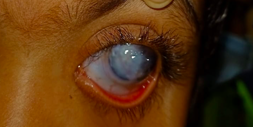
Slit lamp image of the child depicting mild conjunctival congestion, lower tarsal conjunctival hyperaemia, megalocornea, near
Slit lamp image of the child depicting mild conjunctival congestion, lower tarsal conjunctival hyperaemia, megalocornea, near total corneal opacification with deep stromal vascularization suggestive of long standing chronic primary congenital glaucoma
Contributed by Dr. Kirandeep Kaur, MBBS, DNB, FPOS, FICO (UK), MRCS Ed (Ophthalmology). MNAMS

(Click Image to Enlarge)
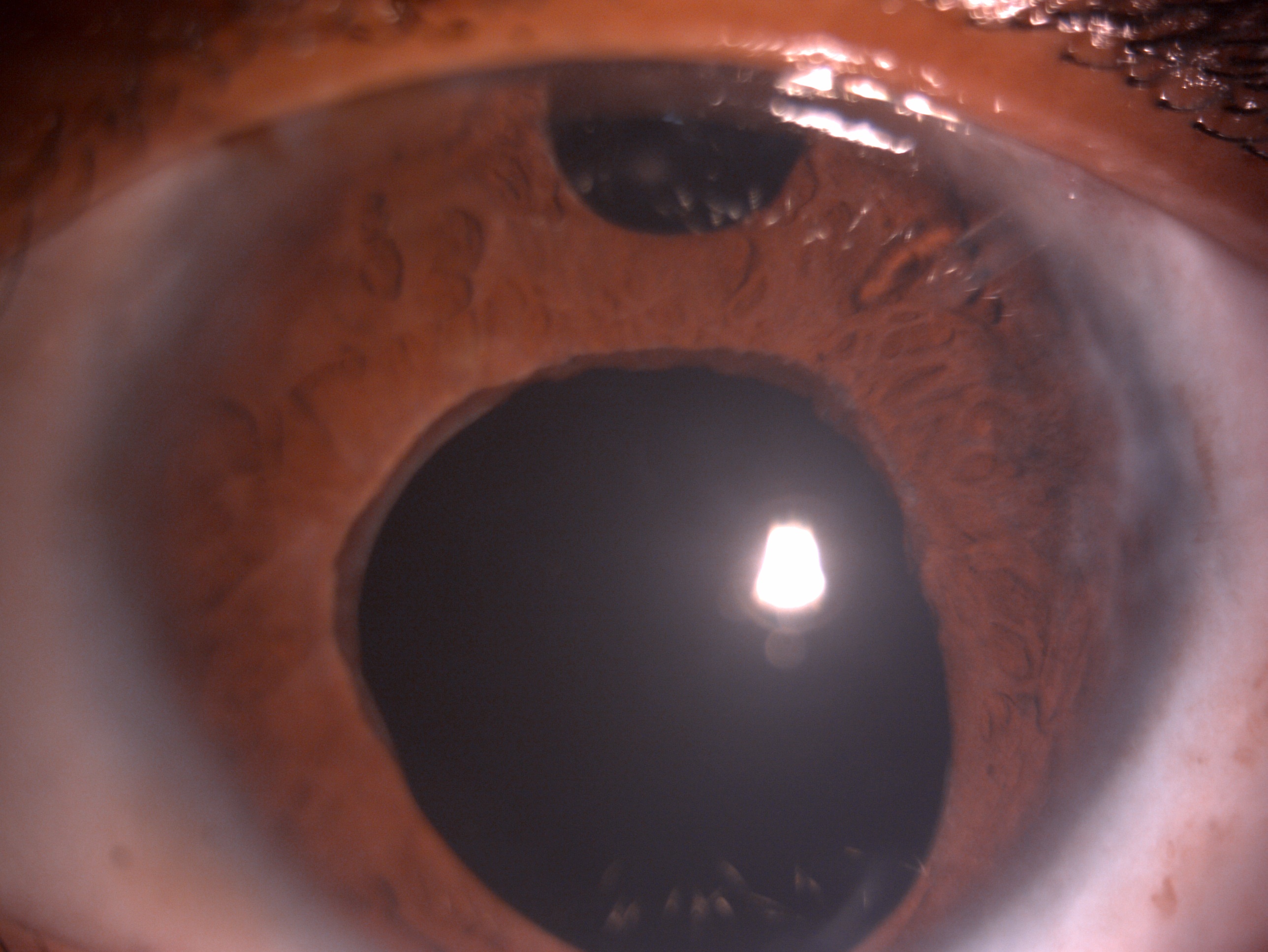
Slit lamp image of the child depicting megalocornea, tube shunt in the anterior chamber, surgical peripheral iridectomy, dila
Slit lamp image of the child depicting megalocornea, tube shunt in the anterior chamber, surgical peripheral iridectomy, dilated ischemic pupil and aphakia post glaucoma drainage device for recalcitrant primary congenital glaucoma refractory to angle and filtration surgery
Contributed by Dr. Kirandeep Kaur, MBBS, DNB, FPOS, FICO (UK), MRCS Ed (Ophthalmology). MNAMS
Details

Editor:

Bharat Gurnani

Updated:

6/11/2023 6:25:35 AM

References


[1]

Badawi AH,Al-Muhaylib AA,Al Owaifeer AM,Al-Essa RS,Al-Shahwan SA, Primary congenital glaucoma: An updated review. Saudi journal of ophthalmology : official journal of the Saudi Ophthalmological Society. 2019 Oct-Dec;     [PubMed PMID: 31920449]


[2]

Hoskins HD Jr,Shaffer RN,Hetherington J, Anatomical classification of the developmental glaucomas. Archives of ophthalmology (Chicago, Ill. : 1960). 1984 Sep;     [PubMed PMID: 6477252]


[3]

Sarfarazi M,Akarsu AN,Hossain A,Turacli ME,Aktan SG,Barsoum-Homsy M,Chevrette L,Sayli BS, Assignment of a locus (GLC3A) for primary congenital glaucoma (Buphthalmos) to 2p21 and evidence for genetic heterogeneity. Genomics. 1995 Nov 20;     [PubMed PMID: 8586416]


[4]

Fan BJ,Wiggs JL, Glaucoma: genes, phenotypes, and new directions for therapy. The Journal of clinical investigation. 2010 Sep;     [PubMed PMID: 20811162]


[5]

Ali M,McKibbin M,Booth A,Parry DA,Jain P,Riazuddin SA,Hejtmancik JF,Khan SN,Firasat S,Shires M,Gilmour DF,Towns K,Murphy AL,Azmanov D,Tournev I,Cherninkova S,Jafri H,Raashid Y,Toomes C,Craig J,Mackey DA,Kalaydjieva L,Riazuddin S,Inglehearn CF, Null mutations in LTBP2 cause primary congenital glaucoma. American journal of human genetics. 2009 May;     [PubMed PMID: 19361779]


[6]

Tamçelik N,Atalay E,Bolukbasi S,Çapar O,Ozkok A, Demographic features of subjects with congenital glaucoma. Indian journal of ophthalmology. 2014 May;     [PubMed PMID: 24881602]


[7]

Alabdulwahhab KM,Ahmad MS, Visual Impairment and Blindness in Saudi Arabia's School for the Blind: A Cross-Sectional Study. Clinical optometry. 2020;     [PubMed PMID: 33117027]

Level 2 (mid-level) evidence

[8]

Yu-Wai-Man C,Arno G,Brookes J,Garcia-Feijoo J,Khaw PT,Moosajee M, Primary congenital glaucoma including next-generation sequencing-based approaches: clinical utility gene card. European journal of human genetics : EJHG. 2018 Nov;     [PubMed PMID: 30089822]


[9]

deLuise VP,Anderson DR, Primary infantile glaucoma (congenital glaucoma). Survey of ophthalmology. 1983 Jul-Aug;     [PubMed PMID: 6353647]

Level 3 (low-level) evidence

[10]

Ohtake Y,Tanino T,Suzuki Y,Miyata H,Taomoto M,Azuma N,Tanihara H,Araie M,Mashima Y, Phenotype of cytochrome P4501B1 gene (CYP1B1) mutations in Japanese patients with primary congenital glaucoma. The British journal of ophthalmology. 2003 Mar;     [PubMed PMID: 12598442]


[11]

BARKAN O, Pathogenesis of congenital glaucoma: gonioscopic and anatomic observation of the angle of the anterior chamber in the normal eye and in congenital glaucoma. American journal of ophthalmology. 1955 Jul;     [PubMed PMID: 14388087]


[12]

Tawara A, Inomata H. Developmental immaturity of the trabecular meshwork in congenital glaucoma. American journal of ophthalmology. 1981 Oct:92(4):508-25     [PubMed PMID: 7294114]


[13]

García-Antón MT,Salazar JJ,de Hoz R,Rojas B,Ramírez AI,Triviño A,Aroca-Aguilar JD,García-Feijoo J,Escribano J,Ramírez JM, Goniodysgenesis variability and activity of CYP1B1 genotypes in primary congenital glaucoma. PloS one. 2017;     [PubMed PMID: 28448622]


[14]

Papadopoulos M,Cable N,Rahi J,Khaw PT,BIG Eye Study Investigators., The British Infantile and Childhood Glaucoma (BIG) Eye Study. Investigative ophthalmology     [PubMed PMID: 17724193]


[15]

Beck AD, Diagnosis and management of pediatric glaucoma. Ophthalmology clinics of North America. 2001 Sep;     [PubMed PMID: 11705150]


[16]

Lewis RA,Christie WC,Day DG,Craven ER,Walters T,Bejanian M,Lee SS,Goodkin ML,Zhang J,Whitcup SM,Robinson MR,Bimatoprost SR Study Group., Bimatoprost Sustained-Release Implants for Glaucoma Therapy: 6-Month Results From a Phase I/II Clinical Trial. American journal of ophthalmology. 2017 Mar;     [PubMed PMID: 28012819]

Level 1 (high-level) evidence

[17]

Ghate D,Wang X, Surgical interventions for primary congenital glaucoma. The Cochrane database of systematic reviews. 2015 Jan 30;     [PubMed PMID: 25636153]

Level 1 (high-level) evidence

[18]

Broughton WL,Parks MM, An analysis of treatment of congenital glaucoma by goniotomy. American journal of ophthalmology. 1981 May;     [PubMed PMID: 7234937]


[19]

SMITH R, A new technique for opening the canal of Schlemm. Preliminary report. The British journal of ophthalmology. 1960 Jun;     [PubMed PMID: 13832124]


[20]

Mendicino ME,Lynch MG,Drack A,Beck AD,Harbin T,Pollard Z,Vela MA,Lynn MJ, Long-term surgical and visual outcomes in primary congenital glaucoma: 360 degrees trabeculotomy versus goniotomy. Journal of AAPOS : the official publication of the American Association for Pediatric Ophthalmology and Strabismus. 2000 Aug;     [PubMed PMID: 10951295]


[21]

McPherson SD Jr,Berry DP, Goniotomy vs external trabeculotomy for developmental glaucoma. American journal of ophthalmology. 1983 Apr;     [PubMed PMID: 6837685]


[22]

Cairns JE, Trabeculectomy. Preliminary report of a new method. American journal of ophthalmology. 1968 Oct;     [PubMed PMID: 4891876]


[23]

Sidoti PA,Belmonte SJ,Liebmann JM,Ritch R, Trabeculectomy with mitomycin-C in the treatment of pediatric glaucomas. Ophthalmology. 2000 Mar;     [PubMed PMID: 10711876]


[24]

Razeghinejad MR,Kaffashan S,Nowroozzadeh MH, Results of Ahmed glaucoma valve implantation in primary congenital glaucoma. Journal of AAPOS : the official publication of the American Association for Pediatric Ophthalmology and Strabismus. 2014 Dec;     [PubMed PMID: 25459201]


[25]

al Faran MF,Tomey KF,al Mutlaq FA, Cyclocryotherapy in selected cases of congenital glaucoma. Ophthalmic surgery. 1990 Nov;     [PubMed PMID: 2270165]

Level 3 (low-level) evidence

[26]

Elhefney EM,Mokbel TH,Hagras SM,AlNagdy AA,Ellayeh AA,Mohsen TA,Gaafar WM, Micropulsed diode laser cyclophotocoagulation in recurrent pediatric glaucoma. European journal of ophthalmology. 2020 Sep;     [PubMed PMID: 31256680]


[27]

Gurnani B,Christy J,Narayana S,Rajkumar P,Kaur K,Gubert J, Retrospective multifactorial analysis of {i}Pythium keratitis{/i} and review of literature. Indian journal of ophthalmology. 2021 May;     [PubMed PMID: 33913840]

Level 2 (mid-level) evidence

[28]

Gurnani B,Narayana S,Christy J,Rajkumar P,Kaur K,Gubert J, Successful management of pediatric pythium insidiosum keratitis with cyanoacrylate glue, linezolid, and azithromycin: Rare case report. European journal of ophthalmology. 2021 Mar 28;     [PubMed PMID: 33779337]

Level 3 (low-level) evidence

[29]

Gurnani B, Kaur K. Pythium Keratitis. StatPearls. 2024 Jan:():     [PubMed PMID: 34424645]


[30]

Balamurugan S, Gurnani B, Kaur K, Gireesh P, Narayana S. Traumatic intralenticular abscess-What is so different? The Indian journal of radiology & imaging. 2020 Jan-Mar:30(1):92-94. doi: 10.4103/ijri.IJRI_369_19. Epub 2020 Mar 30     [PubMed PMID: 32476758]


[31]

Kaur K,Gurnani B,Devy N, Atypical optic neuritis - a case with a new surprise every visit. GMS ophthalmology cases. 2020;     [PubMed PMID: 32269909]

Level 3 (low-level) evidence

[32]

de Silva DJ,Khaw PT,Brookes JL, Long-term outcome of primary congenital glaucoma. Journal of AAPOS : the official publication of the American Association for Pediatric Ophthalmology and Strabismus. 2011 Apr     [PubMed PMID: 21596293]


[33]

Shaffer RN, Prognosis of goniotomy in primary infantile glaucoma (trabeculodysgenesis). Transactions of the American Ophthalmological Society. 1982;     [PubMed PMID: 7182965]


[34]

Kaur K, Kannusamy V, Mouttapa F, Gurnani B, Venkatesh R, Khadia A. To assess the accuracy of Plusoptix S12-C photoscreener in detecting amblyogenic risk factors in children aged 6 months to 6 years in remote areas of South India. Indian journal of ophthalmology. 2020 Oct:68(10):2186-2189. doi: 10.4103/ijo.IJO_2046_19. Epub     [PubMed PMID: 32971637]


[35]

Christy J,Jain N,Gurnani B,Kaur K, Twinkling Eye -A Rare Presentation in Neovascular Glaucoma. Journal of glaucoma. 2019 May 23;     [PubMed PMID: 31135586]


[36]

Gurnani B,Kaur K,Sekaran S, First case of coloboma, lens neovascularization, traumatic cataract, and retinal detachment in a young Asian female. Clinical case reports. 2021 Sep;     [PubMed PMID: 34484773]

Level 3 (low-level) evidence

[37]

Gurnani B,Kaur K,Gireesh P, A rare presentation of anterior dislocation of calcified capsular bag in a spontaneously absorbed cataractous eye. Oman journal of ophthalmology. 2021 May-Aug;     [PubMed PMID: 34345149]


[38]

Gurnani B,Kaur K,Gireesh P, Rare Coexistence of Bilateral Congenital Sutural and Cortical Blue Dot Cataracts. Journal of pediatric ophthalmology and strabismus. 2020 Jan 1;     [PubMed PMID: 31972045]


[39]

Kaur K,Gurnani B,Kannusamy V,Yadalla D, A tale of orbital cellulitis and retinopathy of prematurity in an infant: First case report. European journal of ophthalmology. 2021 Jun 17;     [PubMed PMID: 34137305]

Level 3 (low-level) evidence