Intraocular Foreign Body

Earn CME/CE in your profession:


Continuing Education Activity

Intraocular foreign body (IOFB) is an accidental injury usually resulting from chisel hammering and firearm use. It is a serious ocular condition that can potentially cause permanent vision loss. To avoid the high morbidity associated with this condition, it must be promptly diagnosed and treated. This activity reviews the evaluation and treatment of an IOFB and highlights the role of the interprofessional team in evaluating and treating patients with this condition.

Objectives:

  • Explain the pathophysiology and review the risk factors for IOFB.
  • Summarize the typical presentation of a patient with IOFB.
  • Describe the common physical findings in patients with IOFB.
  • Review the management considerations for patients with IOFB.

Introduction

Ocular injuries are a major cause of blindness.[1] They continue to be a public health problem worldwide.[1][2]  Among these, intraocular foreign bodies (IOFB) are a major cause of morbidity in the young population.[3] IOFB associated ocular trauma is unique and requires skillful investigation with early treatment. IOFB is mostly metallic.[4] They are usually generated during chisel hammering, drilling, gunshot, or bomb blast. Manual laborers are at high risk.[5]

They can lodge in any part of the eye, either in the anterior or posterior segment. Since they are sharp and are generated at high speed, they mostly get lodged in the posterior segment. According to a study, about 2/3rd of IOFBs were found in the posterior segment.[6] With the advancement of surgical techniques, many eyes can be saved, and good final visual acuity can be achieved.[7] Visual prognosis depends upon multiple factors, including size, site, the composition of foreign body, inflammatory response, tissue damage, time since injury, and any associated endophthalmitis and retinal detachment.[8]

Etiology

Hammering is the most common mode of injury.[9] According to a study, the mechanism of injury was hammering in 43%, metal cutting in 19%, chiseling and drilling in 10%, gardening in 5%, fall from height in 5%, and unknown in 14% of cases.[9] Other modes are firearm and blast injuries that are common in war zones.[10]

Epidemiology

In the USA, the incidence of ocular injuries is 2.4 million cases.[9] IOFBs account for 17 to 41% of open globe injuries.[11][12] The majority of IOFBs occur in young working males.[13][14][15][16][17]. Home-related works account for only a minority (13%) of injuries.[6]

Workers and farmers are the two most common occupations because they are involved in hammering, drilling, chiseling, and shoveling. These activities generate more foreign bodies. The annual incidence rate of IOFBs is approximately 0.16 per 100,000 in the United Kingdom.[6] But only 0.77 to 6% of patients with an IOFB wear protective eyewear.[18][19][20] Thus, adequate safety measures at work should be taken.

Pathophysiology

Ocular injury caused by an IOFB depends on its velocity, size, nature, entry site, and impaction.[21] Small and sharp IOFB like iron chips cause a small and linear perforation at the entry site. Such perforations are easy to repair. Large, irregular projectiles like stone particles cause a ragged and large wound at the entry site. They cause significant tissue damage and are difficult to repair. IOFBs generated at high speed generally lodge in the posterior segment of the eyeball. They can also ricochet inside the eye, causing injuries at multiple sites.[14] 

Metallic foreign bodies are the most common type.[20] Iron and copper foreign bodies are highly reactive. Fe and Cu can cause metallosis.[22] They release metallic ions, which deposit in the various ocular tissues.

Ocular damage with iron foreign body results in siderosis bulbi. It is a pigmentary, degenerative process due to chronic retention of an iron IOFB.[23] The iron particles get deposited in structures of epithelial origin such as the cornea, lens, trabecular meshwork, iris, and retina. In the cornea, the pigments get deposited in the stroma or the endothelium.[24] In the iris, iron deposition is noted in the stroma and epithelium, leading to greenish-brown discoloration and iris heterochromia.[25]

The pupil may become fixed and dilated. The pupil may show denervation hypersensitivity.[26] Iron deposition causes pigmented trabecular meshwork and secondary open-angle glaucoma.[27] Cataracts may be seen with anterior subcapsular deposits. Siderosis affects the retinal pigment epithelium resulting in retinal degeneration.[28] 

An inflammatory reaction to a copper foreign body results in chalcosis.[29] The ocular fluid dissolves the metal, which gets deposited in the corneal stroma, Descemet membrane,  endothelium, anterior lens capsule, internal limiting membrane, and superficial retina.[30][31] The clinical manifestation depends upon the copper content. The copper content of more than 85% presents with supportive endophthalmitis and disseminated copper deposits.[29][32][33] Copper content less than 85% presents with localized copper deposits (chalcosis bulbi).[29] Iron ions are deposited intracellularly, and the deposition of copper ions is seen in basement membranes.

Organic foreign bodies such as animal hairs, insect parts, thorn, and vegetable matter are contaminated and cause fulminant endophthalmitis.[34] Foreign bodies such as glass and plastic are inert.[35] They are well-tolerated and do not cause any significant reaction.[36] In blast or firearm injuries, multiple splinters are generated, which can lodge in the various parts of the eye. They are more damaging and difficult to remove.[10] Foreign bodies entering through the cornea cause significant corneal scarring and may have a bad prognosis.[21]

Histopathology

Long-term retention of copper and iron foreign body can cause chalcosis and siderosis, respectively.[37] Histologically, Prussian blue stains the iron blue.[38] Blue staining can be seen in the epithelial structures and areas of scarred trabecular meshwork and scarred retina. On electron microscopy, intracellular sideromes can be seen in the corneal keratocytes, lens epithelium, and outer segments of the photoreceptors.[39] Copper deposits in the ocular tissue can be detected by staining with rhodanine, rubeanic acid, and alizarin blue stains.[29]

History and Physical

A detailed history is essential to know about the mechanism of injury and the nature of the foreign body. Trivial injuries are often difficult to detect. The patient presents with conjunctival congestion and blurring of vision or may be asymptomatic.[40] A careful history that something hit the eye often reveals the diagnosis of an IOFB. History should be taken whether one or both eyes were involved or not. Firearm and blast injuries generate multiple small splinters, which can cause bilateral injuries. Proper history and documentation are essential due to medicolegal issues and large monetary compensation in cases of missed intraocular foreign body.

A meticulous examination is very important. Presenting and best-corrected visual acuity should be recorded. Lids and eyebrows should be examined to look for external signs of injuries and small foreign bodies. Slit-lamp examination is a must. Careful examination should be done to look for conjunctival laceration, hemorrhage, and entry site of IOFB. Pigment under the conjunctiva may indicate uveal prolapse and possible entry site. Corneal perforation should be examined.

The extent of the defect and its location should be recorded. A Seidel test should be performed. Iris should be inspected for transillumination defects and heterochromia (usually seen in longstanding cases). The entry wound of the cornea and iris may help in localizing the foreign body. The lens should be evaluated for pigmentation, focal opacity, cataractous changes, capsule rupture, and phacodonesis.

Gonioscopy is done to look for trabecular pigmentation, scarring, and foreign body, especially in the inferior angle of the anterior chamber. Intraocular pressure should be noted. However, in an obvious open globe injury, any undue pressure on the globe should be avoided as it may lead to extrusion of intraocular contents. Most foreign bodies are located in the posterior segment.

Dilated fundus examination is essential. The retina should be inspected to locate the site of impaction of the foreign body. Often a fibrous capsule forms over it, and there may be surrounding pigmentary changes. Optic atrophy may be present in longstanding cases. In cases of media haze due to cataracts, vitreous hemorrhage, or endophthalmitis, the USG (ultrasonogram) B scan is done to locate the foreign body and measure the size.

Evaluation

Detection and localization of the IOFBs are essential. Anterior segment photography using a slit-lamp or fundus camera helps document the disease and may be helpful in counseling the patient about their clinical condition.

X-ray of orbits (anteroposterior and lateral view) can detect metallic foreign bodies but cannot detect radiolucent objects like wood or glass. It also reveals multiple metallic foreign bodies, if present. X-ray orbit with a limbal ring-rod sutured at the limbus and a proportionate geometric drawing can be used to localize an intraocular IOFB.[41] Another method is the Sweet localization technique which involves frontal and lateral projections.[42]

The patient's X-ray is taken with his eye at primary gaze. Then he is asked to look sideways, and again an X-ray is taken. If the foreign body is intraocular, it will rotate with eye movement. If it rotates in the same direction as the eye, then the IOFB is in the anterior segment. If it rotates in the opposite direction, then it is in the posterior segment.

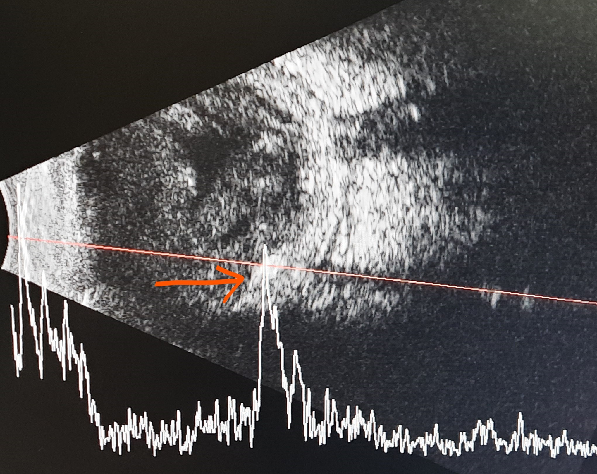
USG B scan is a cheap and useful investigation to detect metallic foreign bodies. The metallic body appears as a hyperechoic structure with acoustic shadowing and a high spike on A-scan. Aluminum, steel, and bottle glass usually show a flashlight artifact, a focused and narrow ring-down artifact.[43] Lead, windshield glass, copper, and silver usually show a headlight artifact which is a broad and dense artifact.[43] 

Ultrasound has varied detection rates for a glass IOFB ranging from 24 to 97%.[44] A glass foreign body can appear as a hyperechoic lesion with a back shadow, or it can show a comet tail (reverberation) artifact.[45] The wooden foreign body usually appears as a hyperechoic structure, and dry wood may show faint reverberations.[43] A plastic foreign body can be of low echogenicity (spectacle) or high echogenicity (polyvinyl chloride, PVC).[43] An air bubble is hyperechoic on B-scan and produces a single-peaked 100% high spike on A-scan.[46] USG can also detect retinal detachment, vitreous hemorrhage, and vitreous exudates. However, the USG B scan should be avoided in an open globe as the pressure from the ultrasonic probe may further increase the damage and may cause extrusion of ocular contents. USG B scans can, however, be done safely after the repair of the open globe.

CT (computed tomography) scan can accurately detect the number, size, shape, and location of a foreign body.[47] A wooden foreign body has low attenuation with no artifact. A plastic IOFB has moderate attenuation with no artifact. A glass foreign body has high attenuation and no artifact. A metallic foreign body has high attenuation with a shadow artifact and a 'scatter' artifact around the object's edge.[43] The CT can give the exact relation of ocular coats with the IOFB, as IOFBs, which are deeply buried in the ocular coats, may be difficult or impossible to remove surgically.[14] 

MRI (magnetic resonance imaging) can detect organic and glass objects with greater sensitivity. MRI is contraindicated in the metallic foreign body as it can dislodge it and cause further damage.[48] 

Ultrasound biomicroscopy (UBM) can detect a foreign body at the angle of the anterior chamber, behind the iris, and ciliary body. UBM uses a 50 MHz frequency and has a penetration of 5mm, which allows for finer resolution.[49][50] As it requires contact, it should be avoided in open globe injuries. The 35 MHz probes can image deeper tissue, including the ciliary body and pars plana.

Anterior segment optical coherence tomography (AS-OCT) is a non-contact imaging modality and helps visualize the anterior segment IOFBs.[51] It has greater patient comfort and compliance than UBM. However, it has poor penetration across the pigmented layer of the iris. Thus IOFBs lying behind the iris might be missed.[52] 

Patients with long-standing iron IOFB present with siderosis. The full-field electroretinogram (ERG) is done to know the status of the photoreceptors' function. In the early stages, increased amplitude of a-wave and b-wave is present (supernormal response).[53] This is because the photoreceptors start recovering from the sudden insult.[54] Then a steady decrease is seen in the b-wave amplitude with a reduction in b: a wave amplitude ratio to less than 1.[53] In end-stage diseases, both waves are extinguished.[55] 

Patients with chalcosis show reduced b-wave amplitude on ERG. A hyper-normal b-wave is not observed. Then a steady decrease is seen in the b wave amplitude. An abnormal ERG  precedes the clinical findings in almost 50% of patients.[56] However, the ERG changes in chalcosis are less severe than in siderosis: ERG waves decrease to 50% and remain stable at that level for many years.[57] Preoperative ERG helps to prognosticate the cases after IOFB removal. Based on the ERG findings, the patient can be counseled about the chances of visual recovery after a successful surgery.[27]

Multifocal ERG may detect subclinical siderosis bulbi even if full-field ERG is normal.[58]

Treatment / Management

The goal of surgery is to prevent further damage and restore the ocular anatomy as far as possible. Functional restoration or visual gain is the secondary goal. The visual prognosis is usually guarded if retinal detachment or endophthalmitis is present. The IOFB should be localized on the CT scan, whether it is intraocular or extraocular or impacting the visual pathway or not.

The injured eye should be protected with a shield. During the examination, the eye should be gently handled. External pressure should be avoided as it may expel the intraocular contents. Tetanus toxoid (intramuscular) injection should be given. The contaminated part should be cleaned with a sterile solution, and small superficial foreign bodies should be removed in the absence of an open globe. Broad-spectrum intravenous antibiotics may be considered as prophylaxis against endophthalmitis. Endophthalmitis is a dreaded complication and is associated with 7 to 13% of IOFB cases.[59] Iron and copper foreign bodies should be removed as soon as possible as they are highly reactive. Stainless steel and aluminum may be better tolerated.[60] Organic matter and stone pieces are contaminated and have a high risk of endophthalmitis.[61] They should be removed as early as possible.

The position of the IOFB should be preoperatively localized in the anterior or posterior segment. The first step is the primary repair of the ocular coats (cornea and/or sclera) of the open globe. Meticulous closure of the perforation site is done first.[62] The anterior chamber should be formed, and the repaired wound should not leak at the conclusion of the surgery.

IOFB in the anterior chamber: The IOFB might be located in the anterior chamber. The perforation site should be inspected. Its extent should be noted. Iris or uveal tissue prolapse is ascertained. The corneal perforation should be repaired with 10-0 monofilament nylon sutures. Scleral perforation is repaired with a 6-0 or 7-0 polyglactin 910 suture. The anterior chamber should be formed with viscoelastic solutions. Any exudates or hyphema in the anterior chamber should be washed first. Small and mobile IOFB can be removed with viscoexpression or an intraocular magnet. If it is immobile due to surrounding fibrosis, then the IOFB should be removed with forceps. 

The IOFB should not be removed through the entry wound.[16] A separate incision should be made for its removal. The anterior chamber aspirate can be sent for microbiological evaluation if there is hypopyon or evidence of infection. A thorough anterior chamber wash should be done to remove all the debris. If the anterior lens capsule is ruptured, the cortical matter is washed. Gentle lens aspiration can be done, or the patient can be taken in second sitting for lens aspiration and intraocular lens implantation.

Intralenticular foreign body: IOFB might be impacted in the crystalline lens. The cataractous lens should be evaluated for the position of IOFB and posterior capsule tear.[63] If the IOFB is impacted in the posterior capsule or a large posterior capsular rent is present, then the whole cataractous lens should be taken out along with the IOFB.[64] Hydrodissection should be avoided as it might cause the posterior capsular tear to enlarge. This might then cause the lens or the IOFB to drop into the vitreous. Thus the possibility of posterior capsular tears should always be kept in mind when devising a surgical plan. If a preexisting posterior capsular tear is possible, a gentle aspiration and nucleus expression is done, and one should be prepared for anterior vitrectomy.[63]

Foreign body in the anterior chamber angle: A foreign body must be ruled out in the angle when not observed on initial examination. Gonioscopy should be done gently.[65] An imaging study should be performed to confirm the diagnosis. CT scan, UBM, and ASOCT help diagnose by accurately estimating the foreign body size and location.[66] The foreign body may be removed by an external approach using a large electromagnet or through an internal approach by forceps or intraocular magnets.[67]

Posterior segment IOFB: Accurate localization is necessary preoperatively. CT scan accurately localizes it in the ocular coat, anterior segment, posterior segment, or extraocular location.[68] Vitreous surgery provides excellent visualization and allows the removal of hemorrhage and exudates. Three standard scleral ports are made. Lensectomy is done if the lens is cataractous, or cataract surgery through the clear corneal/limbal/scleral incision may be performed. If possible, the anterior capsular rim should be preserved to enable a ciliary sulcus intraocular lens implantation. Then core vitrectomy is done. A posterior vitreous detachment is induced. The IOFB is localized. The adhesions and fibrous capsules around it are excised. It is mobilized and brought over the retinal surface. Its shape and size are determined. Accordingly, the decision is taken to remove it through the anterior or pars plana route. If the IOFB is large, it is better to remove it through the anterior route through a corneoscleral tunnel. A large IOFB requires a large pars plana incision which increases the chances of retinal incarceration, vitreous prolapse, and retinal detachment.[69] A small IOFB can be safely taken out through the pars plana. The IOFB, once freed from the surrounding adhesions, can be taken out with the help of an intraocular magnet or intravitreal forceps. PFCL (perfluorocarbon liquids) can be injected over the macula to protect it in the event of a fall of the IOFB in the process of extraction. 

The site of impaction should be laser barraged. The retinal periphery should be inspected for additional breaks. If retinal detachment is present, a complete vitrectomy should be done, followed by fluid air exchange, endolaser photocoagulation, and silicone oil tamponade. If the retina is attached, then the retinal break should be laser barraged. The scleral ports are closed tightly.

Differential Diagnosis

An IOFB is a clinical diagnosis. Sometimes trivial trauma is not reported by the patient. They present after a few years with signs of siderosis. This can resemble a retinal degenerative condition such as retinitis pigmentosa with pale disc and retinal pigmentary epithelial degeneration.[70] 

In chalcosis, the copper deposition will be seen near the limbus in the Descemet membrane, the Kayser-Fleischer ring similar to Wilson disease.[31][71][72][73] Sunflower cataracts may be present.[31] The vitreous may be opacified due to faint dust-like particles.[71] In these cases, a CT scan can detect a small IOFB. These findings will be absent in retinitis pigmentosa.

Sometimes, a fibrous tissue encasing an old retinal IOFB in the periphery can resemble a Toxocariasis granuloma. Ocular Toxocariasis is seen in young children with a history of close contact with a puppy.[74] A detailed history about the mode of injury often rules out this condition.

Prognosis

The prognosis of an IOFB depends upon multiple factors.[75]

  • Timing of presentation: Early presentation and management have a good visual outcome. Patients presenting late develop siderosis (in case of iron foreign body/bodies) and have a poor prognosis.[76]
  • Presenting visual acuity: If the presenting vision is good, then the final prognosis is generally good.[77][78]
  • The extent of injury: Small perforation of the sclera has a good prognosis.[79] Central corneal perforation is associated with scarring in the visual axis and poor vision.[21]
  • Location of the foreign body: Foreign body in the anterior segment has good visual outcome than the posterior segment IOFB.[80][4]
  • Type of injury: Globe rupture and perforating injuries have a bad prognosis.[81]
  • Associated features: The presence of an afferent pupillary defect, hyphema, vitreous hemorrhage, retinal detachment, proliferative vitreoretinopathy, or endophthalmitis has a bad visual outcome.

The prognosis of an IOFB can also be predicted with the help of an Ocular trauma score (OTS). It was proposed by Kuhn et al. in early 2000 to predict the final vision of an injured eye.[82] The variables used in the OTS are initial vision,  globe rupture, perforating injury, retinal detachment (RD), endophthalmitis, and afferent pupillary defect (APD). Each variable is given a score. The initial vision of NPL is given 60 points. Other scores include PL (perception of light)/HM (hand movements)- 70, 1/200 to 19/200- 80, 20/200 to 20/50- 90 and >=20/40- 100 points. Assigned scores for various various features include  globe rupture- -23, endophthalmitis- -17, perforating injury- -14, RD- -11, and APD- -10 points. All six scores are added to get a raw score. The conversion of raw scores into the OTS may predict the probability of final vision.[83]

Complications

Anterior segment injuries can cause corneal scarring, traumatic cataract, iris defect, hypopyon, hyphema, and open-angle glaucoma. IOFB in the posterior segment can cause endophthalmitis, retinal detachment, proliferative vitreoretinopathy, and phthisis bulbi.[84][81][85] Trauma to the optic nerve or siderosis can cause optic neuropathy. Sympathetic ophthalmitis[86] is seen in 0.5% to 2.0% of cases.[87]

The incidence of infectious endophthalmitis has been reported in 7 to 13% of IOFB cases.[59] The mean incidence is 6.8%.[88] A delay in managing more than 24 hours is associated with an increased risk of infectious endophthalmitis.[4] The risk of endophthalmitis in an IOFB has been reported to increase with age.[89] Early vitrectomy helps in gaining functional vision in 25 to 51% of patients.[12][88] In some cases, it helps in eye salvage without improvement of vision. The risk of phthisis should be explained in IOFB cases.[90]

Deterrence and Patient Education

As an IOFB can cause permanent vision loss, preventive measures must be taken by the patients.[81] The patients should be adequately counseled regarding the prognosis of IOFB and the importance of long-term follow-up.

Enhancing Healthcare Team Outcomes

Interprofessional communication can lead to better patient management. The patient will most often present to the primary clinician, and these professionals should be aware of the condition as it is treatable.

Prompt referral to an ophthalmologist is necessary. These patients can then be followed by their primary clinicians, and they should ensure compliance with treatment. The nursing staff will be the first in the department to come in contact with patients on follow-up. They can assess treatment progress, evaluate compliance with medication and lifestyle measures, and report any issues to the primary care clinician. This collaborative, interprofessional approach to care can ensure optimal patient outcomes.


(Click Image to Enlarge)
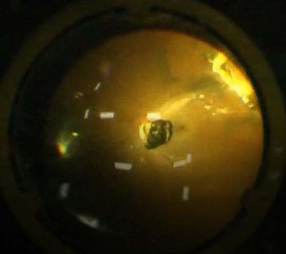
Intraoperative picture of an iron intraocular foreign body lying over the posterior pole.
I performed this surgery. It is my picture.

(Click Image to Enlarge)
Ultrasound B scan of eye showing a metallic foreign body over the retina (orange arrow)
Ultrasound B scan of eye showing a metallic foreign body over the retina (orange arrow). It is hyperechoic, has 100% spike on A scan with a back shadow.
My own picture

(Click Image to Enlarge)
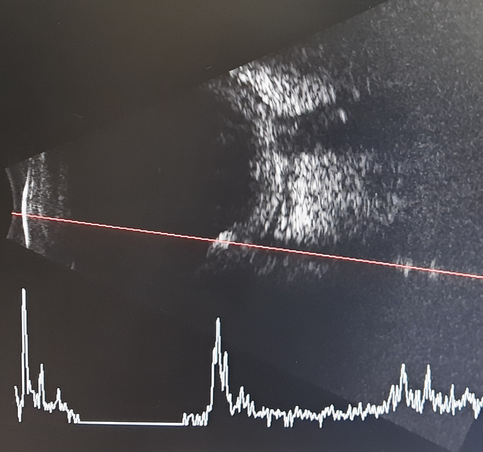
Ultrasound B scan of same foreign body at low gain. The hyperechoicity is persisting at low gain with high spike on A scan.
My own picture
Details

Updated:

8/25/2023 3:04:39 AM

References


[1]

Iftikhar M,Latif A,Farid UZ,Usmani B,Canner JK,Shah SMA, Changes in the Incidence of Eye Trauma Hospitalizations in the United States From 2001 Through 2014. JAMA ophthalmology. 2019 Jan 1;     [PubMed PMID: 30286226]


[2]

Patel SN,Langer PD,Zarbin MA,Bhagat N, Diagnostic value of clinical examination and radiographic imaging in identification of intraocular foreign bodies in open globe injury. European journal of ophthalmology. 2012 Mar-Apr;     [PubMed PMID: 21607931]


[3]

Zhang T,Zhuang H,Wang K,Xu G, Clinical Features and Surgical Outcomes of Posterior Segment Intraocular Foreign Bodies in Children in East China. Journal of ophthalmology. 2018;     [PubMed PMID: 30046460]


[4]

Chaudhry IA,Shamsi FA,Al-Harthi E,Al-Theeb A,Elzaridi E,Riley FC, Incidence and visual outcome of endophthalmitis associated with intraocular foreign bodies. Graefe's archive for clinical and experimental ophthalmology = Albrecht von Graefes Archiv fur klinische und experimentelle Ophthalmologie. 2008 Feb;     [PubMed PMID: 17468878]


[5]

Bourke L,Bourke E,Cullinane A,O'Connell E,Idrees Z, Clinical outcomes and epidemiology of intraocular foreign body injuries in Cork University Hospital, Ireland: an 11-year review. Irish journal of medical science. 2021 Aug;     [PubMed PMID: 33230610]

Level 2 (mid-level) evidence

[6]

Imrie FR,Cox A,Foot B,Macewen CJ, Surveillance of intraocular foreign bodies in the UK. Eye (London, England). 2008 Sep;     [PubMed PMID: 17525772]


[7]

Liggett PE,Gauderman WJ,Moreira CM,Barlow W,Green RL,Ryan SJ, Pars plana vitrectomy for acute retinal detachment in penetrating ocular injuries. Archives of ophthalmology (Chicago, Ill. : 1960). 1990 Dec;     [PubMed PMID: 2256844]


[8]

Azad RV,Kumar N,Sharma YR,Vohra R, Role of prophylactic scleral buckling in the management of retained intraocular foreign bodies. Clinical     [PubMed PMID: 14746594]


[9]

Liu CC,Tong JM,Li PS,Li KK, Epidemiology and clinical outcome of intraocular foreign bodies in Hong Kong: a 13-year review. International ophthalmology. 2017 Feb;     [PubMed PMID: 27043444]

Level 2 (mid-level) evidence

[10]

Justin GA,Baker KM,Brooks DI,Ryan DS,Weichel ED,Colyer MH, Intraocular Foreign Body Trauma in Operation Iraqi Freedom and Operation Enduring Freedom: 2001 to 2011. Ophthalmology. 2018 Nov;     [PubMed PMID: 30037644]

Level 2 (mid-level) evidence

[11]

Pieramici DJ,MacCumber MW,Humayun MU,Marsh MJ,de Juan E Jr, Open-globe injury. Update on types of injuries and visual results. Ophthalmology. 1996 Nov     [PubMed PMID: 8942873]


[12]

Esmaeli B,Elner SG,Schork MA,Elner VM, Visual outcome and ocular survival after penetrating trauma. A clinicopathologic study. Ophthalmology. 1995 Mar;     [PubMed PMID: 7891976]


[13]

Nicoară SD,Irimescu I,Călinici T,Cristian C, Intraocular foreign bodies extracted by pars plana vitrectomy: clinical characteristics, management, outcomes and prognostic factors. BMC ophthalmology. 2015 Nov 2     [PubMed PMID: 26526732]


[14]

Loporchio D,Mukkamala L,Gorukanti K,Zarbin M,Langer P,Bhagat N, Intraocular foreign bodies: A review. Survey of ophthalmology. 2016 Sep-Oct;     [PubMed PMID: 26994871]

Level 3 (low-level) evidence

[15]

Wang W,Zhou Y,Zeng J,Shi M,Chen B, Epidemiology and clinical characteristics of patients hospitalized for ocular trauma in South-Central China. Acta ophthalmologica. 2017 Sep;     [PubMed PMID: 28371405]


[16]

Liu Y,Wang S,Li Y,Gong Q,Su G,Zhao J, Intraocular Foreign Bodies: Clinical Characteristics and Prognostic Factors Influencing Visual Outcome and Globe Survival in 373 Eyes. Journal of ophthalmology. 2019;     [PubMed PMID: 30895158]


[17]

Wang JD,Xu L,Wang YX,You QS,Zhang JS,Jonas JB, Prevalence and incidence of ocular trauma in North China: the Beijing Eye Study. Acta ophthalmologica. 2012 Feb;     [PubMed PMID: 21883988]


[18]

Zhang Y,Zhang M,Jiang C,Qiu HY, Intraocular foreign bodies in china: clinical characteristics, prognostic factors, and visual outcomes in 1,421 eyes. American journal of ophthalmology. 2011 Jul;     [PubMed PMID: 21529762]

Level 2 (mid-level) evidence

[19]

Ehlers JP,Kunimoto DY,Ittoop S,Maguire JI,Ho AC,Regillo CD, Metallic intraocular foreign bodies: characteristics, interventions, and prognostic factors for visual outcome and globe survival. American journal of ophthalmology. 2008 Sep;     [PubMed PMID: 18614135]


[20]

Greven CM,Engelbrecht NE,Slusher MM,Nagy SS, Intraocular foreign bodies: management, prognostic factors, and visual outcomes. Ophthalmology. 2000 Mar;     [PubMed PMID: 10711903]


[21]

Fujikawa A,Mohamed YH,Kinoshita H,Matsumoto M,Uematsu M,Tsuiki E,Suzuma K,Kitaoka T, Visual outcomes and prognostic factors in open-globe injuries. BMC ophthalmology. 2018 Jun 8;     [PubMed PMID: 29884145]


[22]

Casini G,Sartini F,Loiudice P,Benini G,Menchini M, Ocular siderosis: a misdiagnosed cause of visual loss due to ferrous intraocular foreign bodies-epidemiology, pathogenesis, clinical signs, imaging and available treatment options. Documenta ophthalmologica. Advances in ophthalmology. 2021 Apr;     [PubMed PMID: 32949328]

Level 3 (low-level) evidence

[23]

BALLANTYNE JF, Siderosis bulbi. The British journal of ophthalmology. 1954 Dec     [PubMed PMID: 13219251]


[24]

Talamo JH,Topping TM,Maumenee AE,Green WR, Ultrastructural studies of cornea, iris and lens in a case of siderosis bulbi. Ophthalmology. 1985 Dec;     [PubMed PMID: 4088618]

Level 3 (low-level) evidence

[25]

Welch RB, Two remarkable events in the field of intraocular foreign body: (1) The reversal of siderosis bulbi. (2) The spontaneous extrusion of an intraocular copper foreign body. Transactions of the American Ophthalmological Society. 1975;     [PubMed PMID: 1108372]


[26]

Verhoeff FH, SIDEROSIS BULBI. The British journal of ophthalmology. 1918 Nov     [PubMed PMID: 18167834]


[27]

Kannan NB,Adenuga OO,Rajan RP,Ramasamy K, Management of Ocular Siderosis: Visual Outcome and Electroretinographic Changes. Journal of ophthalmology. 2016     [PubMed PMID: 27073692]


[28]

Hope-Ross M,Mahon GJ,Johnston PB, Ocular siderosis. Eye (London, England). 1993     [PubMed PMID: 8224298]


[29]

Rao NA,Tso MO,Rosenthal AR, Chalcosis in the human eye. A clinicopathologic study. Archives of ophthalmology (Chicago, Ill. : 1960). 1976 Aug;     [PubMed PMID: 60098]


[30]

Ravani R,Kumar V,Kumar A,Kumar P,Chawla S,Ghosh S, Fleck-like deposits and swept source optical coherence tomography characteristics in a case of confirmed ocular chalcosis. Indian journal of ophthalmology. 2018 Nov;     [PubMed PMID: 30355890]

Level 3 (low-level) evidence

[31]

Puranik C,Chaurasia S,Ramappa M,Sangwan V,Balasubramanian D, Corneal chalcosis following blast injury. The British journal of ophthalmology. 2012 May     [PubMed PMID: 22317910]


[32]

MISHLER JE,HARLEY RD, Copper within the eye 30 years simulating tumor; report of a case. American journal of ophthalmology. 1952 May     [PubMed PMID: 14923740]

Level 3 (low-level) evidence

[33]

Brown IA, Intraocular foreign bodies. Nature of injury. International ophthalmology clinics. 1968 Spring     [PubMed PMID: 5731762]


[34]

Pereira F,Matieli L,Sacai PY,Salomão SR,Jung LS,Berezovsky A, Electrophysiological findings in delayed discovery of a metallic intraocular foreign body in a child: case report. Documenta ophthalmologica. Advances in ophthalmology. 2019 Dec;     [PubMed PMID: 31286364]

Level 3 (low-level) evidence

[35]

Upshaw JE,Brenkert TE,Losek JD, Ocular foreign bodies in children. Pediatric emergency care. 2008 Jun;     [PubMed PMID: 18562888]


[36]

Bhagat N, Nagori S, Zarbin M. Post-traumatic Infectious Endophthalmitis. Survey of ophthalmology. 2011 May-Jun:56(3):214-51. doi: 10.1016/j.survophthal.2010.09.002. Epub 2011 Mar 12     [PubMed PMID: 21397289]

Level 3 (low-level) evidence

[37]

O'Duffy D,Salmon JF, Siderosis bulbi resulting from an intralenticular foreign body. American journal of ophthalmology. 1999 Feb;     [PubMed PMID: 10030572]


[38]

Meguro R, Asano Y, Odagiri S, Li C, Iwatsuki H, Shoumura K. Nonheme-iron histochemistry for light and electron microscopy: a historical, theoretical and technical review. Archives of histology and cytology. 2007 Apr:70(1):1-19     [PubMed PMID: 17558140]


[39]

Yang Z,Yang XL,Xu LS,Dai L,Yi MC, Application of Prussian blue staining in the diagnosis of ocular siderosis. International journal of ophthalmology. 2014     [PubMed PMID: 25349794]


[40]

Kutlutürk Karagöz I,Söğütlü Sarı E,Kubaloğlu A,Elbay A,Çallı Ü,Pinero DP,Özertürk Y,Yazıcıoğlu T, Characteristics of pediatric and adult cases with open globe injury and factors affecting visual outcomes: A retrospective analysis of 294 cases from Turkey. Ulusal travma ve acil cerrahi dergisi = Turkish journal of trauma     [PubMed PMID: 29350365]

Level 2 (mid-level) evidence

[41]

Poon KY, Use of limbal ring-rod for radiological localisation of ocular foreign body. The British journal of ophthalmology. 1989 Aug     [PubMed PMID: 2765445]


[42]

MINTZ MJ,MATTES MW, Detection of foreign bodies in the anterior chamber of the bulbus oculi. Radiology. 1960 Oct;     [PubMed PMID: 13771120]


[43]

Modjtahedi BS, Rong A, Bobinski M, McGahan J, Morse LS. Imaging characteristics of intraocular foreign bodies: a comparative study of plain film X-ray, computed tomography, ultrasound, and magnetic resonance imaging. Retina (Philadelphia, Pa.). 2015 Jan:35(1):95-104. doi: 10.1097/IAE.0000000000000271. Epub     [PubMed PMID: 25090044]

Level 2 (mid-level) evidence

[44]

Bryden FM,Pyott AA,Bailey M,McGhee CN, Real time ultrasound in the assessment of intraocular foreign bodies. Eye (London, England). 1990;     [PubMed PMID: 2282949]


[45]

Thickman DI,Ziskin MC,Goldenberg NJ,Linder BE, Clinical manifestations of the comet tail artifact. Journal of ultrasound in medicine : official journal of the American Institute of Ultrasound in Medicine. 1983 May     [PubMed PMID: 6864869]


[46]

Fielding JA, The assessment of ocular injury by ultrasound. Clinical radiology. 2004 Apr     [PubMed PMID: 15041449]


[47]

Crowell EL,Koduri VA,Supsupin EP,Klinglesmith RE,Chuang AZ,Kim G,Baker LA,Feldman RM,Blieden LS, Accuracy of Computed Tomography Imaging Criteria in the Diagnosis of Adult Open Globe Injuries by Neuroradiology and Ophthalmology. Academic emergency medicine : official journal of the Society for Academic Emergency Medicine. 2017 Sep;     [PubMed PMID: 28662312]


[48]

Cho WK,Ko AC,Eatamadi H,Al-Ali A,Abboud JP,Kikkawa DO,Korn BS, Orbital and Orbitocranial Trauma From Pencil Fragments: Role of Timely Diagnosis and Management. American journal of ophthalmology. 2017 Aug;     [PubMed PMID: 28554552]


[49]

Pavlin CJ,Foster FS, Ultrasound biomicroscopy. High-frequency ultrasound imaging of the eye at microscopic resolution. Radiologic clinics of North America. 1998 Nov;     [PubMed PMID: 9884687]


[50]

Dada T,Sihota R,Gadia R,Aggarwal A,Mandal S,Gupta V, Comparison of anterior segment optical coherence tomography and ultrasound biomicroscopy for assessment of the anterior segment. Journal of cataract and refractive surgery. 2007 May     [PubMed PMID: 17466858]


[51]

Wylegala E,Dobrowolski D,Nowińska A,Tarnawska D, Anterior segment optical coherence tomography in eye injuries. Graefe's archive for clinical and experimental ophthalmology = Albrecht von Graefes Archiv fur klinische und experimentelle Ophthalmologie. 2009 Apr;     [PubMed PMID: 18766361]


[52]

Mansouri K,Sommerhalder J,Shaarawy T, Prospective comparison of ultrasound biomicroscopy and anterior segment optical coherence tomography for evaluation of anterior chamber dimensions in European eyes with primary angle closure. Eye (London, England). 2010 Feb;     [PubMed PMID: 19444291]


[53]

Acharya I,Raut AA, Siderosis Bulbi StatPearls. 2021 Jan     [PubMed PMID: 33620858]


[54]

Kuhn F,Witherspoon CD,Skalka H,Morris R, Improvement of siderotic ERG. European journal of ophthalmology. 1992 Jan-Mar     [PubMed PMID: 1638168]


[55]

Gupta S,Midha N,Gogia V,Sahay P,Pandey V,Venkatesh P, Sensitivity of multifocal electroretinography (mfERG) in detecting siderosis. Canadian journal of ophthalmology. Journal canadien d'ophtalmologie. 2015 Dec;     [PubMed PMID: 26651311]


[56]

Delaney WV Jr, Presumed ocular chalcosis: a reversible maculopathy. Annals of ophthalmology. 1975 Mar     [PubMed PMID: 1094890]


[57]

Rosenthal AR,Marmor MF,Leuenberger P,Hopkins JL, Chalcosis: a study of natural history. Ophthalmology. 1979 Nov;     [PubMed PMID: 552619]


[58]

Sahay P,Kumawat D,Gupta S,Tripathy K,Vohra R,Chandra M,Venkatesh P, Detection and monitoring of subclinical ocular siderosis using multifocal electroretinogram. Eye (London, England). 2019 Oct;     [PubMed PMID: 31019264]


[59]

Mieler WF,Ellis MK,Williams DF,Han DP, Retained intraocular foreign bodies and endophthalmitis. Ophthalmology. 1990 Nov;     [PubMed PMID: 2255525]


[60]

Valera-Cornejo D,García-Roa M,Ramírez-Neria P,Villalpando-Gómez Y,Romero-Morales V,García-Franco R, The role of various imaging techniques in identifying and locating intraocular foreign bodies related to open-globe injury: three case reports and literature review. Medwave. 2020 Jan 28     [PubMed PMID: 32119652]

Level 3 (low-level) evidence

[61]

Delgado E, Endophthalmitis due to an intra-ocular linear foreign body in a cat. JFMS open reports. 2015 Jan-Jun     [PubMed PMID: 28491351]


[62]

Vlasov A,Ryan DS,Ludlow S,Coggin A,Weichel ED,Stutzman RD,Bower KS,Colyer MH, Corneal and Corneoscleral Injury in Combat Ocular Trauma from Operations Iraqi Freedom and Enduring Freedom. Military medicine. 2017 Mar;     [PubMed PMID: 28291461]

Level 2 (mid-level) evidence

[63]

Arora R,Sanga L,Kumar M,Taneja M, Intralenticular foreign bodies: report of eight cases and review of management. Indian journal of ophthalmology. 2000 Jun     [PubMed PMID: 11116507]

Level 3 (low-level) evidence

[64]

Lin YC, Kuo CL, Chen YM. Intralenticular foreign body: A case report and literature review. Taiwan journal of ophthalmology. 2019 Jan-Mar:9(1):53-59. doi: 10.4103/tjo.tjo_88_18. Epub     [PubMed PMID: 30993070]

Level 3 (low-level) evidence

[65]

Davidson RS,Sivalingam A, A metallic foreign body presenting in the anterior chamber angle. The CLAO journal : official publication of the Contact Lens Association of Ophthalmologists, Inc. 2002 Jan;     [PubMed PMID: 11838993]


[66]

Mete G,Turgut Y,Osman A,Gülşen U,Hakan A, Anterior segment intraocular metallic foreign body causing chronic hypopyon uveitis. Journal of ophthalmic inflammation and infection. 2011 Jun     [PubMed PMID: 21484173]


[67]

Chow DR,Garretson BR,Kuczynski B,Williams GA,Margherio R,Cox MS,Trese MT,Hassan T,Ferrone P, External versus internal approach to the removal of metallic intraocular foreign bodies. Retina (Philadelphia, Pa.). 2000     [PubMed PMID: 10950413]


[68]

Rong AJ,Fan KC,Golshani B,Bobinski M,McGahan JP,Eliott D,Morse LS,Modjtahedi BS, Multimodal imaging features of intraocular foreign bodies. Seminars in ophthalmology. 2019     [PubMed PMID: 31609153]


[69]

Machemer R, A new concept for vitreous surgery. 2. Surgical technique and complications. American journal of ophthalmology. 1972 Dec;     [PubMed PMID: 4646708]


[70]

Lal T,Yu ZX,Guan B,Bender C,Chan CC,Cukras CA,Hufnagel RB, Clinical and Histopathologic Correlates of Asymmetric Retinitis Pigmentosa. JAMA ophthalmology. 2021 Sep 1;     [PubMed PMID: 34351381]


[71]

McGahan MC,Bito LZ,Myers BM, The pathophysiology of the ocular microenvironment. II. Copper-induced ocular inflammation and hypotony. Experimental eye research. 1986 Jun;     [PubMed PMID: 3720874]


[72]

Tripathy K,Tomar AS,Sidhu T,Dada T, A 10-year-old boy with dystonia, expression-less facies, and tremors referred for ophthalmic examination. Oman journal of ophthalmology. 2021 May-Aug     [PubMed PMID: 34345153]


[73]

Sridhar U,Tripathy K, Commentary: Kayser-Fleischer-like rings in patients with hepatic disease. Indian journal of ophthalmology. 2021 May     [PubMed PMID: 33913838]

Level 3 (low-level) evidence

[74]

Cheraghali F,Fadaei Jouybari F,Tohidi F,Ghasemikhah R,Taghipour A,Sharbatkhori M, Seroprevalence, risk factors, and clinical symptoms of Toxocara spp. infection among children 3-15 years old in northern Iran. Comparative immunology, microbiology and infectious diseases. 2021 Jun;     [PubMed PMID: 33819773]

Level 2 (mid-level) evidence

[75]

Ma J,Wang Y,Zhang L,Chen M,Ai J,Fang X, Clinical characteristics and prognostic factors of posterior segment intraocular foreign body in a tertiary hospital. BMC ophthalmology. 2019 Jan 14;     [PubMed PMID: 30642294]


[76]

Dowlut MS,Curragh DS,Napier M,Herron B,McIlwaine G,Best R,Chan W, The varied presentations of siderosis from retained intraocular foreign body. Clinical     [PubMed PMID: 29923333]


[77]

Anderson G,Horvath J, The growing burden of chronic disease in America. Public health reports (Washington, D.C. : 1974). 2004 May-Jun     [PubMed PMID: 15158105]


[78]

Jonas JB, Knorr HL, Budde WM. Prognostic factors in ocular injuries caused by intraocular or retrobulbar foreign bodies. Ophthalmology. 2000 May:107(5):823-8     [PubMed PMID: 10811069]


[79]

Yucel OE,Demir S,Niyaz L,Sayin O,Gul A,Ariturk N, Clinical characteristics and prognostic factors of scleral rupture due to blunt ocular trauma. Eye (London, England). 2016 Dec     [PubMed PMID: 27589050]


[80]

Yang CS,Hsieh MH,Hou TY, Predictive factors of visual outcome in posterior segment intraocular foreign body. Journal of the Chinese Medical Association : JCMA. 2019 Mar     [PubMed PMID: 30913120]


[81]

Konforty N,Lior Y,Levy J,Belfair N,Bilenko N,Lifshitz T,Klemperer I,Knyazer B, [EPIDEMIOLOGY, CLINICAL CHARACTERISTICS, PROGNOSTIC FACTORS, AND VISUAL OUTCOMES IN PATIENTS WITH OPEN OCULAR INJURIES AND INTRAOCULAR FOREIGN BODIES]. Harefuah. 2016 May;     [PubMed PMID: 27526552]


[82]

Kuhn F, Maisiak R, Mann L, Mester V, Morris R, Witherspoon CD. The Ocular Trauma Score (OTS). Ophthalmology clinics of North America. 2002 Jun:15(2):163-5, vi     [PubMed PMID: 12229231]


[83]

Weichel ED,Colyer MH,Ludlow SE,Bower KS,Eiseman AS, Combat ocular trauma visual outcomes during operations iraqi and enduring freedom. Ophthalmology. 2008 Dec;     [PubMed PMID: 19041478]

Level 2 (mid-level) evidence

[84]

Simakurthy S, Tripathy K. Endophthalmitis. StatPearls. 2023 Jan:():     [PubMed PMID: 32644505]


[85]

Tripathy K, Chawla R, Temkar S, Sagar P, Kashyap S, Pushker N, Sharma YR. Phthisis Bulbi-a Clinicopathological Perspective. Seminars in ophthalmology. 2018:33(6):788-803. doi: 10.1080/08820538.2018.1477966. Epub 2018 Jun 14     [PubMed PMID: 29902388]

Level 3 (low-level) evidence

[86]

Chawla R, Kapoor M, Mehta A, Tripathy K, Vohra R, Venkatesh P. Sympathetic Ophthalmia: Experience from a Tertiary Care Center in Northern India. Journal of ophthalmic & vision research. 2018 Oct-Dec:13(4):439-446. doi: 10.4103/jovr.jovr_86_17. Epub     [PubMed PMID: 30479714]


[87]

Rozon JP,Lavertu G,Hébert M,You E,Bourgault S,Caissie M,Tourville E,Dirani A, Clinical Characteristics and Prognostic Factors of Posterior Segment Intraocular Foreign Body: Canadian Experience from a Tertiary University Hospital in Quebec. Journal of ophthalmology. 2021;     [PubMed PMID: 34055400]


[88]

Coleman DJ,Lucas BC,Rondeau MJ,Chang S, Management of intraocular foreign bodies. Ophthalmology. 1987 Dec;     [PubMed PMID: 3431834]


[89]

Thompson JT, Parver LM, Enger CL, Mieler WF, Liggett PE. Infectious endophthalmitis after penetrating injuries with retained intraocular foreign bodies. National Eye Trauma System. Ophthalmology. 1993 Oct:100(10):1468-74     [PubMed PMID: 8414406]


[90]

Jung HC,Lee SY,Yoon CK,Park UC,Heo JW,Lee EK, Intraocular Foreign Body: Diagnostic Protocols and Treatment Strategies in Ocular Trauma Patients. Journal of clinical medicine. 2021 Apr 25     [PubMed PMID: 33923011]