Continuing Education Activity
Morel Lavallee lesions are soft tissue injuries seen in high-velocity trauma and are usually associated with underlying fractures of the pelvis, acetabulum, or proximal femur. Often these injuries are not immediately diagnosed due to the distracting concomitant bony injuries. However, identification of such injuries is important as they may pose as an independent risk factor for surgical site infection. The clinical findings include soft tissue swelling, bruise/ ecchymosis, fluctuance, and compressibility in the swelling. The diagnosis is usually established on physical examination, however, radiological investigations including ultrasonography and CT might help. The management options include nonoperative treatment, percutaneous aspiration, and open debridement. This activity reviews the evaluation and management of Morel Lavalle lesions and highlights the role of the interprofessional team in evaluating and managing patients with this condition.
Objectives:
- Outline the typical etiology of Morel Lavallee lesions.
- Describe the common clinical findings in a patient with Morel Lavallee lesion.
- Identify the imaging findings associated with various duration of Morel Lavallee lesions.
- Explain the treatment considerations taken by the interprofessional team for patients with Morel Lavallee lesions.
Introduction
The Morel-Lavallee lesion was first described in 1863 by a French surgeon named Maurice Morel-Lavallee as a closed degloving injury that occurs post traumatically where deep fascia get separated from the skin and superficial fascia, hence creating a potential space.[1][2][3]
The impact of devastating trauma leads to the injury of lymphatics and blood vessels that lie in the vicinity, resulting in the accumulation of lymph and blood in this potential space. This results in the setting of a chronic inflammatory reaction, which later leads to the formation of a capsulated lesion lined by a fibrous capsule and filled with necrotic fatty tissue, blood products, fibrin, and debris.[2]
The Morel-Lavallee lesion clinically presents as a painful fluctuant swelling at the site of involvement. Concerning the recent literature available, this lesion is also termed as Morel-Lavallée seroma, posttraumatic soft tissue cyst, post-traumatic extravasation, or Morel-Lavallée effusion.[4] This lesion may be missed at the time of initial assessment and can present later, potentially leading to increasing difficulty in management and long-term morbidity.[3][5]
Etiology
The most common causes of Morel-Lavallee lesions are high-velocity trauma, crush injuries, and blunt trauma.[6] Overall, approximately twenty-five percent of all patients who develop Morel-Lavallee lesions have been involved in a road traffic accident.[6][7] This lesion is commonly associated with underlying fractures, especially of the proximal femur, pelvis, and acetabulum. One of the most commonly involved regions is the greater trochanter, accounting for more than sixty percent of the cases.[5]
The primary predisposing factors include the superficial position of the femoral bone in this region along with a large surface area, the strength of the underlying tensor fascia lata, and relative mobility of the subdermal soft tissue.[8] The secondary predisposing factors include a body mass index of 25 kg/m or greater. Other less common sites of involvement include buttocks, scapular region, lumbosacral region, and trunk region. In a small subset of individuals, Morel-Lavallee lesion is associated with direct-blow sports injuries to the knee.[9][10] In rare scenarios, these lesions have also been mentioned following abdominoplasty and liposuction.[11]
Epidemiology
This lesion is not frequently seen in practice, with a prevalence of around 8.3% in acetabular fractures. It shows male predominance with an approximate 2:1 male to female ratio, which might be due to the male predominance seen in polytrauma.[3][5] However, these lesions are often undiagnosed or sometimes diagnosed later, thus underestimating the true prevalence of this condition.
Pathophysiology
The Morel-Lavallee lesion results in the development of a cavity in the pre-fascial plane, which occurs due to the generation of shearing effect in between underlying fascial layers and superficial subcutaneous tissues.[12] The Morel-Lavallee lesion is predominant in areas where the overlying skin is mobile, and the underlying fascia is tough in nature, such as the quadriceps fascia, which lies superior to the knee and the fascia lata in the proximal aspect of the lateral thigh.[6] Due to the shearing force, separation occurs among these layers, which leads to the disruption of lymphatic vessels, locules of subdermal fat, and transaponeurotic capillaries.[5]
These avulsed channels lead to the leakage of blood and lymph into this cavity and result in the collection of a hemolymphatic fluid, and its rate of formation depends on the flow into the cavity and the number of vessels disrupted. With time, the blood components within the cavity start reabsorbing, and hence only the serosanguinous fluid remains in the cavity surrounded by a haemosiderin layer. This haemosiderin layer induces a cascade of inflammation in the surrounding peripheral tissues and results in the formation of a fibrous capsule which prevents the further reabsorption of fluid and leads to the development of a chronic Morel-Lavallee lesion.[3][6]
History and Physical
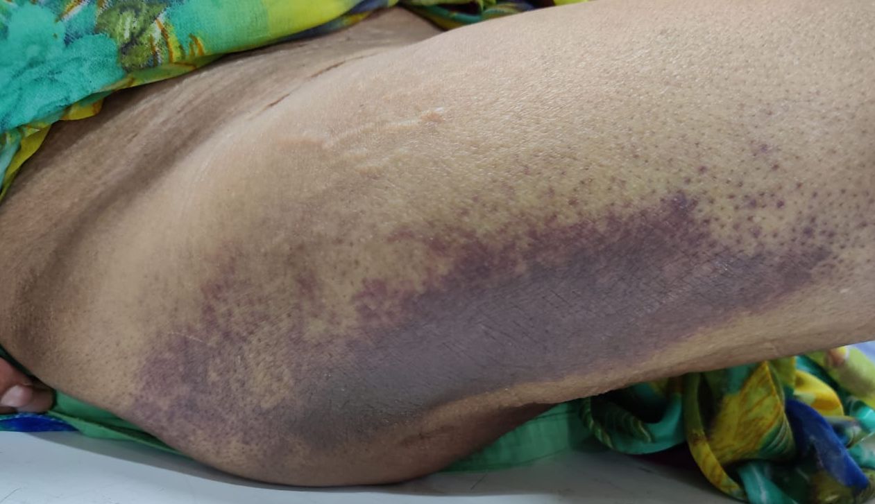
The Morel-Lavallee lesion is commonly associated with an inciting trauma event. However, up to thirty-three percent of cases show delayed presentation.[5] This lesion usually presents as gradually enlarging swelling associated with pain and tautness. The important clinical features that help make a diagnosis and an accurate correlating history are the presence of fluctuance and compression within the lesion.
Clinical findings may resemble a regional contusion. Patients may also experience cutaneous hypoesthesia or anesthesia because of the damage to the subdermal afferent nerves and increased mobility of the skin. Overlying secondary skin changes, including ecchymosis, cracking, drying, abrasions, and even frank necrosis, may also be seen.[2][3][6] Often, these patients present with recurrence of the soft tissue lesion, particularly after minimally invasive treatment modalities.
Evaluation
Whenever managing patients with polytrauma, especially for the regions around the pelvis, the presence of a Morel-Lavallee lesion should be keenly checked, this lesion is usually diagnosed as an incidental finding during the surgical intervention of the fractures, but they can be retrospectively identified on the imaging which was done at the time of admission.[5][13]
The diagnosis is generally made on the physical examination. However, radiological investigations can help when clinical suspicion arises. The investigation of choice for this lesion is Magnetic Resonance Imaging, as it defines various parameters of the injury, including size, shape, contents, and its chronicity, but it is rarely required for the diagnosis. Various types of appearances can be seen depending on the chronicity and content of the lesion. Few studies had reported the appearance of a chronic lesion as homogenous and smooth and acute lesion as heterogeneous and irregular. In the chronic lesion, the formation of the capsule is inevitable, which results in its appearance as a hypointense ring of haemosiderin with fibrous tissue components. The edges of the lesion are tapering in nature and eventually fusing with the surrounding fascial layers.[6]
Ultrasonography is of less value, but it may support the clinical suspicion of this lesion by confirming the location of the lesion as deep to the hypodermis and superficial to the muscle fascia. It can also demonstrate the lesion’s compressibility nature and helps rule out the presence of flow using Doppler imaging, thus ruling out other differentials. Computed tomography has a very limited role in these lesions, especially in refining the differential diagnosis.[3][5]
Mellado and Bercandino proposed a comprehensive classification system for Morel-Lavallee lesions. According to this classification, this lesion can be divided into six types based on the shape of the lesion, characteristics of Magnetic Resonance Imaging, and the presence of the capsule.[14] Although this classification provides a concise manner of categorizing lesions, it does not help in providing any guidance regarding the management or possible potential outcome of each class. Shen et al. proposed a simpler version of classification where he divided the lesion into acute and chronic based on the presence of a capsule. This classification predicts the treatment strategies with the possible potential outcomes.[15]
Treatment / Management
At present, no specific guidelines are mentioned in the current literature regarding the management of Morel-Lavallee lesions. Multiple low evidence studies show variable results of multiple treatment modalities, including conservative management, sclerodesis, percutaneous aspiration, and open surgery.
Conservative Management
In cases of small acute Morel-Lavallee lesions where no capsule is present, nonoperative management can be done. This may involve the application of compression bandaging to soft tissue swelling. However, in chronic cases or large lesions, nonoperative management is not suitable, and surgical intervention is required.[15]
Percutaneous Aspiration
Few studies show effective results after percutaneous aspiration of the Morel-Lavallee lesion. However, the recurrence rate is high, especially in lesions with a volume of more than 50 ml, where multiple aspirations are often required.
Sclerodesis
This treatment modality has been successfully used in Morel-Lavallee lesions, particularly in cases where percutaneous aspiration fails. The common sclerosing agents include doxycycline, erythromycin, vancomycin, tetracycline, bleomycin, absolute ethanol, and talc. The mechanism of action of the majority of sclerodesis agents includes cellular destruction within the periphery of the lesion, which later on results in fibrosis. Overall efficacy of sclerodesis in managing Morel-Lavallée lesions has been reported as 95.7%.
Open Drainage and Mass Resection
The majority of these lesions require open debridement with excision of the pseudocapsule in chronic cases. The end goal in managing Morel-Lavallee lesions is the closure of dead space within the lesion which can be achieved in various ways, including fibrin sealant, quilting sutures, and low suction drains. A single longitudinal incision or multiple small incisions proximally and distally can be used for the open drainage in cases where the overlying skin is viable. If the condition of skin overlying the lesion is necrotic, then dead tissue needs to be debrided, followed by reconstruction of the soft tissue envelope.[15][16] In scenarios where even open drainage has also failed, the last treatment modality is the en masse resection of the lesion with an intact capsule.[3]
Differential Diagnosis
The common differential diagnosis of the Morel-Lavallee lesion includes post-operative seroma, coagulopathy-related hematoma, post-traumatic injuries like fat necrosis, and rarely, post-traumatic early-stage myositis ossificans with diffuse subcutaneous edema. Postoperative seroma holds various pathological similarities with it. As the Morel-Lavallee lesion can clinically, pathologically, and radiographically simulate multiple possible conditions, a prior history of trauma can play a pivotal role in reaching the diagnosis.[2]
Prognosis
The prognosis of Morel Lavallee lesions depends on various factors. Small acute lesions heal themselves without the requirement of operative management and hence have an excellent prognosis. On the other hand, larger lesions pose as an independent risk factor for postoperative surgical site infection for the associated bony injuries, but they may also dictate the timing of surgical management and the surgical approach chosen for the orthopedic injuries. As the lesions become chronic, the formation of the pseudocapsule prevents reabsorption of the contents, leading to undesirable sequelae providing a poor prognosis.[17]
Complications
The complications which are often seen with Morel-Lavallee lesions are either due to its delayed presentation or inaccurate diagnosis. If this lesion is not managed properly, it can result in gradual progressive expansion, leading to the necrosis of the overlying skin because of the pressure effect, which ultimately leaves underlying fractures exposed. Infection is a major concern in such lesions; multiple studies have commented on the contamination of the lesion contents, which may have resulted either due to the inadvertent entry of micro-organisms in the lesion while managing the lesion, especially with the use of sclerosing agents, or during the fixation of an underlying fracture.[15]
Deterrence and Patient Education
The patients suffering from high-velocity injuries, particularly to the hip and pelvic region, should keenly observe for and report any soft tissue swellings noticed near the orthopedic injuries. Also, as the treatment might entail placing an indwelling drain in the lesion for around 2 weeks, proper patient counseling should be done to ensure patient compliance. Moreover, as the less invasive options of percutaneous aspiration are associated with high rates of local recurrence, adequate information needs to be provided to the patients before they can agree on the more invasive option of open debridement. Also, proper rehabilitation and physical therapy are paramount in getting the best functional outcomes after surgical and conservative management of these lesions and the concomitant orthopedic injuries.
Enhancing Healthcare Team Outcomes
The management of Morel Lavalle lesions begins with appropriate evaluation, which requires a thorough and comprehensive clinical examination after adequate history-taking. Also, radiologists help in clinching the diagnosis as well as providing details of the lesion using ultrasonography, CT scan, and MRI. In advanced stages, where the overlying soft tissue envelope is fully necrosed, the help of a plastic surgeon is sought for providing adequate coverage of the underlying muscles and often the underlying orthopedic implants. The role of physical therapy and rehabilitation cannot be undermined in getting the best functional outcomes after the underlying fractures; hence physiotherapists should be informed about the rehab protocol after each case.