Continuing Education Activity
Fibrodysplasia ossificans progressiva is a rare genetic disorder that causes significant disability and morbidity. In this disorder, heterotopic ossification starts in the first decade of life, and a majority of such cases develop inflammatory painful soft tissue swellings. This disability progresses from upper to lower end, proximal to the distal end, and a dorsal to ventral side, i.e., from neck, spine, shoulders to elbow, knee, hip, jaw to wrists, and ankle. The wrists, ankles, elbows, knees, hips, and jaw gradually get involved till 40 years of age. There is no definitive management for this life-threatening disorder to date, and various new drugs are being tested in clinical trials. This activity reviews the presentation, evaluation, and management of fibrodysplasia ossificans progressiva and highlights the role of the interprofessional team in evaluating and managing patients with this condition.
Objectives:
- Describe the etiology of fibrodysplasia ossificans progressiva.
- Review the common presentation of a patient with fibrodysplasia ossificans progressiva.
- Identify the various investigations required for diagnosing fibrodysplasia ossificans progressiva.
- Explain the current treatment options for patients with fibrodysplasia ossificans progressiva, along with the complications anticipated.
Introduction
Fibrodysplasia ossificans progressiva is described as a rare genetic disorder characterized by the organization of heterotopic hard tissues within the soft tissues, such as ligaments, tendons, and skeletal muscle.[1][2][3] It comes under the category of an autosomal dominant disorder. The tissue formed in such patients is not just the mineralized calcium phosphate, but it resembles the new bone formation by osteoblast cells via endochondral ossification. Most of the patients who are suffering from Fibrodysplasia ossificans progressiva can move their joints normally at the time of birth, but disability arises in various joints when they reach their 30’s because gradually heterotopic bones fuse and result in bridge formation with normal bones.[4] Injury to soft tissues can lead to acute heterotopic bone formation in such patients hence invasive procedures, such as injection, surgical operation, and biopsy, are contraindicated.[3][5][6]
Making a diagnosis of fibrodysplasia ossificans progressiva was difficult for a long time as there were no reliable biomarkers for such cases that could be evaluated in urine or peripheral blood. The pathogenesis found in such patients is the gain of function mutation in the ACVR1/ALK2 gene located over chromosome 2 and the involvement of the bone morphogenetic proteins (BMP) signaling pathway.[7] The BMPs lead to the formation of heterotopic bone in the soft tissues.[6] The most common sites affected are the shoulders, neck, and spine. The median age of survival is roughly around 40 years.[8] Death occurs in such cases primarily due to thoracic insufficiency syndrome and related complications. PCR is the main diagnostic modality for the analysis of genetic mutation in FOP.[3][5]
Etiology
Fibrodysplasia ossificans progressiva is a typical monogenic disorder with the recurrent heterozygous gain of function mutation in the ACVR1/ALK2 gene located on chromosome 2, both in the sporadic and inherited cases, and involves the BMP (bone morphogenetic proteins) signaling pathway. This ACVR1/ALK2 gene encodes for a transmembrane serine/threonine (ser/thr) kinase receptor ALK2, which binds with the BMPs present in the bone matrix.[9]
These BMPs induce the development of heterotopic bone in the skeletal muscle. The majority of cases of typical Fibrodysplasia ossificans progressiva show a similar genetic mutation which consists of a change of nucleotide in the codon at position 617 (guanine->adenine) in the ACVR1/ALK2 gene.[3][8] It results in a substitution mutation in the codon at a position 206 (arginine->histidine) within the ALK2 protein.[10]
Some additional mutations are also noticed in exons 4 through 7 within the ACVR1/ALK2 gene, which results in a change in the expression of two domains, including Ser/Thr kinase and the glycine/serine-rich. Both of these domains play an important role in intracellular signaling.[6][7]
Epidemiology
Fibrodysplasia ossificans progressiva shows an incidence of approximately one patient per 2 million people worldwide and is not determined by any race, gender, or geographical distribution.[3][6]
Pathophysiology
The gain of function mutation in the ACVR1/ALK2 gene, which encodes for ALK2 kinases, induces signal transduction by ALK2 in response to ligand binding.[7][11] The extracellular domain of ALK2 (a type I receptor) binds with both the TGF-β family ligands (such as BMP-6, BMP-7, BMP9, and activin B) and with one of the type II receptors [such as BMP receptor type II (BMPR-II), activin receptor type IIA (Acta-IIA) and activin receptor type IIB (Acta-IIB)].[9]
Since these type II receptors are constitutively active ser/thr kinases, phosphorylation of ALK2 occurs at the GS domain consisting of glycine and serine residues in the ternary complex formed by the ligand binding to ALK2 at the cell membrane. This phosphorylated ALK2, in turn, activates the kinase activity and phosphorylates serine and threonine residues in the downstream substrates such as SMAD1, SMAD5, and Smad8/9.[10][12]
These phosphorylated Smad proteins bind to specific DNA sequences and regulate the transcription of the target genes in the nucleus and result in the heterotopic ossification seen in patients with fibrodysplasia ossificans progressiva. Thus, all these mutations induce intracellular signaling where ALK2 acts as the type 1 receptor in co-operation with the type II receptors.[5][6]
Histopathology
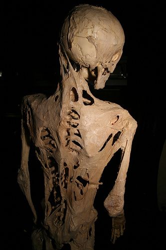
Early lesions of fibrodysplasia ossificans progressiva show a rapid and destructive inflammatory stage characterized by an intense mononuclear and perivascular infiltration by macrophages, mast cells, and lymphocytes. The subsequent migration of these inflammatory cells into the affected skeletal muscle causes its death.[13] This is followed by an intense fibroproliferative phase characterized by angiogenesis, neovascularity, and a very high density of mast cells.[14]
Early lesions of this phase are histologically similar to aggressive juvenile fibromatosis and can not be distinguished. As these lesions mature, the fibroproliferative tissue is converted into cartilage by the process of avascular condensation. This is followed by the final phase of revascularization with osteogenesis in a characteristic process of heterotopic ossification forming new ossicles of heterotopic bone, which appear histologically similar to the mature lamellar bone and often contain marrow elements.[14][15]
This heterotopic bone formation resembles bone formation in embryonic skeletal development and postnatal fracture healing, except for the lack of inflammation in the primary skeletal formation. The fibroproliferative, chondrogenic and osteogenic stages of heterotopic ossification are characterized by multipotent stem-like cells of vascular origin while the late osteogenic phase is characterized by inflammatory cells of hematopoietic origin. All these stages are seen in an active Fibrodysplasia ossificans progressiva lesion, thus suggesting different rates of maturation of different regions of the lesion.[16]
History and Physical
More than 90% of affected individuals show malformations of the great toes at birth but otherwise appear normal with normal joint mobility. In the first decade of life, heterotopic ossification starts, and a majority of the cases of fibrodysplasia ossificans progressiva develop inflammatory soft tissue swellings which are episodic, painful, and may show flare-ups.[5]
Joint disability sets in the third decade of life due to the fusion of the heterotopic bones formed in the soft tissue with the normal bones, thus fixing the joints.[3] The most frequently affected sites are the neck, spine, and shoulders (more than 80% of cases in patients less than 15 years of age). The wrists, ankles, elbows, knees, hips, and jaw gradually get involved till 40 years of age. This disability progresses from upper to lower end, proximal to the distal end, and a dorsal to ventral side, i.e., from neck, spine, shoulders to elbow, knee, hip, jaw to wrists, and ankle. Thus, these patients become wheelchair-bound by the third decade of life, requiring lifelong assistance for daily activities. The median age of survival in these patients is approximately 40 years.[5]
Evaluation
Diagnosis of Fibrodysplasia ossificans progressiva is made mainly by genetic mutations analysis of the involved gene by doing Sanger sequencing of the PCR products.[8] Routine biochemical evaluations of the heterotopic bone seen in Fibrodysplasia ossificans progressiva are usually similar to that of the normal bone, so these are distinguished based on the location of the heterotopic bone. During the disease flare-ups, the serum alkaline phosphatase activity and ESR may be increased along with the elevated urinary basic fibroblast growth factor levels.[5][9]
These increased urinary basic fibroblast growth factor levels coincide with the pre-osseous angiogenic phase of early fibroproliferative lesions. Another important diagnostic tool is Imaging analysis. Plain X-rays can reveal abnormal osteogenesis after heterotopic ossification manifests. CT scan can also show lesions with typical heterotopic ossifications.[17] MRI can reveal preosseous lesions, which appear as soft tissue swelling and skeletal malformations. Diagnosis of fibrodysplasia ossificans progressiva can not be made prenatally.
Treatment / Management
There is no single effective treatment option available for Fibrodysplasia ossificans progressiva.[6] Although symptomatic management for flare-ups can be provided with a short duration of high-dose corticosteroids like prednisone, the frequent use of corticosteroids is not advised. Mast cell inhibitors, NSAIDS, amino bisphosphonates, and COX-2 inhibitors are used for treating later flare-ups.[18][19]
Muscle relaxants can be used in small doses for muscle spasms. Another potential treatment option is to inhibit the activity of the ACVR1/ALK2 gene-related pathway, which inhibits abnormal bone formation. The other strategy would be to inhibit osteoblastic progenitor cell activity, which results in hindering the microenvironment for heterotopic ossificans. With the increasing understanding of the pathogenesis of fibrodysplasia ossificans progressiva, new potential drug targets have been discovered.[20][21]
Many drugs, such as imatinib, a human anti-activin A-neutralizing antibody, dorsomorphin, palovarotene, rapamycin, are in clinical trials, and suitable drugs may be available in the coming future concerning the better understanding of the mechanism of onset of fibrodysplasia ossificans progressiva.[3] Surgery is not generally preferred for such patients.[6]
Differential Diagnosis
The common differential diagnosis of fibrodysplasia ossificans progressiva includes isolated congenital malformations, juvenile bunions, sarcoma, desmoid tumor, aggressive juvenile fibromatosis, lymphedema.
Prognosis
Fibrodysplasia ossificans progressiva has a poor prognosis due to associated complications, including pneumonia and right-sided heart failure. Death in these cases occurs due mainly to thoracic insufficiency syndrome and complications related to it.[3][5][6]
Complications
Common complications associated with this disease include respiratory system infection, joint disabilities, cardiopulmonary complications (thoracic insufficiency syndrome, right-side congestive heart failure), submandibular swelling, temporomandibular joint ankylosis conductive loss of hearing.[5][6][8]
Deterrence and Patient Education
The patients presenting with significant toe deformities associated with fibrodysplasia ossificans progressiva should be promptly identified and evaluated for the disease. As this disorder causes significant morbidity and early mortality, early identification can help prolong the survival of such patients. Once the disorder is diagnosed, the patients should avoid invasive procedures like injections or surgery, which may trigger the new bone formation. Also, proper rehabilitation for joint deformities and stiffness can help in getting the best functional outcomes.
Enhancing Healthcare Team Outcomes
The evaluation of fibrodysplasia ossificans progressiva begins with clinical suspicion for this life-threatening disease. As such, pediatricians and neonatologists should be adequately informed about the clinical signs and the evaluation strategies for this condition. Also, once the condition is diagnosed, physicians and pediatricians can help in the symptomatic management of the inflammatory disease activity. Also, physiotherapist opinion should be sought regarding appropriate means for managing joint stiffness and deformities.