Continuing Education Activity
Neuroretinitis is an uncommon type of optic disc swelling with characteristic retinal macular exudates and must be differentiated from other types of optic disc swelling. This activity reviews the presentation, etiologies, investigation, and treatment of neuroretinitis. This activity also highlights the role that the interprofessional team in managing neuroretinitis when associated with systemic disease.
Objectives:
- Describe the appearance of neuroretinitis.
- Identify the 3 of the most common conditions associated with neuroretinitis.
- Classify the types of neuroretinitis.
- Outline the risk factors that should inform the line of questioning in taking histories from these patients for management by the interprofessional team.
Introduction
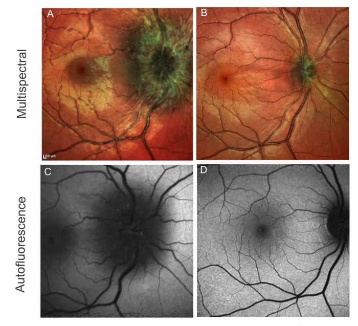
Neuroretinitis (NR) is defined as inflammation of the anterior optic nerve and peripapillary retina. It presents as a triad of vision loss, optic disc swelling, and macular exudates in the formation of a star. It is a term given to the appearance but does not indicate a specific etiology.
Theodor Leber first described neuroretinitis in 1916, and the name 'stellate maculopathy' was coined. As this term denotes, the original site of pathology was thought to be in the macula. However, a subsequent study showed that the fluid leakage at the optic disc precedes the macular star, affecting the macula secondarily.[1] Thus, the term neuroretinitis was born.
The associated infectious and inflammatory etiologies now associated with neuroretinitis have been widely documented, and although the majority of cases self-resolve, it is important to understand where resolution may be minimal or where the disease may be recurrent and therefore require early treatment.
Neuroretinitis is broadly categorized as idiopathic, idiopathic-recurrent, and cat scratch-disease neuroretinitis (CSD-NR). Neuroretinitis may also be categorized based upon the etiology: infectious vs. non-infectious. Idiopathic and idiopathic-recurrent neuroretinitis are usually non-infectious. It is usually unilateral, but bilateral cases have been described, though they should lead the physician to consider other causes.
Etiology
Cat-scratch disease, caused by infection with the Bartonella henselae bacterium, has consistently been shown to be the most common cause of neuroretinitis and accounts for two-thirds of cases.[2][3] Cat-scratch disease can also cause other ocular findings, including Parinaud oculoglandular syndrome, multifocal retinitis, uveitis, retinal vasculitis, and retinal detachment.[4] However, a large study of 107 eyes in 86 patients with confirmed cat-scratch disease found that neuroretinitis was the predominant manifestation in 88%.[5]
Other infectious causes include:[6]
- Lyme disease (Borrelia burgdorferi)
- Syphilis
- Tuberculosis
- Salmonella
- Varicella
- Herpes simplex and zoster
- Measles
- Mumps
- Rubella
- West Nile
- Zika
- Chikungunya
- Influenza
- Hepatitis
- Epstein-Barr virus
- Histoplasmosis
- Coccidioidomycosis
- Actinomycosis
- Toxoplasmosis
- Toxocariasis
- Diffuse unilateral subacute neuroretinitis
- Hepatitis B
- Leptospirosis
- Mumps
- Typhus
Inflammatory causes include:[6]
- Sarcoidosis
- Systemic lupus erythematosus
- Behcet disease
- Polyarteritis nodosa
- Takasayu's arteritis
- Vogt-Koyanagi-Harada
- Inflammatory bowel disease has also rarely been associated with NR.
Fifty percent of cases have no identifiable cause and are labeled idiopathic neuroretinitis.[7]
Epidemiology
The incidence and prevalence of NR have not been determined. Still, it is thought to be underdiagnosed due to the temporal dissociation of disc edema and macular star formation, which follows after 1 to 2 weeks.
Neuroretinitis of all types usually affects young adults between 8 and 40 years with a median of 24 years. CSD-NR affects females more than males with a ratio of 1.8 to 1, and idiopathic and idiopathic-recurrent does not appear to have a sex predilection. There is some suggestion that geographically, neuroretinitis may be more common in the Mid-Western United States, but this is unconfirmed.[8]
Pathophysiology
The use of fluorescein angiography by Gass in 1977 was the first to show that edema in neuroretinitis emanates from the optic disc rather than from within the macula.[1] A study of disease progression in one patient with neuroretinitis used fluorescein angiography and optical coherence tomography to demonstrate the development of fluid spaces within the outer plexiform layer of the retina in the peripapillary region.[9] The aqueous phase of the fluid then passes through the external limiting membrane to collect in the subretinal space. The edema gradually resolves to leave lipid-rich exudates within the outer plexiform layer, which appear in a stellate formation due to the radial arrangement of fibers within this layer.
The mechanism of inflammation and vasculitis is unknown, but it is generally thought it may be due to either direct infection or an autoimmune process.[10] In cat-scratch disease, in particular, a direct vascular invasion method is thought to predominate. This has been suggested due to the evidence of vasculitic leakage in the other posterior segment ocular manifestations of cat-scratch disease, including multifocal chorioretinitis and segmental disc swelling consistent with the perfusion patterns of the ciliary and central arteriolar vessels.[8]
History and Physical
Vision loss in neuroretinitis is usually unilateral but can be bilateral. The first presentation is with disc swelling of any degree. This may or may not be associated with flame hemorrhages at the disc. Other features such as very severe swelling or retinal vascular occlusion should prompt the physician to consider other disease processes. Subretinal and intraretinal fluid may be noted in the peripapillary region, which can be missed but seen on optical coherence tomography (OCT). Fluid from the optic disc tracks directly into the outer nuclear-plexiform layers of the retina and accumulates in the subretinal space giving rise in some cases to a focal macular detachment of the neurosensory retina.[9] With the resolution of the disc edema and retinal/subretinal fluid, lipid-rich exudates are left behind in a stellate formation. This is seen after around 3 weeks. It is thought that the longer the subretinal fluid persists, the more damage may occur to the photoreceptors and thus may be an indicator for treatment initiation.
Vision is often reduced and can range between 20/20 to light perception and can tend to be worse in CSD-NR and recurrent-idiopathic neuroretinitis.[8] The pattern of visual field loss varies greatly but is generally central or ceco-central in the majority of cases. Idiopathic-recurrent neuroretinitis may also involve a nerve fiber bundle giving rise to a central scotoma with an arcuate defect. Pain within the eye or retro-orbital area is uncommonly noted. If severe pain is noted, an alternative diagnosis should be ruled out.
A mild to moderate relative afferent pupillary defect (RAPD) is seen in many cases, and in particular, around two-thirds of CSD-NR. However, the absence or presence of an RAPD does not include or rule out neuroretinitis. Posterior vitreous cells can also be seen, alluding to the inflammatory nature of the disease.
Careful history-taking is essential, and one must inquire about any preceding flu-like prodrome, but also around the risk factors for Lyme disease (tick bite and erythema migrans), sexually transmitted infections, as well as risk factors for tuberculosis. As with every neuro-ophthalmic case, a detailed systemic review and travel history must also be carried out.
Evaluation
Where available, useful laboratory tests include syphilis and tuberculosis testing, along with Bartonella and Borrelia serology.
Optical coherence tomography (OCT) is an exceptionally useful tool to image the optic disc and macula to identify subretinal fluid and intra-retinal edema that may otherwise be missed on clinical examination in the early phases of the process. Findings of flattening of the foveal contour, fluid within the outer plexiform layer, and/or subretinal fluid along with early intraretinal exudates can suggest the diagnosis of neuroretinitis before the appearance of a macular star.[7] Very early on, collections of cells in the vitreous anterior to the optic disc may be seen on OCT before they are visible in the anterior vitreous on slit-lamp biomicroscopy.[11]
This finding is crucial to note in equivocal cases as it will differentiate inflammatory/infective etiologies of disc swelling from non-inflammatory ones such as papilledema and ischemic optic neuropathy. Furthermore, the degree of subretinal and intraretinal fluid, along with the degree of optic disc swelling, can be quantified and monitored with serial OCTs. This can enable the physician to identify the likelihood or not of visual recovery within the first few weeks. Again, it is important to note when subretinal fluid is extensive or persistent as this may indicate a lower likelihood of visual recovery.
Fundus fluorescein angiography is a safe and useful tool to determine the site of leakage, identify other less visible retinal and retinal vascular pathologies such as vasculitis, occlusion, and document diabetic or hypertensive retinopathy. With neuroretinitis, leakage should begin in the optic disc and maybe segmental. It may also identify a single anterior optic disc vessel source. It will also highlight subtle edema within the contralateral optic disc where bilateral pathology may otherwise be overlooked.
MRI is often normal but may show enhancement of the optic disc and may extend a few millimeters posteriorly within the intraorbital portion of the optic nerve.[8] The optic nerve sheath may enhance in rarer cases.[12] This form of imaging should be completed in any case where the diagnosis is not certain and is essential to rule out other optic nerve inflammatory or compressive conditions that may be treatable.
Treatment / Management
There is no consensus regarding optimal treatment, owing to the wide variability in etiologies, relatively uncommon prevalence, and the majority of cases being self-limiting. If a causative organism or disease has been identified, then that requires treatment. It is agreed that most idiopathic cases and most CSD-NR will spontaneously resolve. However, there are no randomized clinical trials to determine the best course of action in these or other cases, and current literature is still in debate.
To treat systemic cat-scratch disease, presenting with fever and lymphadenopathy, a prospective randomized clinical trial showed that a course of azithromycin more rapidly reduces lymph node volume than without treatment.[13]
Treatment regimens analyzed in retrospective series of CSD-NR include antibiotics, steroids, and antibiotics with steroids. Antibiotics commonly used include azithromycin, ciprofloxacin, or doxycycline in combination with rifampin. These have been purported to hasten recovery, but statistical conclusions cannot be drawn due to the vast majority being self-limiting, particularly in immunocompetent patients.
A more recent review of evidence and expert opinion concluded that for patients with severe vision loss and/or moderate to severe systemic, systemic symptoms, treatment with doxycycline or azithromycin with rifampin for a duration of 4-6 weeks may provide benefit, and that routine use of steroids is not recommended.[7]
For recurrent cases of neuroretinitis, long-term immunosuppression with azithromycin may be necessary.
Differential Diagnosis
Other conditions that may cause disc swelling with subretinal fluid or macular star include hypertensive retinopathy (which is usually bilateral), papilledema (usually bilateral or asymmetric), anterior ischemic optic neuropathy, vasculitis, and diabetic papillopathy. Hence, it is essential to check each patient's blood pressure and heart rate, enquire about symptoms of raised intracranial pressure and systemic conditions.
Prognosis
In idiopathic NR and CSD-NR cases, 97% have a final visual acuity of 20/40 or better. However, in idiopathic-recurrent NR, cumulative damage results in a final vision of >20/40 in only 36%.[8][14] Depending upon the severity of the attack, sectoral or diffuse optic disk pallor may ensue, indicating that permanent neuronal loss has occurred.
Complications
In cases of recurrent or severe infectious neuroretinitis, vision may be permanently affected with a reduction in acuity or defect in the visual field. Hence, those with severe vision loss, field defects, or immune compromise are best treated. Other systemic complications depend upon the underlying condition.
Deterrence and Patient Education
There are no proven deterrent advisories for this condition. However, where there is significant vision loss affecting daily life activities, further help can be sought, where available, to provide practical low-visual aids such as magnifiers and lighting equipment to enhance visual function.
Enhancing Healthcare Team Outcomes
The clinical diagnosis of neuroretinitis may be an easy one. However, its management can be complicated. Where an underlying condition is identified, this must be managed by the relevant subspecialty physician, and monitoring of the disease should be carried out to ensure resolution of the ophthalmic presentation follows.
Further advice and input should be sought from internal medicine physicians or infectious diseases specialists where relevant.
In cases of severe vision loss, referral to a low vision clinic to provide holistic support and practical aids for daily living will enable the patient to continue to be as independent as possible.
Management of neuroretinitis requires the efforts of an interprofessional healthcare team, including clinicians, specialists, mid-level practitioners, nurses, and pharmacists, all working and communicating collaboratively to achieve optimal patient outcomes. [Level 5]