PENG Regional Block

Earn CME/CE in your profession:


Continuing Education Activity

The pericapsular nerve group block (PENG) is a regional anesthetic technique that was described in 2018, developed primarily in total hip arthroplasties (THA) for postoperative analgesia with motor sparing benefits. This activity reviews the pericapsular nerve group block, a supplemental analgesic technique for hip surgery and hip fractures, and explains the role of an interprofessional team in improving care for patients with these conditions.

Objectives:

  • Describe the technique of nerve block.
  • Identify the indications for the PENG block.
  • Summarize the risks associated with any nerve block.

Introduction

The pericapsular nerve group block (PENG) is a regional anesthetic technique described in 2018, developed primarily in total hip arthroplasties (THA) for postoperative analgesia with motor sparing benefits. The block is thought to provide more complete analgesia to the hip by depositing local anesthetic within the myofascial plane of the psoas muscle and superior pubic ramus.[1] Furthermore, the blocking and understanding of the terminal nerves that innervate the hip joint have also been described in patients with chronic hip pain.

The indications for THA often include degenerative hip disease and traumatic hip fractures. These indications for surgery are relatively common in the elderly population and are associated with significant morbidity and mortality.[2] Operative intervention, such as THA, has also been associated with significant pain.[3] Historically, the most commonly performed peripheral nerve blocks include lumbar plexus block, a femoral nerve block, or a fascia iliaca compartment block to manage post-operative analgesia.[4] With the understanding that additional articular branches (i.e., from the sciatic nerve) these blocks will provide incomplete analgesia to the hip and may also predispose the patient to fall due to weakness of the quadriceps muscles.[5] Therefore the ideal block technique should provide complete analgesia of the hip joint and without muscle weakness.

Anatomy and Physiology

The hip joint is a di-arthrodial articulation that connects the femur and the pelvis. This anatomical configuration provides mechanical stability and multidimensional motion. The joint capsule itself is responsible for a majority of the pain experienced in the hip. Furthermore, the joint is largely supplied by the femoral, sciatic, and obturator nerves. The joint capsule itself is a complex arrangement of articular branches receiving contributions from the aforementioned nerves.  Sakamoto and colleagues identified two articular branches to the hip joint derived from the femoral nerve, namely from the pectineus and iliopsoas muscles.[6] Additionally, they identified articular contributions from the obturator nerve, consistent with previous findings that these articular branches primarily innervated the inferior and anteromedial aspect of the hip joint.[7][8] 

Although less studied, the posteromedial portion of the joint capsule also likely has contributions from the nerve to the quadratus femoris and sciatic nerves.[9] The proximal location of these nerves may explain why patients experience incomplete analgesia from techniques such as the femoral nerve block alone. Also, blocking the proximal innervations (femoral, sciatic, obturator, etc..) of all the terminal branches that innervate the hip joint would also result in significant weakness of the leg.

Indications

The PENG has been described for postoperative pain control for surgery at the hip joint or for the management of post-traumatic pain associated with fractures of the proximal femur/ femoral head.[5][10]

Contraindications

There are currently no unique contraindications that are specific to the PENG block. Therefore, similar guidelines applicable to most peripheral nerve block would apply and include:

  • Lack of patient consent
  • Skin infection at the site of injection
  • Systemic bacteremia or sepsis
  • Anticoagulation and antithrombotic medications precautions as detailed by the American society of regional anesthesia for peripheral nerve blocks[11]

Equipment

The recommended equipment includes the following: 

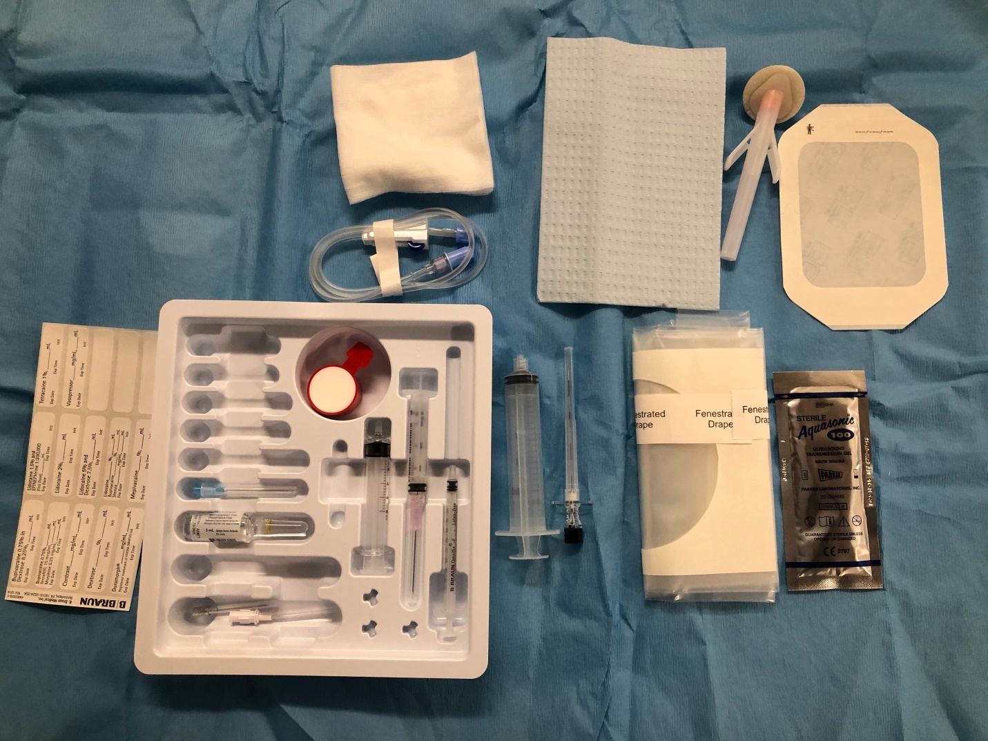
  • Standard nerve block tray (figure 1)
  • Chlorhexidine gluconate or povidone-iodine
  • One 20-mL syringe containing a local anesthetic
  • Ultrasound machine with curvilinear transducer (2.5 Mhz to 7.5 Mhz ), sterile sleeve, and gel
  • Injection pressure monitor
  • Sterile gloves

Personnel

A practitioner with expertise in regional anesthesia. Additional support such as nursing staff with experience in administering sedation is beneficial.

Preparation

The performing provider must obtain informed consent. A pre-procedure timeout is conducted, and the patient is placed in the supine position. The procedural leg is slightly abducted. Mild to moderate sedation may be administered (50 -100 mcg of fentanyl and 1-2 mg midazolam intravenously). Chlorhexidine gluconate 2% or povidone-iodine is applied to the skin (iodine must remain on the skin for three minutes). Utilizing the aseptic technique, the sterile conduction gel is applied to the skin, and the sterile ultrasound probe cover is placed around the ultrasound probe.

Technique or Treatment

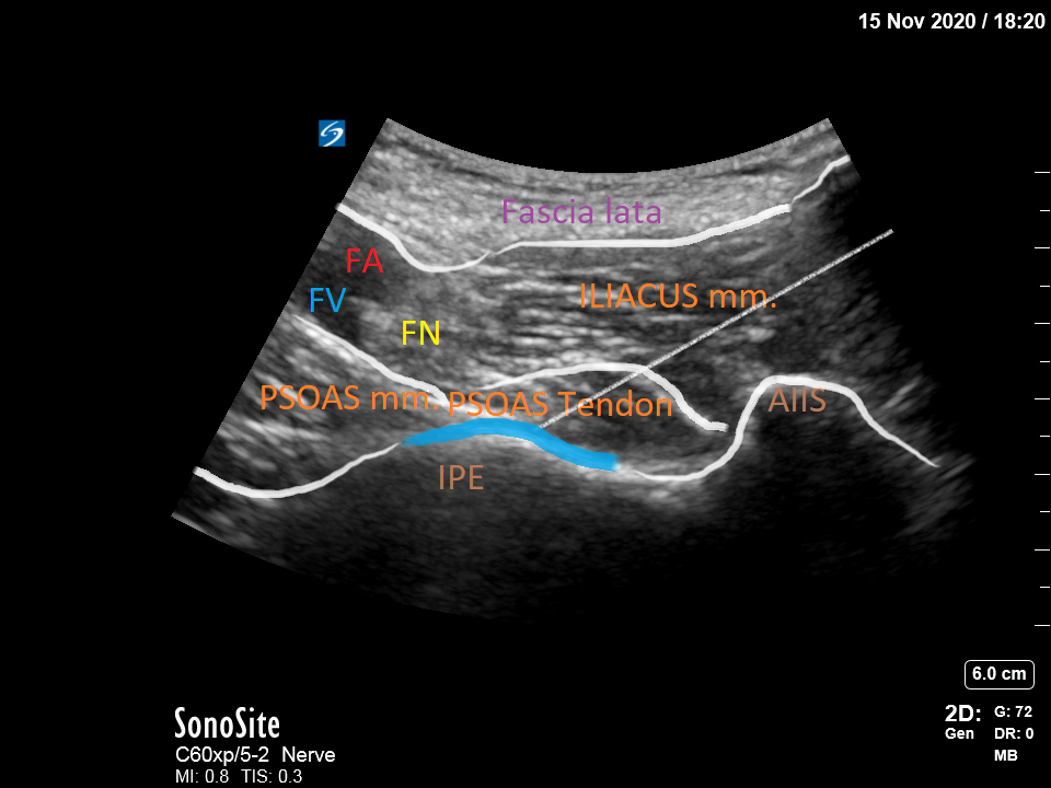
With the patient in the supine position, the ultrasound probe is placed on a transverse plane over the anterior superior iliac spine (ASIS). Once the ASIS is identified, the transducer is aligned with the pubic ramus and rotated at approximately 45 degrees, parallel to the inguinal crease. The transducer is then slid medially along this axis until the anterior inferior iliac spine (AIIS), iliopubic eminence (IPE), and the psoas tendon is clearly identified, serving as anatomic landmarks. 

Sliding the probe distally or gently tilting the caudal will expose the head of the femur. Returning to the initial starting position, a standard 20-22 gauge 100mm needle is inserted in-plane, from lateral to medial, in the plane between the psoas tendon and the pubic ramus. 15-20ml of a long-lasting local anesthetic ((i.e., 0.5% ropivacaine) is then deposited in this plane, lifting the psoas tendon. Care should be taken to avoid puncturing the psoas tendon.

Complications

General complications resulting from peripheral nerve blockade include infection, bleeding, neuropathy, and systemic toxicity from large volume local anesthetic deposition. Quadriceps muscle weakness may occur secondary to the femoral nerve blockade.

Clinical Significance

The hip joint has a complex innervation, and the pain following hip fractures or total hip arthroplasties is particularly severe. An appropriate plan for perioperative analgesia is challenging, but a multimodal approach including acetaminophen, cox-2 selective NSAIDs, regional anesthesia, and periarticular infiltration techniques improves patient outcomes.[12][13] 

The ultrasound-guided PENG block allows for coverage of the hip joint, targeting the proximal articular branches that innervate the joint capsule. This proximal approach via ultrasound guidance can confer several advantages over a femoral nerve block by providing more complete analgesia to the hip joint. Additionally, the motor function of the involved extremity should be spared. The PENG block can be used alone as a primary analgesic or in conjunction with other forms of anesthesia during surgery or in the perioperative period. For lateral surgical incisions, a supplemental lateral femoral cutaneous nerve block provides additional coverage.[13]

Enhancing Healthcare Team Outcomes

The PENG block is typically performed by an anesthesiologist with experience in regional anesthesia, usually with the assistance of a block nurse. Interprofessional communication skills are essential, as the nurse assists with sterile technique, preparation of the ultrasound by handing the probe to the performing provider. Ensuring that medications for sedation and analgesia are available, as well as preparation of the equipment, are some of these duties.

It is recommended that a regional anesthesia cart equipped with emergency medications and airway management equipment be readily available. Nurses are responsible for being able to quickly retrieve such items in the event of an emergent complication. All involved team members from the nursing staff, trainees, and performing providers should be familiar with the adverse effects of local anesthetics.


(Click Image to Enlarge)
<p>Standard Nerve Block Tray (PENG block)</p>

Standard Nerve Block Tray (PENG block)


Contributed by Beric Berlioz MD, Mayo Clinic, Florida


(Click Image to Enlarge)
Relevant sonoanatomy for PENG block (FA= femoral artery; FV = femoral vein; FN= femoral nerve AIIS = antero inferior iliac sp
Relevant sonoanatomy for PENG block (FA= femoral artery; FV = femoral vein; FN= femoral nerve AIIS = antero inferior iliac spine; IPE = iliopubic emminece
Contributed by Beric Berlioz, MD
Details

Editor:

Elird Bojaxhi

Updated:

1/29/2023 9:17:32 AM

References


[1]

Girón-Arango L, Peng PWH, Chin KJ, Brull R, Perlas A. Pericapsular Nerve Group (PENG) Block for Hip Fracture. Regional anesthesia and pain medicine. 2018 Nov:43(8):859-863. doi: 10.1097/AAP.0000000000000847. Epub     [PubMed PMID: 30063657]


[2]

Lee DJ, Elfar JC. Timing of hip fracture surgery in the elderly. Geriatric orthopaedic surgery & rehabilitation. 2014 Sep:5(3):138-40. doi: 10.1177/2151458514537273. Epub     [PubMed PMID: 25360345]


[3]

Keene GS, Parker MJ, Pryor GA. Mortality and morbidity after hip fractures. BMJ (Clinical research ed.). 1993 Nov 13:307(6914):1248-50     [PubMed PMID: 8166806]


[4]

Scala VA,Lee LSK,Atkinson RE, Implementing Regional Nerve Blocks in Hip Fracture Programs: A Review of Regional Nerve Blocks, Protocols in the Literature, and the Current Protocol at The Queen's Medical Center in Honolulu, HI. Hawai'i journal of health     [PubMed PMID: 31773105]


[5]

Acharya U, Lamsal R. Pericapsular Nerve Group Block: An Excellent Option for Analgesia for Positional Pain in Hip Fractures. Case reports in anesthesiology. 2020:2020():1830136. doi: 10.1155/2020/1830136. Epub 2020 Mar 12     [PubMed PMID: 32231802]

Level 3 (low-level) evidence

[6]

Sakamoto J, Manabe Y, Oyamada J, Kataoka H, Nakano J, Saiki K, Okamoto K, Tsurumoto T, Okita M. Anatomical study of the articular branches innervated the hip and knee joint with reference to mechanism of referral pain in hip joint disease patients. Clinical anatomy (New York, N.Y.). 2018 Jul:31(5):705-709. doi: 10.1002/ca.23077. Epub 2018 Apr 16     [PubMed PMID: 29577432]


[7]

Kampa RJ, Prasthofer A, Lawrence-Watt DJ, Pattison RM. The internervous safe zone for incision of the capsule of the hip. A cadaver study. The Journal of bone and joint surgery. British volume. 2007 Jul:89(7):971-6     [PubMed PMID: 17673597]


[8]

Short AJ,Barnett JJG,Gofeld M,Baig E,Lam K,Agur AMR,Peng PWH, Anatomic Study of Innervation of the Anterior Hip Capsule: Implication for Image-Guided Intervention. Regional anesthesia and pain medicine. 2018 Feb;     [PubMed PMID: 29140962]


[9]

Martin HD, Khoury AN, Schroder R, Gomez-Hoyos J, Yeramaneni S, Reddy M, James Palmer I. The effects of hip abduction on sciatic nerve biomechanics during terminal hip flexion. Journal of hip preservation surgery. 2017 Jul:4(2):178-186. doi: 10.1093/jhps/hnx008. Epub 2017 Apr 11     [PubMed PMID: 28630740]


[10]

Kukreja P, Avila A, Northern T, Dangle J, Kolli S, Kalagara H. A Retrospective Case Series of Pericapsular Nerve Group (PENG) Block for Primary Versus Revision Total Hip Arthroplasty Analgesia. Cureus. 2020 May 19:12(5):e8200. doi: 10.7759/cureus.8200. Epub 2020 May 19     [PubMed PMID: 32572357]

Level 2 (mid-level) evidence

[11]

Horlocker TT, Vandermeuelen E, Kopp SL, Gogarten W, Leffert LR, Benzon HT. Regional Anesthesia in the Patient Receiving Antithrombotic or Thrombolytic Therapy: American Society of Regional Anesthesia and Pain Medicine Evidence-Based Guidelines (Fourth Edition). Regional anesthesia and pain medicine. 2018 Apr:43(3):263-309. doi: 10.1097/AAP.0000000000000763. Epub     [PubMed PMID: 29561531]

Level 1 (high-level) evidence

[12]

Kehlet H, Multimodal approach to control postoperative pathophysiology and rehabilitation. British journal of anaesthesia. 1997 May;     [PubMed PMID: 9175983]


[13]

Højer Karlsen AP, Geisler A, Petersen PL, Mathiesen O, Dahl JB. Postoperative pain treatment after total hip arthroplasty: a systematic review. Pain. 2015 Jan:156(1):8-30. doi: 10.1016/j.pain.0000000000000003. Epub     [PubMed PMID: 25599296]

Level 1 (high-level) evidence