Facial fractures occur less commonly in children than adults but may result in significant morbidity and lifelong consequences. Children's unique anatomical and developmental characteristics necessitate nuanced evaluation and management approaches. As facial fractures often accompany broader trauma, a comprehensive understanding of associated injuries is crucial, with particular attention to potential airway compromise, head trauma, and concomitant injuries. Pediatric facial trauma's diverse etiologies make interprofessional collaboration between facial surgery, neurosurgery, and pediatric specialists essential for optimal care. Adherence to the Advanced Trauma Life Support guidelines ensures systematic evaluation and timely intervention, emphasizing the importance of early recognition and treatment of life-threatening conditions.
This activity for healthcare professionals is designed to enhance learners' proficiency in evaluating and managing pediatric facial fractures. Participants gain deeper insights into the pediatric facial skeleton's unique anatomical features and growth patterns compared to adults, strengthening their skills in creating individualized diagnostic and treatment approaches for affected patients. After participation, learners become prepared to collaborate effectively within an interprofessional team to improve outcomes for young patients with facial fractures.
Objectives:
Identify the signs and symptoms indicative of facial fractures in pediatric patients.
Create a clinically guided diagnostic strategy for a young patient with a suspected facial fracture.
Develop a personalized management plan for a pediatric patient with facial trauma.
Collaborate within an interprofessional team to formulate short- and long-term management strategies to enhance outcomes for children with facial fractures.
Introduction
Trauma is a significant cause of morbidity and mortality in the pediatric population.[1] The head is the most common site of trauma. Facial fractures in very young children are rare due to their greater facial elastic cartilage content and cranial-to-facial volume ratio than adults. While facial fractures are infrequent in this age group, these injuries can be severe enough to produce lifelong consequences.[2]
Facial growth dictates age-specific fracture patterns different from adults mainly due to secondary dentition eruption and paranasal sinus pneumatization. Isolated facial fractures can occur in pediatric patients. However, the potential for concurrent injuries must be considered in the setting of acute trauma, particularly in the head, eyes, brain, neck, and airway.[3][4][5][6][7][8]
Anatomy, Development, and Vulnerabilities of the Pediatric Facial Skeleton
The bones forming the facial framework include the paired maxillae, mandibles, zygomae, and nasal and frontal bones. The maxilla forms part of the cheek and the upper jaw and its tooth sockets. The mandible forms the lower jaw and is the only movable bone in the skull. The zygoma contributes to the cheeks' prominence and supports the orbit. The nasal bones form the nose's bridge and are susceptible to fractures due to their relative thinness. The frontal bone comprises the forehead and upper part of the orbit, protecting the brain from the anterior side. Cranial bones protecting the rest of the brain include the parietal bones superolaterally, temporal bones laterally, occipital bones posteriorly, and the sphenoid and ethmoids inferiorly.
The neonatal skull is much larger than the face, with an 8:1 volume ratio compared to adults' 2:1 proportions. The forehead protrudes over the face more in infants than adults. With growth, the face expands to comprise a greater relative area of the head until adult proportions are reached in the teenage years.[9][10] Thus, head trauma in young children is more likely to affect the skull than the face.
Pediatric facial bones have more elastic cartilage than adult bones, making them resilient and more likely to compress than fracture after traumatic impact. Consequently, children develop fewer facial fractures from mechanisms that can easily break adult bones. If they occur, pediatric facial fractures tend to be minimally displaced and do not assume the classic adult fracture patterns, such as Le Fort injuries.[11]
Nasal fractures are the most common facial fractures in children overall due to the nasal bridge's prominence and minimal surrounding structural support. Mandible fractures are the 2nd most common, accounting for nearly half of pediatric facial fractures. Fracture location relates to the sinus' age-dependent development and, to a lesser degree, the dentition stage.[12] During development, facial bones thicken before becoming fully pneumatized and thinning into the final adult configuration. Active growth and early pneumatization make children's facial bones thicker and more resistant to fractures than more developed, thin adult bones.
Maxillary sinus pneumatization occurs at birth but may continue until around age 7. Thus, the midface at this time is thicker and more elastic than the upper face. Blunt trauma to the midface in this age group often transmits impact forces superiorly toward the thinner frontal bone, increasing the likelihood of orbital roof fractures, which are less frequent in older children. Mixed dentition forms and progresses in the midface between ages 6 and 12, adding further stability and strength to the region while maxillary pneumatization slows. However, orbital floor thinning during this period makes orbital floor and wall injuries, including blowout fractures, more common with increasing age in this group.
Beyond age 12, the maxillary sinuses become fully pneumatized, midfacial bones thin, cartilages ossify, and frontal sinuses continue to thicken and develop.[13][14] Mid- or upper-face blunt trauma transmits impact forces downward, away from the thick, elastic frontal sinus toward the thin, adult-like upper maxilla. Hence, orbital floor fractures are more prevalent in adolescents than younger children.
Etiology
Facial fractures in children are typically caused by blunt trauma, eg, from falls, sports injuries, motor vehicular crashes (MVCs), assault, and child abuse.[15] Penetrating facial injuries, particularly gunshot wounds, also occur. These incidents are declining in frequency in the United States.[16]
Epidemiology
Pediatric trauma causes approximately 12,000 deaths and over 8 million emergency department visits annually in the United States.[17] Less than 15% of individuals with facial fractures are children. Most pediatric facial injuries are limited to soft tissues, with only 10% to 15% resulting in craniomaxillofacial fractures.[18][19] However, most facial trauma presentations are associated with concurrent severe injuries outside of the face. Many minor facial traumas are treated at home and are, therefore, likely underreported. In contrast, fractures cause significant pain and swelling and may be more accurately reported than facial soft tissue injuries.
Adolescent boys are twice as likely to present with facial fractures than adolescent girls. Facial fractures are rare under the age of 6, although skull fractures occur more frequently than facial fractures in this age group. Half of all fracture presentations are seen in patients aged 10 to 18. Roughly half of pediatric facial fractures arise from MVCs. Bicycle accidents and sports injuries comprise most of the remaining trauma etiologies in school-age children. Infants and toddlers are more likely to sustain injuries from falls. Assault-related facial injuries are less common and often involve adolescent boys. Fracture locations are age-dependent due to the effect of physical activities on etiology and aging-related bone growth and development patterns.
Nasal fractures are generally thought to be the most common but likely underreported, as evaluation of these injuries at trauma centers is not mandatory. The facial bone most often reported to be involved in traumatic injuries across age groups is the mandible, affecting 40% to 60% of pediatric patients with facial fractures and increasing in incidence with age.[20] Alveolar ridge fractures are more common in young patients, affecting 60% of children aged under 6 with fractures and becoming less common with increasing age. Orbital and midface fractures are the next most common across all age groups. Frontal bone fractures are associated with intracranial injuries in 35% to 64% of cases, and cerebrospinal fluid (CSF) leaks in 18% to 36%. CSF leakage can occur in younger patients, with the frontal bone absorbing the impact before full pneumatization when it still has a "crumple zone."
Nasoorbitoethmoid fractures are uncommon, representing only 1% to 8% of all pediatric facial fractures. Fracture patterns described in adults, such as Le Fort midfacial injuries, are rare in pediatric patients. Such injuries are generally only seen in older adolescents and account for less than 2% of pediatric facial fractures.
History and Physical
Significant force is necessary to fracture a child's facial skeleton. Hence, finding facial fractures in this age group should prompt a search for potentially associated injuries. A facial bone injury may be accompanied by airway compromise, spinal or neck fractures, or traumatic brain damage. Patients with traumatic facial injuries should thus be evaluated according to the Advanced Trauma Life Support (ATLS) guidelines. Significant midfacial and nasal bleeding and tongue retrodisplacement from bilateral mandible fractures are common reasons for airway compromise. The clinician should return for a more detailed facial structure evaluation once life-threatening and other severe injuries are identified and stabilized.
History
An accurate account of the events, including changes in mental status, sensory and motor function, range of motion, vision, and associated symptoms, is critical when evaluating trauma. A history from parents, coaches, or first responders is usually necessary, particularly with younger patients. Children rarely have underlying medical conditions that contribute to the traumatic etiology or cause complications, such as anticoagulation. Nonetheless, routine historical questions should still be pursued for their potential impact on management. Allergies, vaccinations (particularly tetanus), and timing of the last meal must be elicited, especially if considering emergency surgery.
Patients may report swelling or stiffness in the head, neck, jaw, eyes, or nose, though not all children can communicate a complete history of the traumatic event or relay all of their symptoms. The presence or a recent history of epistaxis does not increase the likelihood of facial fractures. On the other hand, the sensation that something is stuck in the facial or oral area, persistent diplopia, subjective malocclusion or loose teeth, and facial paresthesias should raise concern for facial fractures.
Physical Examination
Many facial injuries are visually apparent to the patient and the examiner. Still, a careful physical examination is critical in children. A calm, relaxed patient often accommodates a thorough examination.
A facial examination should be systematic. The exact approach is not as important as long as all aspects are examined. One method is to attempt the exam in 3 dimensions: superior to inferior, lateral to medial, and superficial to deep.The physical examination's diagnostic yield can be improved when younger patients are held by their parents and pain, distraction, and anxiety are addressed appropriately.[21][22] A cranial nerve evaluation should complete every thorough head and neck physical examination.
Musculoskeletal and skin examination
Wound evaluations must determine the extent of damage in the muscles, tendons, vessels, nerves, and ducts. Facial nerve palsy after blunt trauma is suspicious for a temporal bone fracture.[23][24] Pain and stiffness are expected to limit joints' range of motion after trauma. Bony tenderness and soft tissue swelling are suggestive but nonspecific for facial bone fractures. However, crepitus near a sinus more strongly correlates with an underlying fracture. Similarly, step-offs or mobility of the facial bones on palpation suggests the presence of a fracture.
Eye examination
Periorbital swelling can develop and hinder a complete facial examination. Hence, the eye examination should occur early if direct eye trauma has occurred. Extraocular muscle range of motion impairment suggests entrapment, possibly from an orbital floor fracture. Pediatric patients are more likely to experience "white-eyed" blowout fractures, wherein the orbital floor breaks and swings inferiorly into the maxillary sinus, permitting intraorbital contents to herniate through the fracture. Herniated soft tissues are entrapped and may strangulate when the fractured bone returns to its original position.
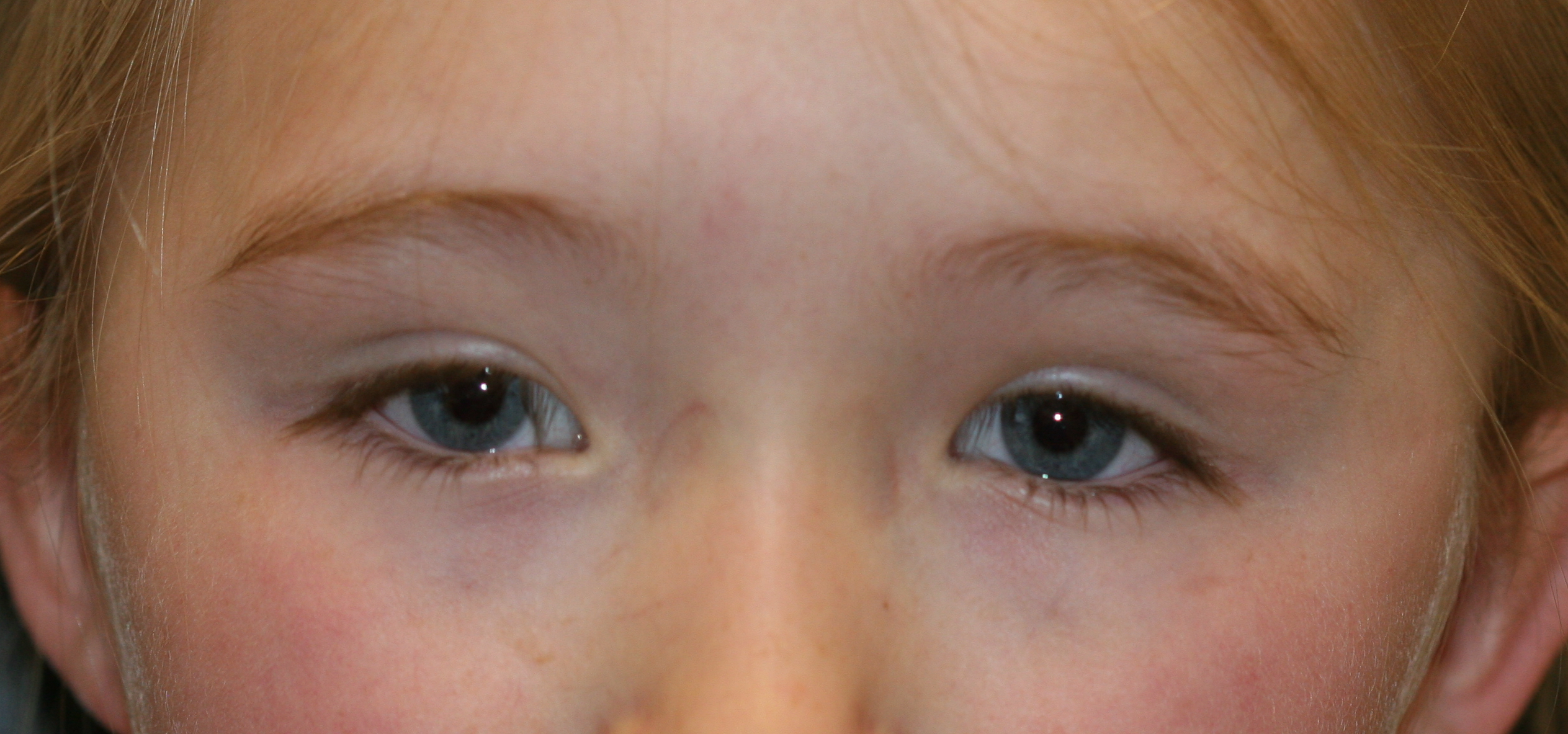
Young patients may not demonstrate significant erythema, ecchymosis, or edema. Hence, the term "white-eyed blowout fracture" is often used to describe orbital floor trauma in this age group. However, children may require urgent surgical intervention, particularly when eye movement causes bradycardia, nausea, or syncope due to the oculocardiac reflex.[25] Globe, retina, and optic nerve injuries may need to be addressed before open reduction and internal fixation (ORIF) of facial fractures to prevent worsening visual impairment. Telecanthus on examination should raise concern for a nasoorbitoethmoid fracture (see Image. Telecanthus). The pediatric ocular evaluation should be performed by a pediatric ophthalmologist whenever possible.
Mouth and intraoral examination
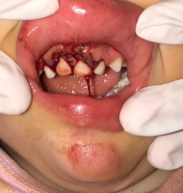
Oral examination should focus on the upper and lower jaws, teeth, and temporomandibular joint (TMJ). Bony tenderness, trismus, malocclusion, dental laxity on palpation, gingival ecchymosis, and lacerations may be signs of a mandibular or maxillary fracture. Tooth mobility may arise in several scenarios: loose teeth, alveolar ridge fracture, and maxillary or mandibular fractures with mobile segments (see Image. Alveolar Fracture of the Anterior Segment). Mandibles frequently fracture in 2 locations due to their curved shape. Therefore, finding a mandible fracture should raise suspicion for a second. Tongue retrodisplacement may narrow the airway if 2 mandibular fractures produce a large flail segment that includes the central portion. This condition is potentially fatal and must be addressed immediately.
Evaluation
Laboratory tests are unnecessary for pediatric patients who have sustained isolated facial fractures without suspected intracranial involvement. However, if concerns persist regarding surgical management, testing may be indicated according to institutional preoperative protocols.
The preferred imaging modality for evaluating suspected facial trauma at any patient age is computerized tomography (CT). Magnetic resonance imaging (MRI), typically used to assess soft-tissue structures, is less sensitive than CT to subtle fractures and should not be used as a primary imaging modality for assessing facial fractures. Plain film and panoramic x-rays may be used for dental evaluation but do not reliably identify facial fractures. Isolated soft tissue injuries do not benefit from imaging. Imaging is also futile in clinically identified isolated nasal bone fractures, which should be treated empirically.
Treatment / Management
Stabilization and Initial Treatments
Patients presenting with unstable vital signs, severe hemorrhage, or signs of cardiorespiratory arrest (eg, unresponsiveness, pulselessness, and apnea) should be immediately resuscitated. Airway, breathing, circulation, disability, and exposure must be addressed without delay. Resuscitation measures should follow the ATLS guidelines. Immobilization and treatment of injuries, especially profusely bleeding sites, should proceed while stabilizing the patient.
As with most traumatic injuries, pediatric patients with facial fractures benefit from ice, rest, and pain control. Once fractures are identified, the appropriate specialists should be consulted for further management and treatment recommendations. These specialists may include pediatric specialists in facial surgery (otolaryngology, oral-maxillofacial surgery, and plastic surgery, depending on local resources), ophthalmology, neurosurgery, anesthesia (for advanced airway stabilization), psychiatry (if self-harm is suspected), and any other specialty team clinically indicated for consultation. Social work and law enforcement may be required when nonaccidental trauma is suspected.
Child-specific anxiolysis and pain control approaches may be required for younger patients. The first-line option is having a parent hold the patient in their lap and attempt to soothe and restrain the child as necessary. Failing this, various pharmacologic agents are available. Oral sucrose solution may be required for neonates and young infants. Older children may be calmed with cold teething toys or popsicles.
Oral medications may include acetaminophen and ibuprofen. Carefully dosed opioids are often the most helpful for managing pain from facial fractures. Nitrous oxide is an effective anxiolytic and analgesic that may be considered in this patient group.[26] Fentanyl, ketamine, and midazolam may all be administered intranasally to avoid traumatic intravenous access.[27] Topical lidocaine, epinephrine, and tetracaine mixtures can provide analgesia without injections, which is helpful when evaluating or managing lacerations that may accompany facial fractures.[28]
Antibiotics and Vaccination
The patient’s tetanus vaccination status should be confirmed when dental injuries or open wounds are present. Evidence does not suggest that antibiotics provide any mortality benefit or reduce the rate of complications, such as infections or poor healing.[29][30] However, antibiotics are commonly prescribed for open fractures. Contaminated lacerations often accompany fractures with sinus or intraoral involvement, as happens after an MVC or animal bite. Antibiotic coverage for intraoral flora, including amoxicillin-clavulanate or clindamycin, is usually sufficient in such cases.
Fracture Management
Most facial fractures in children can be managed conservatively without surgery in consultation with a pediatric facial surgeon. Markedly deformed structures can remodel during the healing and normal growth processes as children age. A trial of watchful waiting and observation may be a reasonable approach.
Facial and jaw restructuring may be accomplished with orthodontic manipulation, particularly for injuries causing malocclusion.[2][9] Surgery is avoided whenever feasible, but delayed surgical intervention may be advised for cosmetic or structural purposes if it was not initially warranted for the injury. Plates made from bioresorbable polymers like poly-L-lactic acid and polyglycolic acid, rather than titanium, are suggested for ORIF of non-load-bearing fractures. These plates have minimal impact on bone growth, but not all facial surgeons agree with this recommendation.[31]
Frontal Bone Fractures
Frontal fractures are uncommon before frontal sinus pneumatization, which typically occurs at ages 5 to 6. Older patients with frontal sinus fractures may have them reduced and rigidly fixated if causing substantial displacement or cosmetic deformity. However, bone remodeling during growth can help fractures heal, though with noticeable deformities over time.
Mucocele formation can be prevented after nasofrontal drainage compromise by obliterating the sinus with fat or a pericranial flap after exenterating the mucosa. Watchful waiting may be employed in less severe nasofrontal duct injury cases. However, posterior table fractures are typically addressed jointly by otolaryngology and neurosurgery, cranializing the frontal sinus and stopping CSF leaks.
Forehead lacerations may provide sufficient access to the frontal bone to permit ORIF in some cases. An incision through the upper eyelid may be adequate for small, inferior fractures. However, most cases require a coronal incision to expose the fractures well enough for fixation.
Nasoorbitoethmoid Fractures
Nasoorbitoethmoid fractures are, fortunately, rare in children because the region is not as prominent as the forehead. Operative intervention is indicated in pediatric fractures of this type if traumatic telecanthus, medial canthus blunting, or horizontal palpebral fissure shortening is present. However, these injuries may be difficult to find in young children, who have a relatively more comprehensive nasal root that may exceed the intercanthal distance in breadth. A coronal incision is typically used if surgery is necessary. For frontal bone fractures, wires or permanent sutures can fixate the bone segments bearing the medial canthi in their anatomical positions. The classic complication of this procedure is persistent telecanthus. The recommendation is to overcorrect when tightening the sutures or wires to narrow the intercanthal distance.
Nasal Fractures
Nasal fractures in adolescent patients may be reduced under general anesthesia, similar to how they are managed in adults.[32] Rhinoplasty may be performed once the patient is older should closed reduction prove inadequate in the long term. However, many surgeons prefer to avoid septoplasty for actively growing children, as it can affect midfacial development. Septal hematomas should always be ruled out in cases of nasal trauma because of their potential to cause septal perforation, saddle nose deformity, or potential growth abnormalities. Incision and drainage should be performed if necessary. Identifying cosmetic deformity after trauma in very young children is difficult.
Orbital Fractures
The primary indication for urgent orbital floor and medial wall fracture repair is extraocular muscle entrapment, most commonly of the inferior rectus muscle. Surgery is indicated urgently when entrapment causes an oculocardiac reflex. Otherwise, orbital floor repair may be performed 24 to 48 hours after the injury to release entrapped soft tissue and prevent muscle fibrosis and long-term eye mobility problems (see Video.Forced Duction Testing in a Pediatric Patient).[33][34]
If muscle entrapment is not a concern, the repair of orbital floor defects greater than 1 cm2, 50% of the area of the floor, or leading to enophthalmos of greater than 2 mm may be delayed for several days until edema has subsided. These injuries require orbital floor reconstruction to prevent long-term enophthalmos. Portions of the medial wall may also need reconstruction. Materials commonly employed include porous polyethylene, titanium mesh, a combination of the 2, or a thin sheet of nylon or polydioxanone, the latter of which is resorbable over several months. Split calvarial bone grafting is another option that some surgeons prefer, particularly on healthcare mission trips when commercially available orbital floor prostheses are unavailable.
Orbital floor access is provided via a subciliary or transconjunctival incision, which may be used for either a preseptal or postseptal approach. Fracture visualization may also be improved with a rigid Hopkins rod endoscope. Complete fracture exposure allows for implant placement. The implant is positioned with overlap along the fracture's entire margin to prevent its displacement into the maxillary sinus. A forced duction test is then performed to ensure no residual eye entrapment. The globe position is also evaluated to ensure sufficient exophthalmos, facilitating edema resolution. The globe then protrudes into a position symmetric with the other side. A CT scan should confirm the correct implant placement.
Zygomaticomaxillary Complex Fractures
These fractures may be managed conservatively, with or without a soft diet, unless visible facial deformity or functional limitations, such as trismus or malocclusion, are present. Fracture reduction, if indicated, may involve several incisions. A lateral brow incision can address the zygomaticofrontal suture, while a transconjunctival incision is used for the infraorbital rim. An upper gingivolabial sulcus incision provides access to the maxilla and the zygomaticomaxillary and nasomaxillary buttresses. Gillies or Keen incisions, made in the temporal hair tuft or intraorally, respectively, allow for depressed zygomatic arch segment reduction. Each incision is strategically chosen to provide optimal access and facilitate the reduction of specific fracture components.
Orbital rim or floor involvement warrants orbital floor evaluation after a zygomaticomaxillary complex reduction to ensure that a bony segment distraction has not produced a significant defect and additional intervention is not indicated. Rigid screw fixation should be monocortical, particularly near the alveolar ridge, to avoid injury to the unerupted secondary dentition.[35]
Mandible Fractures
Fractures in different mandibular regions are managed differently. Surgical techniques vary according to the patient's dental development stage.[36] Most mandible fractures in pediatric patients, including those that typically require surgery in adults (eg, condylar fractures), can be managed conservatively with a soft diet and pain control. Maxillomandibular fixation (MMF) or guiding elastics may be necessary in some children.
MMF duration is generally shorter (2 to 3 weeks) than recommended for adults to avoid TMJ ankylosis. Applying MMF in edentulous young children may involve circummandibular wiring and wiring through the piriform aperture to secure splints for stabilization.[37] Primary teeth may be used for MMF. However, wiring arch bars to loose teeth must be avoided in patients aged 6 to 12. Monocortical screws are preferred if rigid fracture fixation is required to minimize the risk of injury to unerupted secondary teeth.
Fractures most likely requiring surgical reconstruction include those causing condyle displacement into the middle cranial fossa, limiting mandibular movement, bilateral fractures reducing mandibular height and causing an open bite, mobile anterior segments getting displaced by masticatory muscle action, and mobile alveolar ridge fractures needing wiring or braces. Orthodontics may be necessary if malocclusion persists after healing, regardless of fracture location.
Home Medications and Diet
Injuries involving the upper or lower jaw or teeth should prompt recommendations for a soft or liquid diet and avoiding food temperature extremes. Liquid-form medication prescriptions are often necessary. Patients with injuries involving the sinuses or nasal cavity should be counseled to avoid nose-blowing, sneezing with a closed mouth, drinking through a straw, exercising strenuously, and swimming.
Follow-Up
Follow-up for most pediatric fractures typically involves a pediatric facial surgeon. The timing of follow-up should be discussed with the specialist, although within 1 week is usually reasonable if no urgent operative indications are apparent. Patients with dental injuries should be referred to a pediatric dentist. The patient’s caregivers must be provided with the images and radiological reports obtained in the emergency department to minimize redundant imaging and unnecessary radiation exposure. These images and reports also allow outside clinicians to assess the changes after the initial injury.
Differential Diagnosis
Facial fractures in children are typically only produced by dramatic impacts and are, therefore, associated with severe concomitant injuries. Of primary concern are airway consequences from severe facial trauma and neurological damage from head injuries. The likely etiologies of pediatric facial fractures include the following:
Nonaccidental trauma, such as child abuse or neglect
Physical or sexual assault
Risk-taking behaviors
Lack of protective equipment use or availability
Suicide attempt or other self-injurious behavior
In addition to specific bony facial fractures, the following conditions must also be considered in children with facial fractures:
Bony injuries
Mandible dislocation
Sinus involvement in fractures
Skull or cervical spine fractures
Cartilage injuries, including nose and ear
Tooth avulsions or fractures
Soft tissue wounds and injuries
Lacerations and contusions
Penetrating wounds and retained foreign bodies
Septal or auricular hematomas
Eye injuries, including globe rupture, retrobulbar hematoma, and corneal damage
Ductal and glandular injuries
Neurovascular damage
Proper differentiation and management of facial injuries can significantly impact functional and aesthetic recovery and prevent long-term sequelae.
Prognosis
The prognosis of pediatric facial trauma is generally favorable. However, extensive injuries may result in long-term deformity, possibly requiring surgical repair. Reassuringly, pediatric bone and cartilage are adept at remodeling, and most patients heal well with minimal discernable evidence of injury.
Complications
The possible pediatric facial fracture complications include the following:
Growth abnormality or long-term disfigurement
Infection, especially if with dental or sinus involvement or penetration of foreign objects
Long-term dental effects
Psychosocial sequelae
Poor visual acuity, especially if entrapped orbital muscles were not released immediately
Persistent paresthesia or muscle weakness if peripheral nerves are damaged
Posttraumatic or chronic facial pain
Timely assessment and accurate diagnosis of facial fractures are essential to identifying potential complications early and initiating appropriate management.
Deterrence and Patient Education
Pediatric facial trauma is typically accidental, but several measures can reduce the frequency and severity of these injuries. Such actions include evaluating for possible child abuse, suicidal tendencies, and risky social behavior, including high-impact sports and recreational activities. Adults and children alike see a decreased rate of injury and death when using age- and size-appropriate restraints in motor vehicles.[38][39] Additionally, the use of personal protective equipment, especially helmets with face shields or visors, should be encouraged during sports and other activities with the potential for high-energy impacts, such as all-terrain vehicle riding, snowmobiling, skiing, skateboarding, and bicycling.[40][41]
Pearls and Other Issues
Recognizing the pediatric skull and facial skeleton's unique anatomical and developmental characteristics is essential when managing pediatric facial fractures. Pediatric facial bones are more pliable and less dense than adult bones. Additionally, the presence of growth plates and fontanelles adds complexity to fracture patterns and can influence treatment strategies. Interprofessional collaboration between pediatricians, maxillofacial surgeons, and otolaryngologists ensures comprehensive care in the acute phase. Treatment plans should be tailored to the child's age, developmental stage, and fracture characteristics. Close monitoring of growth and development is crucial to identify and address potential complications, such as growth disturbances or malocclusion.
Enhancing Healthcare Team Outcomes
Pediatric facial fractures are uncommon despite the high incidence of pediatric facial trauma. Cases involving fractures are typically evaluated in the emergency room and involve emergency medicine clinicians and nurses, a facial surgeon, a radiologist, a trauma surgeon, a pediatrician, a social worker, emergency medical service personnel, and other specialty services. Facial fractures in children are often conservatively managed, although an organized team approach can prevent missing subtle or dangerous injuries and improve patient outcomes. Every patient with facial trauma should be evaluated according to ATLS guidelines to ensure concomitant injuries, particularly life-threatening ones, are identified and treated appropriately.
(Click Image to Enlarge)
Telecanthus. This 6-year-old girl has developed a telecanthus 6 months after sustaining facial fractures from a motor vehicle accident. Telecanthus is characterized by an increased distance between the eyes' inner corners.
Contributed by BCK Patel, MD, FRCS
(Click Image to Enlarge)
Alveolar Fracture of the Anterior Segment. This image shows an alveolar fracture of the anterior segment with displaced primary teeth.
Borse N, Sleet DA. CDC Childhood Injury Report: Patterns of Unintentional Injuries Among 0- to 19-Year Olds in the United States, 2000-2006. Family & community health. 2009 Apr-Jun:32(2):189. doi: 10.1097/01.FCH.0000347986.44810.59. Epub
[PubMed PMID: 19305217]
Alcalá-Galiano A, Arribas-García IJ, Martín-Pérez MA, Romance A, Montalvo-Moreno JJ, Juncos JM. Pediatric facial fractures: children are not just small adults. Radiographics : a review publication of the Radiological Society of North America, Inc. 2008 Mar-Apr:28(2):441-61; quiz 618. doi: 10.1148/rg.282075060. Epub
[PubMed PMID: 18349450]
Iida S, Matsuya T. Paediatric maxillofacial fractures: their aetiological characters and fracture patterns. Journal of cranio-maxillo-facial surgery : official publication of the European Association for Cranio-Maxillo-Facial Surgery. 2002 Aug:30(4):237-41
[PubMed PMID: 12231205]
Imahara SD, Hopper RA, Wang J, Rivara FP, Klein MB. Patterns and outcomes of pediatric facial fractures in the United States: a survey of the National Trauma Data Bank. Journal of the American College of Surgeons. 2008 Nov:207(5):710-6. doi: 10.1016/j.jamcollsurg.2008.06.333. Epub 2008 Aug 9
[PubMed PMID: 18954784]
Woodruff G, Palmer L, Fontane E, Kalynych C, Hendry P, Thomas AC, Crandall M. Nine years of pediatric gunshot wounds: A descriptive analysis. Preventive medicine reports. 2022 Aug:28():101890. doi: 10.1016/j.pmedr.2022.101890. Epub 2022 Jul 5
[PubMed PMID: 35832637]
Zempsky WT, Cravero JP, American Academy of Pediatrics Committee on Pediatric Emergency Medicine and Section on Anesthesiology and Pain Medicine. Relief of pain and anxiety in pediatric patients in emergency medical systems. Pediatrics. 2004 Nov:114(5):1348-56
[PubMed PMID: 15520120]
Gordin E, Lee TS, Ducic Y, Arnaoutakis D. Facial nerve trauma: evaluation and considerations in management. Craniomaxillofacial trauma & reconstruction. 2015 Mar:8(1):1-13. doi: 10.1055/s-0034-1372522. Epub
[PubMed PMID: 25709748]
Singer AJ, Stark MJ. LET versus EMLA for pretreating lacerations: a randomized trial. Academic emergency medicine : official journal of the Society for Academic Emergency Medicine. 2001 Mar:8(3):223-30
[PubMed PMID: 11229943]
Malekpour M, Bridgham K, Neuhaus N, Widom K, Rapp M, Leonard D, Baro S, Dove J, Hunsinger M, Blansfield J, Shabahang M, Torres D, Wild J. Utility of Prophylactic Antibiotics in Nonoperative Facial Fractures. The Journal of craniofacial surgery. 2016 Oct:27(7):1677-1680
[PubMed PMID: 27391655]
Burlini D, Conti G, Amadori F, Bardellini E, De Giuli C. Management of paediatric maxillofacial fractures: conventional methods and resorbable materials. European journal of paediatric dentistry. 2015 Mar:16(1):24-8
[PubMed PMID: 25793949]
Dulley B, Fells P. Long-term follow-up of orbital blow-out fractures with and without surgery. Modern problems in ophthalmology. 1975:14():467-70
[PubMed PMID: 1177910]
Hegab A. Management of mandibular fractures in children with a split acrylic splint: a case series. The British journal of oral & maxillofacial surgery. 2012 Sep:50(6):e93-5. doi: 10.1016/j.bjoms.2011.11.003. Epub 2011 Dec 6
[PubMed PMID: 22153178]
Murphy RX Jr, Birmingham KL, Okunski WJ, Wasser TE. Influence of restraining devices on patterns of pediatric facial trauma in motor vehicle collisions. Plastic and reconstructive surgery. 2001 Jan:107(1):34-7
[PubMed PMID: 11176598]
Tyroch AH, Kaups KL, Sue LP, O'Donnell-Nicol S. Pediatric restraint use in motor vehicle collisions: reduction of deaths without contribution to injury. Archives of surgery (Chicago, Ill. : 1960). 2000 Oct:135(10):1173-6
[PubMed PMID: 11030874]