Continuing Education Activity
Dental caries is the most prevalent chronic disease worldwide. The condition courses its early stages silently. The first clinical sign is a white spot, known as an initial lesion, which can be reversed or arrested through appropriate treatment. This activity reviews the management of initial caries lesions by remineralization therapies and the role of the healthcare team in evaluating these lesions based on the patient's risk. It also describes the pathophysiology of dental caries, fluoride and non-fluoride remineralization therapies, prognosis, and differential diagnosis.
Objectives:
- Explain the pathophysiology of dental caries.
- Describe the evaluation of initial caries.
- Summarize the different remineralization techniques based on fluoride and non-fluoride therapies.
- Review the importance of modern caries management based on the evaluation of caries risk.
Introduction
Dental caries is the most prevalent chronic disease worldwide.[1] It is a biofilm-mediated, diet-modulated, multifactorial, and non-transmissible disease. Its dynamic pathogenesis involving mineral loss of dental hard tissues results in demineralization and remineralization phases. Biological, behavioral, psychosocial, and environmental factors are also associated with the development of the condition.[2][3]
A carious lesion is the clinical sign of the disease; however, it doesn’t determine its onset.[2] That is why the extensive scientific evidence throughout these years proves that the traditional treatment of dental caries (focused on detecting cavitated lesions and restoring them) is not the solution to this problem. The treatment must be based on a deep understanding of the caries process, acknowledging that early stages can be prevented, reversed, or arrested primarily through managing etiological factors (e.g., diet) and enhancing those that favor remineralization (e.g., fluoride therapy).[4]
Function
Pathophysiology of Dental Caries
The pathophysiology of dental caries is characterized by a dynamic process that alternates periods of demineralization and remineralization.[5] The disease will progress or reverse according to which factors prevail. Pathological factors such as fermentable carbohydrates, cariogenic bacteria, and decreased salivary flow will favor demineralization, therefore, disease progression. By contrast, protective factors like antibacterial agents, remineralization techniques, and management of xerostomia will benefit the disease regression.[6]
Under normal circumstances, the concentration of calcium and phosphate in oral fluids (saliva and biofilm fluid) is supersaturated compared to their concentration in the enamel. These ions are continually deposited on the enamel surface or redeposited on enamel areas where they were previously lost. But when bacteria convert sugars from the diet into acids, the biofilm fluid becomes undersaturated compared to the enamel, leading to demineralization (mineral loss). Even though pH levels remain low (acidic) for a certain time, when the amount of sugar available goes down, the pH of the biofilm fluid returns to neutrality (rises) and becomes sufficiently saturated with calcium, phosphate, and fluoride so that demineralization stops and redeposition of mineral starts. Due to the biofilm calcium and phosphate, redeposition of minerals can occur or by the direct action of salivary calcium and phosphate soon after the biofilm is removed by toothbrushing.[7]
When repeated events of mineral dissolution happen, and the number of subsurface minerals lost surpasses the ones gained over a long period, the disease will show its first clinical sign: a white spot.[8] The progression of the white spot and the next step of the disease, a cavitated caries, can be prevented, reversed, or arrested by controlling the caries process. Still, the pH fluctuations in the biofilm are due to bacterial metabolism; therefore, loss and redeposition of minerals are inevitable. That is why we have to think about the initial phases of caries as a natural process that cannot be avoided.[9]
All the tooth surfaces are susceptible to caries throughout an individual's lifetime. But, the disease will not occur unless teeth are exposed to cariogenic biofilm and frequent sugar consumption. The modern concept of caries also considers other factors, such as sociodemographic situation, access to dental care, and fluoride exposure, which are significant but not decisive to develop the disease. For all of this, dental caries should be described as a complex biofilm-mediated affection linked mostly to frequent ingestion of sugars and poor oral hygiene combined with inadequate fluoride exposure.[3]
Evaluation
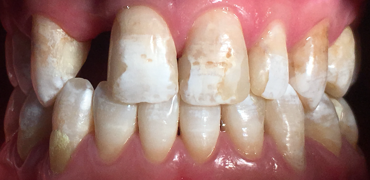
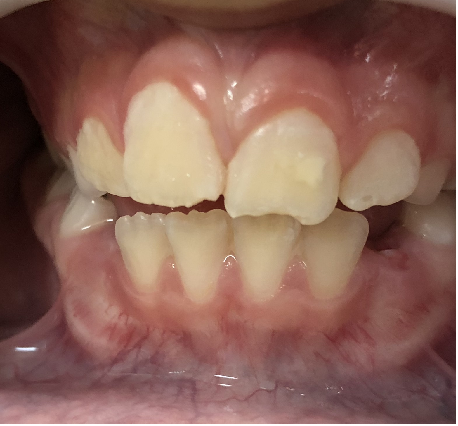
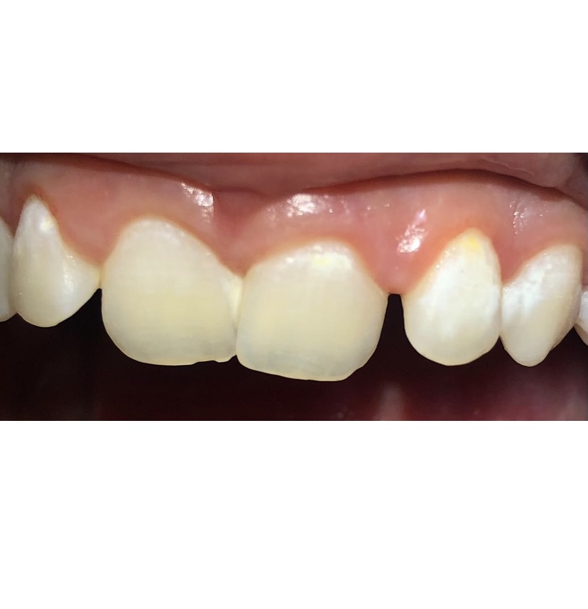
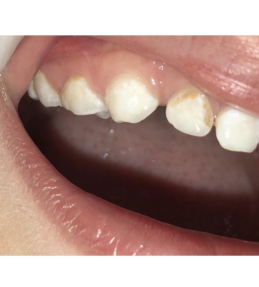
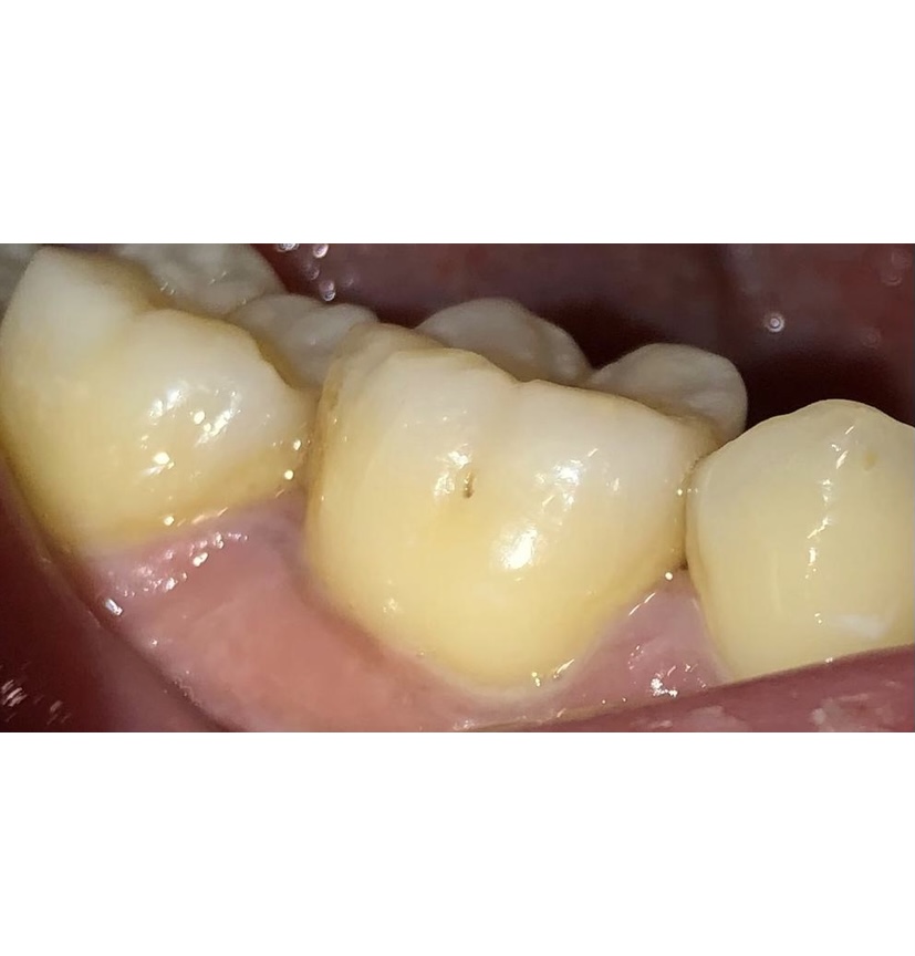
We can classify non-cavitated caries lesions as active and inactive, each with different characteristics.
Active lesion: The enamel surface is whitish/yellowish, opaque, and typically located close to the gingival margin. It is commonly covered with plaque, with no clinically detectable loss of substance. It feels rough to the touch when the curved part of the explorer is moved softly among the surface - using a sharp explorer to evaluate the lesion's surface is contraindicated because it can break down the enamel.[10]
Inactive lesion: The enamel surface is darker and shiny but intact. It feels smooth and hard to the touch; despite that, it is not recommended to evaluate these lesions with an explorer.[11]
Furthermore, as part of the evaluation, patients are classified into two groups considering their risk to develop caries:
High risk: patients present one or more risk factors like frequent consumption of dietary carbohydrates (more than four times of sugar per day), inadequate exposure to fluoride, poor oral hygiene, and salivary dysfunction. Although, the best indicator that patients will develop future caries is their past caries history.[3]
Low risk: patients that present protective factors like a healthy diet, tooth brushing with a fluoride toothpaste at least twice a day, professional application of topical fluorides, and normal salivary function.
Initial lesions should be managed considering the patient's risk of developing caries (patient-level) and the lesion's activity status (tooth level).
Issues of Concern
Treatment
The treatment for initial caries lesion (even when active) is managed through non-operative care, including remineralization therapy, behavioral changes, and using fluoride-containing products. Remineralization is aimed at stopping the progression of the lesion or, ideally, reversing it.[3]
Behavioral changes mean educating patients regarding the importance of diet, oral hygiene, and regular dental checkups. We as dentists should explain the rationale behind reducing sugar consumption and the action of saliva. The indication of a toothbrush and hygiene technique should be done based on an individual assessment. Generally, oral hygiene recommendations include tooth brushing twice a day (after breakfast and after dinner). Still, another time can be added if the patient has a high caries risk, usually after lunch. We should always motivate our patients to maintain the new habits gained (behavioral changes) over time with regular checkup appointments - at least twice a year.[12] Besides behavioral changes that depend on the patient's motivation, another way of managing initial lesions includes fluoride techniques that can increase the rate and magnitude of remineralization.
Fluoride Mechanism of Action
Fluor (F) must be present in the right place and at the right time to interfere with demineralization and remineralization events. However, even low ppm values are sufficient to fulfill its effect. When pH drops up to 4.5, fluorapatite (FA) is formed while hydroxyapatite (HA) is dissolved. If F is present, the enamel dissolution decreases because the enamel recovers those lost minerals as fluorapatite. Furthermore, the action of F is complemented by its natural effect on remineralization, enhancing the redeposition of calcium and phosphate present in the biofilm fluid when the pH rises.[7]
Fluoride used at high concentrations, more than 2500 ppm, can penetrate the dental biofilm, deliver fluoride to the tooth surface, and concentrate in incipient lesions. Thus, fluoride decreases enamel demineralization and increases remineralization. A higher fluoride concentration also prolongs the fluoride retention in the oral cavity by forming a fluoride reservoir (calcium fluoride-like deposits) on the tooth surface and in dental biofilms. It is worth noting that very high fluoride levels have a transitory bactericidal effect. However, they usually require frequent professional applications, which is impractical for both patients and professionals.[3]
Fluoride Therapy
The method with the highest level of evidence to prevent, reverse, or arrest the early stages of dental caries is topical fluoride - the topic effect of fluoride is superior to its systemic action. Fluorides can be delivered topically either as gel or varnish by a dental professional or in the form of toothpaste or mouthwash at-home settings.[2][3]
Self-applied (at-home) Topical Fluoride
Fluoride toothpaste: Tooth brushing with fluoride toothpaste significantly reduces dental caries prevalence in primary and permanent dentition.[13] The number of toothpaste brands available on the market today is undeniable. Regardless of their taste or texture, the latest research on the matter shows that concentrations must be at least 1000 ppm or higher to exploit the advantages of fluoride. The indication of toothpaste must be based not only on the age of the patients but also on the risk of developing caries (Table I).[3]
Professionally Applied topical Fluoride
Mouth rinses: The fluoride compound most commonly used in mouth rinses is sodium fluoride (NaF). They are available in two presentations: for daily use, with 230 ppm of fluoride, and weekly use, with 900 ppm (0.09%). The latter is indicated for patients over 11 years old with a high risk of caries.[3] As it happens with fluoride toothpaste, fluoride mouth rinses are associated with a considerable reduction in caries prevalence in permanent teeth.[14]
The most commonly used agents for professionally applied fluoride treatments are 5% sodium fluoride varnish and acidulated phosphate fluoride.
Gels: They contain acidified phosphate fluoride at a concentration of 1.23%, which means 12,300 ppm of fluoride ion and acid pH (3.5).
Before applicating the gel, dental cleaning is advised to take better advantage of its benefits. Some topical fluoride gels are marketed with recommended treatment times of less than four minutes. As for the frequency of application, it will depend on the patient's risk. In a low-risk patient, twice per year would be sufficient, while in a high-risk patient, four times per year.[13]
Fluoride gel is principally indicated in patients older than six years old. Under this age, the risk of experiencing adverse effects when accidentally swallowing the gel, particularly nausea and vomiting, outweighs the potential benefits of using this agent.[15]
Varnishes: More than 30 fluoride-containing varnish products are on the market today, and they have varying compositions and delivery systems. 5% sodium fluoride varnish (NaFV) and 2.26% sodium fluoride varnish are the most commonly used.
Because of the low risk of experiencing harm in children younger than six, 2.26% fluoride varnish is the only topical fluoride agent recommended for this age group, even though other topical fluorides may be beneficial.[15] The application should be made twice per year in primary and permanent dentition. However, in patients with an elevated caries risk, the varnish should be applied every three months.[13]
Non- fluoride Remineralization Agents
New phosphate-based agents with anti-caries potential have been studied as a method of promoting remineralization of initial caries lesions. The calcium- and phosphate-based delivery systems increase the saturation of these ions in the oral cavity. Casein phosphopeptide-amorphous calcium phosphate (CPP-ACP) can release calcium and phosphate in the dental biofilm to maintain a supersaturated state, favoring the remineralization process.[8]
Cyclophosphates, other phosphate-based agents, act as a barrier against acid diffusion into the dental substrate and as nucleators of calcium phosphate apatite-like. One example is sodium trimetaphosphate (TMP).[8]
The action of TMP and CPP-ACFP potentiates the effect of toothpaste containing fluor in the remineralization of the enamel, especially for those patients with high risk and caries activity.[8]
Regardless of the remineralizing agent chosen (with or without fluoride), the ideal properties should be as follows:
- Rapidly precipitate mineral deposition on the tooth surface with a partial loss of minerals (non-cavitated lesions).
- Rapidly transform deposited minerals into a more stable apatite.
- Rapidly diffuses across the surface and into subsurface lesions to complete remineralization.
- It needs to achieve this even in a patient with high caries risk.[4]
Clinical Significance
Differential Diagnosis
White spot is a popular term to name a non-cavitated or initial caries lesion. However, it refers to the lesion's color and has no bearing on its activity. That is why it may be confused with other types of dental white lesions, such as dental fluorosis or molar incisor hypomineralization (MIH).[2] The principal differences between these pathologies are their etiology and location. As mentioned previously in this article, the etiology of the initial lesions is dental caries. They are commonly located on plaque accumulation areas: the gingival or cervical margins and the pits and fissures of the molars.
Dental fluorosis results from increased fluoride ingestion during enamel formation, leading to defects in the enamel seen as white spots or striations. Such defects are most frequently located on the vestibular face of the teeth. The primary etiology of dental fluorosis is consuming increased fluoride levels from the drinking water, a public health problem in some regions of the world. However, inappropriately fluoride supplement consumption may also cause dental fluorosis.[16]
Molar-incisor hypomineralization (MIH) is the hypomineralization of systemic origin of one to four permanent first molars, frequently associated with affected incisors. It is a qualitative defect of the enamel that happens when ameloblasts are impaired in late amelogenesis stages: mineralization or maturation. It presents as white, yellow, or brown demarcated enamel opacities located, mainly on the vestibular face of the teeth (in the middle of the incisal third), with a clear distinction between the affected and sound enamel.[17] Although the etiology of the lesions is not defined, they have been related to birth prematurity, digestive system disease, asthma, frequent high fever, renal failure, and dioxins.
Prognosis
Dental caries prognosis depends on the patient's health, maintenance of oral hygiene, and the extent and severity of the lesion. Ideally, full recovery of an initial caries lesion would be achieved with the described non-operative measures and are preferred. Despite this, a minimal intervention therapy can also be used, like pit and fissure sealants using resin-based or glass ionomer agents). Dentists should only consider restoring if the initial lesion progresses.[3]
Enhancing Healthcare Team Outcomes
The treatment of initial caries lesions by demineralization is a significant advance in the clinical management of dental caries, where the main goal is to preserve tooth structure and arrest or reverse the process to prevent the lesion from cavitating. The dentist must acquire a more conservative look, motivating their patients to practice prevention: regularly visiting the dental office, brushing teeth at least twice a day with fluoride toothpaste, adequate exposure to fluorides, and a diet low in fermentable carbohydrates.[3]